R E S E A R C H
Open Access
Reprogramming non-human primate somatic
cells into functional neuronal cells by defined
factors
Zhi Zhou
1, Kazuhisa Kohda
1, Keiji Ibata
1, Jun Kohyama
1, Wado Akamatsu
1, Michisuke Yuzaki
1,
Hirotaka James Okano
2, Erika Sasaki
1,3and Hideyuki Okano
1*Abstract
Background:The common marmoset (Callithrix jacchus) is a New World primate sharing many similarities with humans. Recently developed technology for generating transgenic marmosets has opened new avenues for faithful recapitulation of human diseases, which could not be achieved in rodent models. However, the longer lifespan of common marmosets compared with rodents may result in an extended period forin vivoanalysis of common marmoset disease models. Therefore, establishing rapid and efficient techniques for obtaining neuronal cells from transgenic individuals that enablein vitroanalysis of molecular mechanisms underlying diseases are required. Recently, several groups have reported on methods, termed direct reprogramming, to generate neuronal cells by defined factors from somatic cells of various kinds of species, including mouse and human. The aim of the present study was to determine whether direct reprogramming technology was applicable to common marmosets. Results:Common marmoset induced neuronal (cjiN) cells with neuronal morphology were generated from common marmoset embryonic skin fibroblasts (cjF) by overexpressing the neuronal transcription factors:ASCL1,
BRN2,MYT1LandNEUROD1. Reverse transcription-polymerase chain reaction of cjiN cells showed upregulation of neuronal genes highly related to neuronal differentiation and function. The presence of neuronal marker proteins was also confirmed by immunocytochemistry. Electrical field stimulation to cjiN cells increased the intracellular calcium level, which was reversibly blocked by the voltage-gated sodium channel blocker, tetrodotoxin, indicating that these cells were functional. The neuronal function of these cells was further confirmed by electrophysiological analyses showing that action potentials could be elicited by membrane depolarization in current-clamp mode while both fast-activating and inactivating sodium currents and outward currents were observed in voltage-clamp mode. The 5-bromodeoxyuridine (BrdU) incorporation assay showed that cjiN cells were directly converted from cjFs without passing a proliferative state.
Conclusions:Functional common marmoset neuronal cells can be obtained directly from embryonic fibroblasts by overexpressing four neuronal transcription factors underin vitroconditions. Overall, direct conversion technology on marmoset somatic cells provides the opportunity to analyze and screen phenotypes of genetically-modified common marmosets.
Keywords:Common marmoset, Direct reprogramming, Induced neuronal cells, Transcription factor, Regenerative medicine, Disease modeling, Cell-fate plasticity, Transdifferentiation
* Correspondence:[email protected]
1
Department of Physiology, Keio University School of Medicine, 35 Shinanomachi, Shinjuku-ku, Tokyo 160-8582, Japan
Full list of author information is available at the end of the article
Background
The common marmoset (Callithrix jacchus) is a New World primate that has recently attracted considerable attention as a non-human primate model for biomed-ical research [1]. Specific features of the common mar-moset are its small size, ease of handling, high fertility, early sexual maturity, its similarity of physiological properties with humans, drug metabolism, and neuro-physiological functions [1]. Thus far, transgenic mice modeling human neurodegenerative diseases have con-tributed to disease research and drug development. However, none of them have succeeded in faithfully re-capitulating the full spectrum of disease pathologies ob-served in humans [2,3]. In a recent report by our group, transgenic marmosets with germline transmission were successfully generated for the first time by lentiviral vector-mediated gene transfer [4]. For these reasons, our novel transgenic non-human primate models may be suitable for studying human diseases, particularly those that are neurodegenerative, such as Alzheimer’s and Parkinson’s disease. These in vivo models are ex-pected to faithfully recapitulate pathophysiology of hu-man diseases, and thus provide for the missing link between mouse and human disease research with sub-sequent drug development. However, results of studies from these models may require an extended period be-cause of the longer lifespan of common marmosets compared with mice [5]. Moreover, detailed in vitro analyses using primary neuronal cultures of the affected area of the common marmoset transgenic models are not realistic.
Recent studies using human neuronal cells derived from either pluripotent stem cells or somatic cells have succeeded in modeling human neurological disorders
in vitro [6,7]. These results prompted us to develop a convenient and rapid method for obtaining common marmoset neuronal cells from accessible somatic cells. Therefore, we focused on somatic cell reprogramming technology, including induced pluripotent stem (iPS) cell and direct conversion technology [8,9]. However, few studies have succeeded in generating common mar-moset iPS cells from neonatal skin fibroblasts, fetal liver cells, and adult bone marrow-derived cells [10-12]. More-over, only a few protocols exist for obtaining functional common marmoset neuronal cells from pluripotent stem cells [13]. Furthermore, little attention has been given to the direct conversion technology of common marmoset dermal fibroblasts into neuronal cells thus far. Therefore, in the present study, we aimed to generate common mar-moset neuronal cells directly from dermal fibroblasts. Our results provide the first line of evidence for the generation of electrophysiologically functional neuronal cells from common marmoset somatic cells by defined neuronal transcription factors.
Results and discussion
Validation of the lentivirus-mediated overexpression of neuronal transcription factors
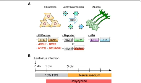
Recently, generation of induced neuronal (iN) cells was reported using mouse and human dermal cells [9,14]. In the present study, we used a set of neuronal transcrip-tion factors for human iN cells on common marmoset embryonic skin fibroblasts (cjF) isolated from embryonic day 91 (E91) embryos to determine whether common marmoset somatic cells could be converted into neur-onal cells. We first confirmed transgene expression in the mouse fibroblast cell line, NIH3T3, by infecting these cells with lentiviral vectors coding the neuronal transcription factors: ASCL1, BRN2, MYT1L, and
NEU-ROD1 [14], under the control of tetracycline response element (TRE) together with reverse tetracycline transacti-vator (rtTA)-expressing vector. Upregulation of transgenes in NIH3T3 cells after doxycycline (dox) treatment was confirmed by immunocytochemistry (Additional file 1).
Generation of neuron-like cells from common marmoset somatic cells
[7,19,20]. Therefore, the induction conditions of cjiN cells may be optimized in the future study to enhance the con-version efficiency. Nevertheless, our results show that the four-factor set of neuronal transcription factors is suffi-cient to convert cjFs to cjiN cells.
The duration of dox treatment was critical for neuronal conversion
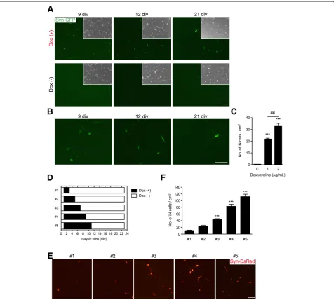
Next, we investigated the required duration of exogenous neuronal transcription factor expression to drive neuronal transdifferentiation. Therefore, we examined the exposure time of dox and the cjiN induction efficiency (Figure 2D). Treatment with dox from 1–3 div promoted the conver-sion of cjFs into synapsin reporter-positive cells with neur-onal morphology (Figure 2E). Similarly, a previous study has shown that the endogenous neuronal transcriptional factor network is activated 48 h after dox treatment [14]. Our findings also revealed that the induction efficiency at 23 div depended on the exposure time of dox (Figure 2F). The treatment of cjFs with dox from 1–11 div followed by
a culture without dox treatment from 11–23 div generated 112.1 ± 28.1 cjiN cells/cm2(Figure 2F). However, cells cul-tured with dox from 1–3 div followed by a culture without dox treatment from 3–23 div generated only 10.6 ± 5.4 cjiN cells/cm2 (Figure 2F). The efficiency of the former group was significantly (P***< 0.001, n = 4) higher (more than 10-fold) compared with the latter group (Figure 2F). These results indicate that a longer expression of exogen-ous neuronal transcriptional factors is required for effi-cient lineage conversion of somatic cells.
cjiN cells expressed a subset of neuronal genes and proteins
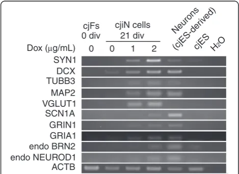
To characterize cjiN cells as neuronal cells, we per-formed reverse transcription-polymerase chain reaction (RT-PCR) analysis using bulk RNA samples, including the remaining synapsin reporter-negative cells to explore the expression of neuronal genes (see Table 1 for the pri-mer sets used). We detected an upregulation of neuronal genes (Figure 3), which included cytoskeletal markers
(MAP2,DCXandTUBB3), synaptic vesicle markers (SYN1 and VGLUT1), and cation channel-related genes (SCN1A,
GRIN1andGRIA1) in cjiN cells at 21 div (Figure 3). How-ever, expression of these neuronal genes was not detected in cjFs at 0 div (Figure 3). These results were in line with the concentration-dependent induction efficiency of dox
in Figure 2C. We also detected endogenous expression of
BRN2andNEUROD1in cjiN cells at 21 div (Figure 3), in-dicating that ectopic expression of neuronal transcription factors activated the endogenous neuronal program. This effect may have caused neuronal transdifferentiation from somatic cells [9,14,21]. Thus, ectopic neuronal differentiation
Figure 2Generation of iN cells. (A, B)Transgene-dependent conversion of common marmoset fibroblasts into neuronal cells, in which synapsin reporter activation and morphological changes were dependent on the exogenous transcription factor and time. Cells became synapsin reporter-positive accompanied by a gradual morphological changes into neuronal cells, both effects of which were not found in cells cultured in neural medium without dox at 21 div.(B)Magnified images of Figure 1A showing synapsin reporter-positive cells with morphological changes from fibroblasts to neuronal cells.(C)Counts of synapsin reporter-positive cells with neuronal morphology showed a dox-dependent increase in their number (positive cells with fibroblast morphology were excluded from the counts).P***< 0.001,P##< 0.01 (one-way ANOVA followed by
Tukey’s test).(D-F)The effect of sustained dox treatment on the production of cjiN cells.(D)Cells were treated with dox at 1 div until 3, 5, 7, 9 or 11 div and then maintained without dox until 23 div.(E)Synapsin reporter-positive cells with neuronal morphology were observed in all treatment group.(F)Cell counts revealed that a longer treatment time of dox increased the number of cjiN cells. Although dox treatment from 1–3 div sufficiently promoted cjiNs, a longer treatment time increased their number.P***< 0.001 (one-way ANOVA followed by Tukey’s test).
signals are likely to work together with the endogenous neuronal program to efficiently convert non-neuronal cells into neuronal cells [21].
To further characterize the property of cjiN cells, we per-formed immunocytochemistry for the neuronal markers. The results showed that most synapsin reporter-positive cells also expressed (>88%) the pan-neuronal marker, microtubule-associated protein 2 (MAP2) (data not shown). This result indicated that the synapsin reporter using the sequence of human synapsin I promoter was also func-tional in common marmoset cells, and was thus a reliable reporter for neuronal conversion, as previously reported [15,16]. In cjiN cells at 43 div, MAP2-negative cells were found to be negative for glial fibrillary acidic protein and only weakly positive forα-smooth muscle actin (data not shown), raising the possibility that they were partially reprogrammed cells. The MAP2- and synapsin reporter-positive cells also showed immunoreactivity for the synaptic
vesicle marker, synaptophysin, at 38 div (Figure 4A). How-ever, immunostaining for synaptophysin and PSD95 re-vealed that these cells were unlikely to make synapse structures at 38 div (data not shown). Although the cjiN cells expressed neuronal marker genes and proteins, they did not appear to be mature enough to make syn-aptic contacts themselves. However, a future study may facilitate synaptic formation by improving induction ef-ficiency and by co-culturing with astrocytes [9,14].
The majority of cjiN cells were glutamatergic
To characterize the neurotransmitter phenotype of iN cells, we examined the expression of neurotransmitters in MAP2-positive cjiN cells. Immunostaining at 38 div for vesicular glutamate transporter 1 (vGlut1) and gamma aminobutyric acid (GABA) revealed the presence of both excitatory glutamatergic and inhibitory GABAergic neuronal cells, respectively (Figure 4B). Counts of vGlut1 and GABA-positive cjiN cells showed a signifi-cantly (P***< 0.001, n = 4) greater number of vGlut1-positive cells (39.2 ± 1.9/cm2) than GABA-positive cells (16.4 ± 2.8 cells/cm2) (Figure 4C). These results are in accordance with previous studies showing that the ma-jority of iN cells are excitatory cells [7,14,22]. Our find-ings thus indicate that iN induction may be feasible in the future for the in vitro analysis of the transgenic common marmoset model of Alzheimer’s disease, in which forebrain excitatory neurons are expected to be affected. Moreover, our cjiN cell induction protocol is likely advantageous over the previously reported neur-onal differentiation protocol which used common mar-moset embryonic stem (ES) cells and iPS cells, because the ES/iPS cell-derived neural precursor cells showed caudal identity [13]. Thus far, several groups have suc-ceeded in generating reprogrammed neuronal cells with specific neuronal subtypes, such as dopaminergic neurons and motor neurons [23-26], which implicate the cell fate plasticity of terminally differentiated somatic cells.
Table 1 Primer sets used in RT-PCR analysis
Gene name Forward primer Reverse primer Product size (bp)
SYN1 acggagactaccgcagtttg cgatctgctccagcattgca 459
DCX ctgtgcgtgtgcttctgaac tcagctggagacttgcttcg 346
TUBB3 catagaccccagtggcaactacg caccctccgtgtagtggcccttgg 241
MAP2 cctgtgttaagcggaaaacc agagactttgtcctttgcctgt 86
VGLUT1 tcaataacagcacgacccac tcccggaatttgagtgacaatg 131
SCN1A attggcaattccgtgggagc cccacacagcacgcggaaca 204
GRIN1 accccaagatcgtcaacatcg ggctaaccagaatggcgtaga 213
GRIA1 cgagctttcctgttgatacat tctgccacttgtaatggtcgatg 99
endo BRN2 aattaaggaaaaaggaaagcaact caaaacatcattacacctgct 71
endo NEUROD1 gttattgtgttgccttagcacttc agtgaaatgaattgctcaaattgt 77
ACTB ggcatccacgaaactaccttt acactgagtacttgcgctcg 202
Our success in reprogramming common marmoset som-atic cells into excitatory and inhibitory neuronal cells using defined iN factors may therefore provide great promise in the future for generating specific subtypes of neuronal cells with specific sets of neuronal transcription factors.
cjiN cells were functional as matured neurons
To further confirm the successful conversion of cjFs into functional neuronal cells, we performed calcium imaging analysis. cjiN cells cultured with dox were incubated with the calcium indicator, Fluo-4 AM [13], followed by response recordings. The intracellular calcium level ([Ca2+]i) in cjiN cells at 15 div was increased in cjiN cells
perfused with 80 mM of KCl, which was then decreased by washout (Additional file 2). Furthermore, electrical field stimulation on cjiN cells at 28 div increased [Ca2+]i,
which was reversibly blocked by the voltage-gated so-dium channel blocker, tetrodotoxin (0.2μM) (Figure 5), suggesting that the increase in [Ca2+]iwas likely evoked
by action potentials through voltage-gated sodium chan-nels. These results showed that cjiN cells derived from cjFs were functionally comparable to common marmoset ES cell-derived neuronal cells [13], and thus strongly sug-gest that reprogramming of common marmoset somatic
cells generates functional neuronal cells. Moreover, electrophysiological analyses of cjiN cells at 29–42 div revealed that action potentials were elicited by mem-brane depolarization in current-clamp mode in 17 out of 21 cjiN cells (81.0%) (Figure 6A and Additional file 3). Among these 17 cjiN cells, 7 cjiN cells (41.2%) generated a single action potential and the remaining 10 cjiN cells (58.8%) generated repetitive action potentials (Figure 6A and Additional file 3). In the voltage-clamp mode, both fast-activating and inactivating sodium currents and out-ward currents were observed (Figure 6B). The resting membrane potentials of cjiN cells ranged between -26 and -55 mV, with a mean ± SEM of -38.1 ± 2.0 mV (Figure 6C and Additional file 3). This mean value was sig-nificantly (P***< 0.001) lower than that of cjFs (-16.4 ± 0.6 mV) with a range between -13.1 and -19.0 mV (Figure 6C and Additional file 3). Overall, our patch clamp recordings showed that cjiN cells are functional neuronal cells.
cjiN cells were directly converted from cjFs without passing through a proliferative state
To determine whether cjiN cells were directly converted from cjFs without passing through a proliferative neural
Figure 5Increase in the intracellular calcium level upon electrical field stimulation. (A)cjiN cells (synapsin reporter-positive with a neuronal morphology).(B, C)Intracellular calcium level ([Ca2+]
i) in cjiN cells was measured by the intensity of Fluo-4 fluorescence. Electrical field stimulation
(40 Hz for 5 seconds) induces a robust elevation of [Ca2+]
iin some synapsin reporter-positive cells. The increase in [Ca2+]iwas reversibly suppressed by
progenitor-like cell state, 5-bromodeoxyuridine (BrdU; 10μM) was added to the media at 1, 3 or 5 div until 24 div (Figure 7A), and the percentage of double-positive cells for BrdU and MAP2 among the MAP2 single-positive cells was determined (Figure 7B, C). The result showed that while 85.3 ± 5.4% of MAP2-positive cells in-corporated BrdU when treated from 1–24 div, only 30.1 ± 9.0% (P**< 0.01) and 1.5 ± 1.5% (P***< 0.001) of MAP2-positive cells incorporated BrdU when treated from 3–24 and 5–24 div, respectively (Figure 7C). This result indi-cates that most of the cells that were destined to be cjiN cells became postmitotic at the early phase of the repro-gramming process, suggesting that cjiN induction is a dir-ect process unless cells pass through a proliferative neural
progenitor-like cell state, from which neuronal cells can be differentiated. In the present study, however, no Sox2-positive cells were found during 2–5 div, while MAP2-positive cells were present at 21 div (data not shown), indicating that the induction of neural progenitor-like cells is unlikely during 1-5 div. Therefore, these results show that cjiN cells are directly converted from cjFs without passing through proliferative neural progenitor cells.
Conclusions
In the present study, we established anin vitromethod to convert common marmoset somatic cells into functional neuronal (i.e. cjiN) cells. The majority of the cjiN cells were
Figure 6Neuronal electrophysiological property of cjiN cells. (A)cjiN cells elicited repetitive action potential by membrane depolarization in current-clamp mode.(B)cjiN cells showed both fast-activating and inactivating sodium currents and outward currents in voltage-clamp mode.
(C)cjiN cells exhibited significantly lower resting membrane potentials than cjFs.P***< 0.001 (Mann-WhitneyU-test).
Figure 7Cells destined to be cjiN cells became postmitotic at the early phase of the reprogramming process. (A)Time course for the BrdU incorporation experiment. BrdU (10μM) was added into the culture medium at 1, 3 or 5 div until cells were fixed at 24 div.(B, C)
vGlut1-positive excitatory neuronal cells and expressed the neuronal marker genes: TUBB3, DCX, MAP2, SYN1,
VGLUT1, SCN1A, GRIN1 and GRIA1. Importantly, cjF-derived cjiN cells exhibited functional neuronal properties and responded to exogenous stimulation. Overall, these findings suggest that direct conversion technology may be beneficial in rapid and robust screening of neuronal phenotypes of transgenic common marmoset models of human diseases and analyzing underlying molecular mechanisms of diseases.
Methods Animals
All animal experiments were approved by the Institu-tional Animal Care and Use Committee of the Central Institute for Experimental Animals (CIEA), and was per-formed in accordance with CIEA and Keio University guidelines.
Cell culture
Common marmoset embryonic fibroblasts, NIH3T3 cells and 293T cells were cultured in Dulbecco’s modified Eagle’s medium supplemented with 10% fetal bovine serum, 100 U/mL of penicillin and 100μg/mL of strepto-mycin (10% FP medium) at 37°C with 5% CO2incubation. Molecular cloning and lentivirus production
cDNA entry clone of humanASCL1[GenBank: NM_004316] was purchased from DNAFORM (clone ID: 100006383, Japan). cDNAs of human BRN2 [GenBank: NM_0056 04.3], MYT1L[GenBank: NM_015025.2] and NEUROD1 [GenBank: NM_002500.4] were cloned into pENTR-D-TOPO vector (Invitrogen, USA). Then cDNAs were inserted into a self-inactivation human immunodeficiency virus-1-based lentivirus construct, CSIV-TRE-RfA (CSIV-TRE-RfA-CMV-KT was kindly provided by Dr. Hiroyuki Miyoshi (RIKEN BRC, Japan), and then modified by Dr. Takuji Maeda (Nagoya University, Japan)), by LR reaction (Invitrogen, USA). Similarly, reverse tetracycline trans-activator (rtTA) gene was inserted into CSII-EF1α -RfA-TK-HygR construct [27]. The human synapsin I reporter constructs, pCSC-hSynI-GFP [16] and pHIV7-hSynI-DsRed [18], were kindly provided by Dr. Fred H. Gage, Salk Institute, USA, and Dr. Alysson R. Muotri, University of California, USA, respectively. Besides, CSIV-hSynI-GFP-IRES2-NeoR and CSIV-hSynI-DsRed-CSIV-hSynI-GFP-IRES2-NeoR were constructed in-house. These reporters were constructed using CSIV-TRE-RfA-CMV-KT, pCSC-hSynI-GFP, pHIV7-hSynI-DsRed and pIRESneo3 (Clontech, USA) with PCR-and restriction enzyme-based method. For lentivirus production, 293T cells were transfected with lentivirus plas-mid, pCAG-HIVgp and pCMV-VSV-G-RSV-Rev [28] (kindly provided by Dr. Hiroyuki Miyoshi, RIKEN BRC, Japan). After 16-20 h, supernatant was replaced by fresh
media followed by 48-72 h incubation. The virus containing media were then collected and 0.45μm-filterated followed by ultracentrifugation. The concentrated virus was sus-pended in PBS and used in subsequent experiments.
Induction of common marmoset iN cells
Common marmoset embryonic fibroblasts were seeded directly on culture ware at 1 × 10⁴cells/cm2. Twenty-four hours later, the cells were infected with lentivirus in 10% FP media containing polybrene (8μg/mL) (Sigma-Aldrich, USA). After 16–20 h in media containing lentivirus, the cells were switched into fresh 10% FP medium containing doxycycline (dox) (2 μg/mL) to drive transgene expres-sion. Procedures for experiments determining the suffi-cient concentration and duration of dox are shown in the main text. After 48 h in 10% FP media with dox, the media was replaced with dox-containing neural media composed of N2B27 media [20], brain-derived neuro-trophic factor (BDNF) (10 ng/mL, R&D systems, USA) and neurotrophin-3 (NT-3) (10 ng/mL, R&D systems, USA). For calcium imaging, 8-(4-Chlorophenylthio) ad-enosine 3′, 5′-cyclic monophosphate (8-CPT, 100 μM, Sigma-Aldrich, USA), one of the cAMP analogs [29], was also supplemented to promote neuronal matur-ation. The media was changed every 2–3 days during culture period. BrdU incorporation assay was performed as previously described [9] incubating cells with BrdU (10μΜ, BD, USA) until cells were fixed.
Immunocytochemistry
37°C and then rinsed with PBS three times before primary antibody incubation.
RNA isolation and reverse transcription-polymerase chain reaction (RT-PCR)
Total RNA was isolated with RNeasy Micro Kit with DNase I treatment (QIAGEN, Germany) and was used to synthesize cDNA with ReverTraAce qPCR RT Kit (TOYOBO, Japan) according to the manufacturer’s in-struction. RT-PCR was conducted using Ex Taq HS (TAKARA, Japan) according to the manufacturer’s instruc-tion. Common marmoset ES (cjES) cells and cjES-derived neurons were used as control [13]. The primer sets used are listed in Table 1.
Calcium imaging and electrical stimulation
Calcium imaging analyses were performed as described previously [13]. To load the calcium imaging dye, cells were incubated with 1μM Fluo-4 AM (Invitrogen, USA) in imaging solution consisting of 117 mM NaCl, 2.5 mM KCl, 2 mM CaCl2, 2 mM MgSO4, 25 mM HEPES and
30 mM D-(+)-glucose, (pH 7.4), at 37°C for 20 minutes, followed by washing for 30 minutes in imaging solution. Coverslips were placed on a custom-made field stimula-tion chamber and mounted on the stage of a Nikon Eclipse microscope with a 20× (NA 0.45) objective. Cells were perfused at 2 ml/minute with the imaging solution at room temperature with or without 0.2μM tetrodo-toxin (TTX; Alomone Labs Ltd., Israel). Images were acquired at 2 Hz (500 millisecond exposure time) with a cooled CCD camera (Andor iXon, DU897). Extracel-lular field stimulation was performed with two parallel platinum wires at 25 V/cm. Each stimulation was a train of 500 microsecond pulses at 40 Hz for 5 seconds. Images were analyzed with ImageJ software (NIH, Bethesda, MD).
Electrophysiology
Electrophysiological recordings were performed as de-scribed previously [30]. Synapsin reporter-positive cjiN cells were identified under an inverted microscope (Dia-phot-TMD 200; Nikon, Japan) and whole cell patch clamp recordings were done using Axopatch 200B (Axon Instru-ments, USA) at room temperature. The extracellular solu-tion composed of 117 mM NaCl, 2.5 mM KCl, 2 mM CaCl2, 2 mM MgCl2, 15 mM D-Glucose and 20 mM
HEPES (pH 7.4 adjusted with NaOH, 304 mOsm) was continuously perfused during recordings. Patch pipettes had a resistance of 5-6 MΩfilled with the intracellular so-lution containing 130 mM K-gluconate, 1 mM CaCl2,
1 mM MgCl2, 10 mM EGTA, 10 mM Sucrose and 20 mM
HEPES (pH adjusted with KOH, 305 mOsm). In voltage-clamp recordings, iN cells were held at -70 mV and voltage steps (10 mV, 300 msec) were applied to elicit
voltage-activated currents. Action potentials were evoked by injecting step currents (20-40 pA, 500 msec) in the current-clamp mode. Data were digitized at 10 kHz with a 2 kHz low-pass filter. Liquid junction potential was corrected.
Statistical analysis
All data were expressed as means ± SEM. The statistical significance of differences was analyzed by Student’st-test, Mann-Whitney U-test or one-way ANOVA followed by Tukey’s test using Graph Pad Prism5 software. Differences ofP< 0.05 were considered statistically significant.
Additional files
Additional file 1:Doxycycline-dependent transgene induction in NIH3T3 cells.Immunocytochemistry against Ascl1, Brn2, Myt1L and NeuroD1 in a mouse fibroblast cell line, NIH3T3 cells, that were lentivirally transduced with iN factors and treated with doxycycline from 1–4 div revealed doxycycline-dependent transgene expressions. Scale bar; 100μm.
Additional file 2:KCl perfusion increased the intracellular calcium level. (A)cjiN cells (synapsin reporter-positive with a neuronal morphology).(B, C)Intracellular calcium level ([Ca2+]
i) in cjiN cells was
measured by the intensity of Fluo-4 fluorescence. KCl (80 mM) perfusion caused a robust elevation of [Ca2+]
iin some synapsin reporter-positive
cells. This increase was reversibly suppressed by washout. Scale bar; 50μm.
Additional file 3:Electrophysiological parameters in cjiN cells at 29–42 div.
Abbreviations
cjiN cells:Common marmoset induced neuronal cells; cjF: Common marmoset embryonic fibroblast; RT-PCR: Reverse transcription-polymerase chain reaction; dox: Doxycycline; rtTA: Reverse tetracycline transactivator.
Competing interests
H.O. is a paid scientific consultant to San Bio, Inc., Eisai Co., Ltd., and Daiichi Sankyo Co., Ltd.
Authors’contributions
ZZ conceived the concept of this study, designed the experiments, performed experiments and analyzed data. KI, KK and MY performed calcium imaging assays and electrophysiology, and ZZ, KI, KK and MY analyzed data. JK, WA, HJO and HO coordinated the study. ES prepared and provided common marmoset embryonic fibroblasts. HO provided financial support for the experiments. ZZ, JK and HO wrote the paper. All authors read and approved the final manuscript.
Acknowledgements
Author details 1
Department of Physiology, Keio University School of Medicine, 35 Shinanomachi, Shinjuku-ku, Tokyo 160-8582, Japan.2Division of Regenerative
Medicine, Jikei University School of Medicine, 3-25-8 Nishi-Shinbashi, Minato-ku, Tokyo 105-8461, Japan.3Center of Applied Developmental
Biology, Central Institute for Experimental Animals, 3-25-12 Tonomachi, Kawasaki-ku, Kawasaki, Kanagawa 210-0821, Japan.
Received: 12 December 2013 Accepted: 28 March 2014 Published: 3 April 2014
References
1. Mansfield K:Marmoset models commonly used in biomedical research.
Comp Med2003,53:383–392.
2. Gotz J, Ittner LM:Animal models of Alzheimer’s disease and frontotemporal dementia.Nat Rev Neurosci2008,9:532–544. 3. Lim K-L, Ng C-H:Genetic models of Parkinson disease.Biochim Biophys
Acta2009,1792:604–615.
4. Sasaki E, Suemizu H, Shimada A, Hanazawa K, Oiwa R, Kamioka M, Tomioka I, Sotomaru Y, Hirakawa R, Eto T, Shiozawa S, Maeda T, Ito M, Ito R, Kito C, Yagihashi C, Kawai K, Miyoshi H, Tanioka Y, Tamaoki N, Habu S, Okano H, Nomura T:Generation of transgenic non-human primates with germline transmission.Nature2009,459:523–527.
5. Nishijima K, Saitoh R, Tanaka S, Ohsato-Suzuki M, Ohno T, Kitajima S:Life span of common marmoset (Callithrix jacchus) at CLEA Japan breeding colony.Biogerontology2012,13:439–443.
6. Imaizumi Y, Okada Y, Akamatsu W, Koike M, Kuzumaki N, Hayakawa H, Nihira T, Kobayashi T, Ohyama M, Sato S, Takanashi M, Funayama M, Hirayama A, Soga T, Hishiki T, Suematsu M, Yagi T, Ito D, Kosakai A, Hayashi K, Shouji M, Nakanishi A, Suzuki N, Mizuno Y, Mizushima N, Amagai M, Uchiyama Y, Mochizuki H, Hattori N, Okano H:Mitochondrial dysfunction associated with increased oxidative stress andα-synuclein accumulation in PARK2 iPSC-derived neurons and postmortem brain tissue.Mol Brain2012,5:35. 7. Qiang L, Fujita R, Yamashita T, Angulo S, Rhinn H, Rhee D, Doege C, Chau L,
Aubry L, Vanti WB, Moreno H, Abeliovich A:Directed Conversion of Alzheimer’s Disease Patient Skin Fibroblasts into Functional Neurons.
Cell2011,146:359–371.
8. Takahashi K, Tanabe K, Ohnuki M, Narita M, Ichisaka T, Tomoda K, Yamanaka S:
Induction of pluripotent stem cells from adult human fibroblasts by defined factors.Cell2007,131:861–872.
9. Vierbuchen T, Ostermeier A, Pang ZP, Kokubu Y, Südhof TC, Wernig M:
Direct conversion of fibroblasts to functional neurons by defined factors.
Nature2010,463:1035–1041.
10. Wu Y, Zhang Y, Mishra A, Tardif SD, Hornsby PJ:Generation of induced pluripotent stem cells from newborn marmoset skin fibroblasts.Stem Cell Res2010,4:180–188.
11. Tomioka I, Maeda T, Shimada H, Kawai K, Okada Y, Igarashi H, Oiwa R, Iwasaki T, Aoki M, Kimura T, Shiozawa S, Shinohara H, Suemizu H, Sasaki E, Okano H:Generating induced pluripotent stem cells from common marmoset (Callithrix jacchus) fetal liver cells using defined factors, including Lin28.Genes Cells2010,15:959–969.
12. Wiedemann A, Hemmer K, Bernemann I, Göhring G, Pogozhykh O, Figueiredo C, Glage S, Schambach A, Schwamborn JC, Blasczyk R, Müller T:
Induced pluripotent stem cells generated from adult bone marrow-derived cells of the nonhuman primate (Callithrix jacchus) using a novel quad-cistronic and excisable lentiviral vector.Cell Reprogram2012,
14:485–496.
13. Shimada H, Okada Y, Ibata K, Ebise H, Ota S-I, Tomioka I, Nomura T, Maeda T, Kohda K, Yuzaki M, Sasaki E, Nakamura M, Okano H:Efficient Derivation of Multipotent Neural Stem/Progenitor Cells from Non-Human Primate Embryonic Stem Cells.PLoS One2012,7:e49469.
14. Pang ZP, Yang N, Vierbuchen T, Ostermeier A, Fuentes DR, Yang TQ, Citri A, Sebastiano V, Marro S, Südhof TC, Wernig M:Induction of human neuronal cells by defined transcription factors.Nature2011,476:220–223. 15. Adler AF, Grigsby CL, Kulangara K, Wang H, Yasuda R, Leong KW:Nonviral
direct conversion of primary mouse embryonic fibroblasts to neuronal cells.Mol Ther Nucleic Acids2012,1:e32.
16. Kim J-E, O'Sullivan ML, Sanchez CA, Hwang M, Israel MA, Brennand K, Deerinck TJ, Goldstein LSB, Gage FH, Ellisman MH, Ghosh A:Investigating synapse formation and function using human pluripotent stem cell-derived neurons.Proc Natl Acad Sci U S A2011,108:3005–3010.
17. Kucharova K, Hefferan MP, Patel P, Marsala S, Nejime T, Miyanohara A, Marsala M, Drummond JC:Transplantation of rat synapsin-EGFP-labeled embryonic neurons into the intact and ischemic CA1 hippocampal region: distribution, phenotype, and axodendritic sprouting.Cell Transplant2011,
20:1163–1178.
18. Marchetto MCN, Carromeu C, Acab A, Yu D, Yeo GW, Mu Y, Chen G, Gage FH, Muotri AR:A model for neural development and treatment of Rett syndrome using human induced pluripotent stem cells.Cell2010,
143:527–539.
19. Yoo AS, Sun AX, Li L, Shcheglovitov A, Portmann T, Li Y, Lee-Messer C, Dolmetsch RE, Tsien RW, Crabtree GR:MicroRNA-mediated conversion of human fibroblasts to neurons.Nature2011,476(7359):228–231. 20. Ladewig J, Mertens J, Kesavan J, Doerr J, Poppe D, Glaue F, Herms S, Wernet P,
Kögler G, Müller F-J, Koch P, Brüstle O:Small molecules enable highly efficient neuronal conversion of human fibroblasts.Nat Methods2012,9:575–578. 21. Wapinski OL, Vierbuchen T, Qu K, Lee QY, Chanda S, Fuentes DR, Giresi PG,
Ng YH, Marro S, Neff NF, Drechsel D, Martynoga B, Castro DS, Webb AE, Südhof TC, Brunet A, Guillemot F, Chang HY, Wernig M:Hierarchical mechanisms for direct reprogramming of fibroblasts to neurons.
Cell2013,155:621–635.
22. Yang N, Ng YH, Pang ZP, Südhof TC, Wernig M:Induced neuronal cells: how to make and define a neuron.Cell Stem Cell2011,9:517–525. 23. Pfisterer U, Kirkeby A, Torper O, Wood J, Nelander J, Dufour A, Björklund A,
Lindvall O, Jakobsson J, Parmar M:Direct conversion of human fibroblasts to dopaminergic neurons.Proc Natl Acad Sci U S A2011,108:10343–10348. 24. Caiazzo M, Dell’Anno MT, Dvoretskova E, Lazarevic D, Taverna S, Leo D,
Sotnikova TD, Menegon A, Roncaglia P, Colciago G, Russo G, Carninci P, Pezzoli G, Gainetdinov RR, Gustincich S, Dityatev A, Broccoli V:Direct generation of functional dopaminergic neurons from mouse and human fibroblasts.Nature2011,476(7359):224–227.
25. Kim J, Su SC, Wang H, Cheng AW, Cassady JP, Lodato MA, Lengner CJ, Chung C-Y, Dawlaty MM, Tsai L-H, Jaenisch R:Functional integration of dopaminergic neurons directly converted from mouse fibroblasts.Cell Stem Cell2011,9(5):413–419.
26. Son EY, Ichida JK, Wainger BJ, Toma JS, Rafuse VF, Woolf CJ, Eggan K:
Conversion of mouse and human fibroblasts into functional spinal motor neurons.Cell Stem Cell2011,9:205–218.
27. Naka H, Nakamura S, Shimazaki T, Okano H:Requirement for COUP-TFI and II in the temporal specification of neural stem cells in CNS development.
Nat Neurosci2008,11:1014–1023.
28. Miyoshi H, Blömer U, Takahashi M, Gage FH, Verma IM:Development of a self-inactivating lentivirus vector.J Virol1998,72:8150–8157.
29. Matsui T, Takano M, Yoshida K, Ono S, Fujisaki C, Matsuzaki Y, Toyama Y, Nakamura M, Okano H, Akamatsu W:Neural stem cells directly differentiated from partially reprogrammed fibroblasts rapidly acquire gliogenic competency.Stem Cells2012,30:1109–1119.
30. Tsuji O, Miura K, Okada Y, Fujiyoshi K, Mukaino M, Nagoshi N, Kitamura K, Kumagai G, Nishino M, Tomisato S, Higashi H, Nagai T, Katoh H, Kohda K, Matsuzaki Y, Yuzaki M, Ikeda E, Toyama Y, Nakamura M, Yamanaka S, Okano H:
Therapeutic potential of appropriately evaluated safe-induced pluripotent stem cells for spinal cord injury.Proc Natl Acad Sci U S A
2010,107:12704–12709.
doi:10.1186/1756-6606-7-24
Cite this article as:Zhouet al.:Reprogramming non-human primate