1
Neuroanatomical Correlates of Cognitive Dysfunction in
Obstructive Sleep Apnoea
Thesis submitted to Imperial College, London for the degree of Doctor of
Philosophy in the Faculty of Science
Martin Glasser, MBBS
CID: 00234721
Clinical and Academic Unit of Sleep and Breathing
National Heart and Lung Institute, Imperial College London
Royal Brompton Hospital, Fulham Road, London
A Thesis submitted for the degree of Doctor of Philosophy
January 2016
2
Copyright Declaration
The copyright of this thesis rests with the author and is made available under a Creative Commons Attribution Non-Commercial No Derivatives licence. Researchers are free to copy, distribute or transmit the thesis on the condition that they attribute it, that they do not use it for commercial purposes and that they do not alter, transform or build upon it. For any reuse or redistribution, researchers must make clear to others the licence terms of this work
3
Declaration of Originality
I, Martin Glasser, hereby declare that this thesis contains the results of my own work except where otherwise acknowledged. The studies presented in this thesis were conceived and designed with the assistance of my supervisors, Professor Morrell and Professor Simonds and Dr Ivana Rosenzweig. In addition, I acknowledge the invaluable assistance of Dr Alison McMillian who randomised the patients in Study 2 (Chapter 4) and collected the sleep data in Study 3 (Chapter 5). Peter Drivas set up the scanning sequences for Study 2 (Chapter 4) on the Royal Brompton scanners and captured the images. The physicists, radiographers and radiologists at Charing Cross Hospital were similarly involved in Studies 1 and 3 (Chapter 3 and 5).
The MRI scans for Study 1 (Chapter 3) had already been collected prior to my re-analsyis in the study. Statistical support was received from Dr Milan Milosovic for studies 1 and 2 (Chapter 3 and 4). Chi Cheng Tsai assisted in the statistical analysis of study 3 (Chapter 5).
Information derived from the work of others and discussed in this thesis is referenced in the text and listed in the bibliography.
The results of studies contained within this thesis have previously been presented. A list of publications arising from this work follows.
4
PUBLICATIONS ARISING FROM THIS THESIS
Published Original Research Papers, Reviews and Editorials
M. Glasser, N. Bailey, A. McMillan, E. Goff, M.J. Morrell, Sleep apnoea in older people. Breathe 2011; 7: 248-256 (Review)
Morrell MJ, Glasser M. The brain in sleep-disordered breathing: a vote for the chicken? Am J Respir Crit Care Med. 2011 May 15;183(10):1292-4. (Editorial)
Morrell MJ, Glasser M, McMillan A, Rosenzweig I. CPAP for the treatment of cognitive dysfunction in obstructive sleep apnoea. Neuropsychiatry News. 2012 Winter.
Rosenzweig I, Kempton MJ, Crum WR, Glasser M, Milosevic M, Beniczky S, Corfield DR, Williams SC, Morrell MJ. Hippocampal hypertrophy and sleep apnea: a role for the ischemic preconditioning? PLoS One. 2013 Dec 13;8(12):e83173
Rosenzweig I, Glasser M, Polsek D, Leschziner GD, Williams SC, Morrell MJ. Sleep apnoea and the brain: a complex relationship. Lancet Respir Med. 2015 May;3(5):404-14. (Review)
Rosenzweig I, Glasser M, Crum WR, Kempton MJ, Milosevic M, McMillan A, Leschziner GD, Kumari V, Goadsby P, Simonds AK, Williams SC, Morrell MJ. Changes in Neurocognitive Architecture in Patients with Obstructive Sleep Apnea Treated with Continuous Positive Airway Pressure. EBioMedicine. 2016 May;7:221-9. doi: 10.1016/j.ebiom.2016.03.020. Epub 2016 Mar 25
Abstracts
Glasser M, Rosenzweig I, McMillan A, Drivas P, Satkunam K, Man WDC, Simonds AK, Morrell MJ. Neuroanatomical Correlates Of Cognitive Dysfunction In Obstructive Sleep Apnoea: An Ongoing Study. A109. Sleep disordered breathing: Cardiovascular, metabolic and Neurocognitive outcomes. May 1, 2013, A2319-A2319. First published online May 09, 2013 as doi:10.1164/ajrccm-conference.2013.187.1_MeetingAbstracts.A2319
Other publications completed during the Thesis
Carlisle T, Carthy ER, Glasser M, Drivas P, McMillan A, Cowie MR, Simonds AK, Morrell MJ Upper airway factors that protect against obstructive sleep apnoea in healthy older males. . Eur Respir J. 2014 Sep;44(3):685-93
5
Prizes
NIHR BRU/BRC Experimental Training Camp 2011, MPHRP Project Winner American Thoracic Society Abstract Scholarship Award, 2013
6
Abstract
Obstructive sleep apnoea (OSA) has been reported to be associated with brain hypotrophy and cognitive dysfunction; however, whether these normalise after treatment is unclear. The overall aim of this thesis is to investigate the relationship between OSA and brain structure using FreeSurfer (a new automated technique that reliably measures brain structures). I have investigated changes in brain morphology and the newly described phenomenon in OSA of ischaemic preconditioning. Chapters 4 and 5 will also assess brain structural response to CPAP, and investigate the association between brain structure and cognitive function in OSA.
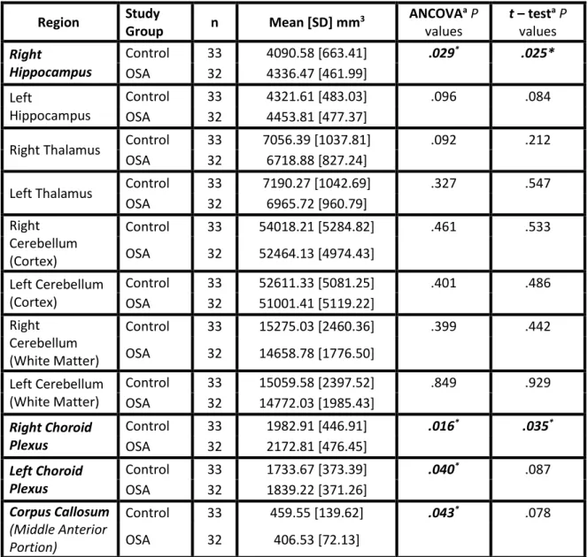
Chapter 3 reports an observational study investigating brain structure. FreeSurfer analysis of magnetic resonance imaging (MRI) found OSA patients had hypertrophy in the right hippocampus (p=0.03) and right choroid plexus (p=0.02) but hypotrophy of the corpus callosum (p=0.04) compared to healthy controls.
Chapter 4 reports a randomised controlled trial of CPAP in OSA. At baseline hypotrophy was seen in the corpus callosum (p=0.03) and pallidum (p=0.03) of OSA patients compared to healthy controls. Hypertrophic changes in the right thalamus were seen in the CPAP group after 1 month (p=0.06), associated with improvement in verbal memory (p=0.04).
Chapter 5 reports a randomised controlled trial of CPAP in older patients with OSA. A significant decrease in left fimbria volume was seen in the CPAP group (p=0.01). A significant increase in the left presubiculum volume was seen in the best supportive care group (p=0.03). No hippocampal hypertrophy was seen in the CPAP group.
In summary, young and middle-aged OSA patients had evidence of brain hypotrophy, but also areas of hypertrophy that may signify dendritic sprouting and increased connectivity as a result of ischaemic preconditioning. This allows recovery of brain hypotrophy after CPAP treatment. This was not seen in older OSA patients suggesting an age-related difference which may have implications for OSA treatment in older people.
7
Acknowledgements
I would like to thank my PhD supervisors, Professor Mary Morrell and Professor Anita Simonds as well as my honorary third supervisor Ivana Rosenzweig for their invaluable support and assistance. I know I haven’t always made it easy for you, but you have made it as easy as it could be for me.
I am also grateful for the support and knowledge of Professor Mike Polkey and Dr Matt Hind and all the staff of the Sleep and Ventilation Department at The Royal Brompton Hospital.
I am grateful to Dr Will Man and Dr Karnan Satkunam for their help with recruiting patients from Harefield Hospital and Queen Elizabeth Hospital Woolwich respectively.
Nia Voase and the staff at the Respiratory BRU were always happy to lend a hand and made setting up a new lab an enjoyable experience. Charlotte Goward and Lyn Bingham also inspired me to take up running and became my race partners. Sometimes on a cold, wet night I hate you for it, but I’d never run 13 miles without stopping before I met you, so thank you both.
My colleagues in the sleep lab Alison McMillan, Tom Carlisle, Lydia Pannicia, Julia Kelly, Neil Ward, Zarrin Sheikh, Annette Woods, Glenn McGuire, Jay Jaye and Agela Atalla were all a pleasure to work with, and my office mates Michelle Chatwin and Adam Rochester made being at work fun.
I especially wish to express my gratitude to all of the patients who agreed to take part in my study. I would like to acknowledge the financial support of NIHR Respiratory Disease Biomedical Research Unit at the Royal Brompton and Harefield NHS Foundation Trust who provided a grant to fund this research.
My parents have always encouraged my search for knowledge and professional qualifications and this is perhaps the pinnacle of that quest. Their support, financial, emotional and with childcare has been limitless and I hope I can be as good a role model to my children as you both continue to be to me. Azriella, Tsofia and Ashira no matter how difficult a day I am having you always make me smile. I love you more than you will ever know and you make me so proud.
Finally, I would like to dedicate this thesis to my wife and best friend, Lucinda. You are everything I wish I could be; abundantly kind, excessively giving, and meticulously organised. You have been my bedrock throughout this whole process and I could not have done it without you.
8
Table of contents
Section TitleDeclaration of Originality
Publications arising from this Thesis Abstract Acknowledgements Table of contents Abbreviations Chapter 1 – Introduction 1.1 Overview
1.2 Obstructive sleep apnoea 1.2.1 The aetiology of OSA 1.2.2 The prevalence of OSA 1.2.3 The sequelae of OSA 1.2.4 The treatment of OSA
1.3 Cognitive function and obstructive sleep apnoea 1.3.1 Impaired cognitive function in OSA
1.3.2 Treatment of cognitive function in OSA
1.3.3 Sleep deprivation and cognitive function in OSA 1.4 Ischaemic conditions and neuro-inflammation in OSA 1.4.1 Ischaemic preconditioning
1.4.2 Neuro-inflammation
1.5 Description and structure of thesis
Chapter 2 – General Methods
2.1 Participant and laboratory information 2.2 Nocturnal polysomnography
2.2.1 Assessment of sleep: EEG, EOG and EMG
2.2.2 Measurement of airflow using nasal pressure transducer
2.2.3 Measurement of respiratory effort using respiratory inductance plethysmorgraphy 2.2.4 Measurement of arterial oxygen saturation using pulse oximetry
2.2.5 Measurement of heart rate using ECG 2.2.6 Measurement of leg movement using EMG 2.3 Assessment of sleepiness
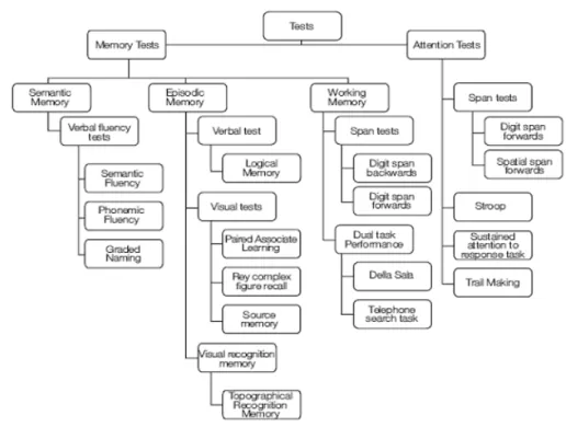
2.3.1 Epworth Sleepiness Scale 2.4 Assessment of cognitive function 2.4.1 Logical memory
2.4.2 Trail making 2.4.3 Spatial span 2.4.4 Digit span 2.4.5 ACER
2.5 Assessment of brain structure 2.5.1 Magnetic resonance imaging 2.5.2 Images analysis
2.6 Statistical analysis
Chapter 3 – The association between OSA and hippocampal hypertrophy
3.1 Summary 3.2 Introduction
3.2.1 Neurogenesis and the role of intermittent hypoxia in OSA 3.3 Methods
3.3.1 Measurements and analysis
9
3.3.3 Statistical testing 3.4 Results
3.5 Discussion
3.5.1 Hypertrophic changes and relation to previous studies 3.5.2 Automated FS analysis
3.5.3 Hypotrophic changes
3.5.4 Correlations with cerebellar cortex volumes 3.5.5 Limitations
3.5.6 Conclusions Appendix
Chapter 4 - Changes in neurocognitive function and brain morphology in OSA patients treated with CPAP
4.1 Summary 4.2 Introduction 4.3 Methods
4.3.1 Participants and design 4.3.2 Measurements and analysis 4.3.3 Statistical analyses
4.4 Results
4.4.1 Changes in brain morphology 4.4.2 Changes in cognitive function 4.4.3 Post hoc (secondary) analyses 4.5 Discussion
4.5.1 Excessive daytime somnolence 4.5.2 Thalamocortical Circuitry in OSA
4.5.3 Neurocognitive architecture of working and episodic memory in OSA 4.5.4 Limitations
4.5.5 Conclusions
Chapter 5 – Changes in brain morphology in older people with OSA following CPAP treatment
5.1 Summary 5.2 Introduction
5.2.1 The aging population
5.2.2 The prevalence of obstructive sleep apnoea in older people 5.2.3 Aetiology of sleep apnoea in older people
5.2.4 Cognitive function in older people with OSA 5.2.5 Sleepiness in older people with OSA
5.2.6 Changes in brain morphology in older people with OSA 5.3 Aims
5.4 Methods 5.4.1 Trial design
5.4.2 Recruitment and Screening 5.4.3 Data collection
5.4.4 Brain MRI analysis 5.4.5 Additional analysis 5.4.6 Sample size 5.5 Statistical methods 5.6 Results
5.6.1 Baseline data
5.6.2 Brain MRI: Hippocampal volume analysis 5.6.3 CPAP adherence
5.7 Discussion
5.7.1 Left fimbria volume decrease 5.7.2 Left presubiculum volume increase
10
5.7.3 Absence of brain hypertrophy after CPAP 5.7.4 Limitations of the analysis
5.7.5 Conclusion
Chapter 6 – General Discussion
6.1 Summary of findings 6.2 Future research 6.3 Conclusion
11
Abbreviations
ACER – Addenbrooke’s Cognitive Examination – revised AD – Alzheimer’s disease
AHI – Apnoea Hypopnoea Index ANCOVA – Analysis of Covariance
ASV - Bilevel Ventilation and Adaptive Servoventilation Auto-CPAP – Autotitrating CPAP
BDNF - Brain Derived Neurotrophic Factor BRU - NIHR Respiratory Biomedical Research Unit BSC – Best Supportive Care
CPAP – Continuous Positive Airway Pressure DBM - Deformation based morphometry DG - Dentate Gyrus
ECG – Electrocardiogram EEG – Electroencephalogram EMG - Surface Electromyogram EOG – Electro-oculogram ESS – Epworth Sleepiness Scale FS – FreeSurfer
ICV - Intracranial Volume IH – Intermittent hypoxia LM – Logical Memory
MMSE – Mini Mental Score Examination MRI – Magnetic Resonance Imaging NREM – Non-rapid Eye Movement OSA – Obstructive Sleep Anoea
OSAHS – Osbstuctive Sleep Apnoea-hypopnoea syndrome PLM – Periodic Limb Movements
RF – Radiofrequency
SDB – Sleep Disordered Breathing SWA – Slow Wave Activity
12 TMT – Trail Making Test
VAS – Visual Analogue Scale VBM - Voxel Based Morphometry
13
Chapter 1
Introduction
14
1.1
OVERVIEW
Obstructive sleep apnoea (OSA) is the name given to repetitive episodes of sleep-related upper airway collapse, which occlude the airway and lead to hypoxaemia plus hypercapnaemia. These periods of sleep apnoea are frequently terminated by arousal from sleep, which can lead to symptoms of sleepiness, as well as cardiovascular disease and neurocognitive impairment. The cognitive impairment associated with OSA is the particular focus of the studies presented in this thesis.
Chapter 1 outlines the aetiology, pathophysiology and epidemiology of OSA, as well as a brief discussion of some of its sequelae. The neurological consequences of OSA will be reviewed focusing on cognitive impairment. The chapter will also describe the cognitive domains that are thought to be affected by OSA, as well as the functional outcomes e.g. driving impairment. Possible mechanisms of cognitive impairment will be considered. These will focus on a) attention deficits as a result of sleepiness, b) changes in brain structure as a result of either intermittent hypoxia or sleep fragmentation. The different methods of assessing cognitive function, brain structure and sleepiness will be included in Chapter 2. The final section of Chapter 1 will list the specific aims of the thesis.
1.2 OBSTRUCTIVE SLEEP APNOEA
OSA is at the severe end of a disease spectrum referred to as ‘sleep disordered breathing’ (Fig 1.1). Snoring and nocturnal airflow limitation (sometimes called ‘upper airway resistance syndrome’) is at the mild end, and complete obstruction resulting in cessation of airflow (apnoea) is at the other. Partial obstruction of the upper airway is in the middle of the disease severity spectrum, and results in reduction of airflow termed ‘hypopnoea’. The definition of what constitutes a hypopnea varies but it is often considered that a 50% reduction in airflow, in combination with a 4% decrease in arterial oxygen saturations or an arousal from sleep is classed as a hypopnoea (see Chapter 2 for more details). OSA severity is described by the apnoea-hypopnoea index (AHI). This is the mean number of apnoeas and hypopnoeas per hour of sleep. OSA is defined as 5 events or more per hour. Mild OSA is usually an AHI less than 15 events per hour. Moderate OSA is defined as an AHI between 15 and 30 events per hour, and an AHI greater than 30 events per hour is considered severe (Fig 1.1). Obstructive sleep apnoea-hypopnoea syndrome (OSAHS) is defined as the presence of OSA in combination with excessive daytime sleepiness.
15
Figure 1.1: The spectrum of disease referred to as ‘sleep disordered breathing’. UAR: Upper airway resistance, OSA: Obstructive sleep apnoea, AHI: Apnoea/Hypopnoea Index
1.2.1
The aetiology of OSA
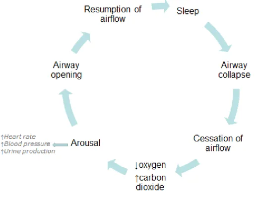
The aetiology of OSA is complex and multi-factorial. In simple terms the cycle of OSA is produced by upper airway occlusion leading to apnoea, which in turn produces changes in blood gases, and a subsequent arousal from sleep that triggers the airway re-opening and the resumption of airflow (Fig 1.2). This section will give an overview of the factors that contribute to the development of OSA.
16
Upper airway narrowing in OSA: The most common site of upper airway collapse in patients with OSA is the retroglossal airway; (fig 1.3). However, narrowing can occur at different points along the airway and any condition that leads to upper airway narrowing will predispose to OSA (Dempsey et al., 2010), including craniofacial abnormalities (Mixter et al., 1990; Pijpers et al., 2004), enlargement of upper airway soft tissue structures (Hwang et al., 2013) and external compression due to obesity (Pahkala et al., 2013) or oedema (Redolfi et al., 2009). However, it is important to note that people with anatomical narrowing of the upper airways are able to maintain airway patency during wakefulness, and it is only during sleep that airway obstruction occurs.
Figure 1.3 Midsagital MRI in a normal subject (left) and a patient with Severe OSA (Apnoeic – Right). Highlighted are the 4 regions of the pharynx, the upper airway soft tissues and craniofacial structures that can contribute to OSA. Fat tissue is shown in white. Note that the OSA patient has more fat tissue around the neck, and in the tongue. He/She also has a smaller retroplatal and retroglossal airway (Dempsey et al., 2010).
Hypoventilation during sleep: The respiratory minute volume in healthy individuals has been shown to be up to 15% lower during sleep, compared to wakefulness (Douglas et al., 1982). A number of factors contribute to this reduction. During wakefulness there is a ventilatory drive from the cortex called the ‘Wakefulness Drive to Breathe’. This cortical drive is absent during sleep (Fink, 1961; Morrell
et al., 1993; Wellman et al., 2004). Furthermore, a simultaneous sleep-related reduction in chemosensitivity also occurs (Dempsey & Skatrud, 1986). These two factors reduce the ventilatory drive to the respiratory pump muscles, which produces a relative hypercapnia during sleep (Dempsey & Skatrud, 1986; Leevers et al., 1994).
Reduced muscle tone during sleep: During sleep muscle tone is reduced throughout the body. Reduced diaphragm and accessory respiratory muscle tone contributes to the reduction in minute volume that has been recorded during sleep (Orem et al., 2002). The reduced tone in the upper airway
17
dilator muscles results in an increased resistance to airflow in the pharynx. This hypotonia can produce upper airway collapse in predisposed individuals as a result of negative inspiratory pressure generation in the thorax, and positive extra-luminal pressure, produced by gravitational forces, and adipose tissue. The critical collapsing pressure of the upper airway (termed Pcrit) is ultimately determined by the transmural pressure (Dempsey, 2010).
Apnoeas caused by upper airway obstruction are terminated by an arousal from sleep that leads to increased tone in the airway dilator muscles and a return to waking-state ventilatory control; hence regular breathing. The physiological conditions that induced the airway obstruction return on resumption of sleep, and OSA patients can experience multiple cycles of sleep-apnoea throughout the night (Fig 1.2).
1.2.2
The prevalence of OSA
The prevalence of OSA appears to be rising. This is likely to be due to the aging population, and an obesity epidemic in developed countries. Both obesity and aging are high risk factors for OSA. This section describes the way in which the prevalence of OSA has increased over the past 20 years. The most widely quoted figures for the prevalence of OSA are taken from the Wisconsin Sleep Cohort (Young et al., 1993). During the period between 1988 and 1994 the prevalence of moderate-severe OSA (AHI ≥ 15 events/hour) in the Cohort was estimated to be 8.8% in males aged 30-70. By comparison, during the period between 2007 and 2010 the estimated prevalence had risen to 13%. OSA is less common in women, but a similar increase has occurred from 4% in the period between 1988 and 1994, to 6% in the period between 2007 and 2010 (Young et al., 1993; Peppard et al., 2013). As mentioned above the most likely reason for the increasing prevalence of OSA is the increasing prevalence of obesity. In 1993, 15% of the UK adult population was obese (BMI >30 Kgm2). In 2011 this had risen to 25% (Health and Social Care Information Centre; http://www.hscic.gov.uk).
OSA is also more common with increasing age. The Wisconsin Sleep Cohort data showed that 10% of men aged 30-49.9 years had moderate-severe OSA, compared to 17% of men aged 50-70 years. This age distribution is even more pronounced in women where the prevalence increases from 3% in 30-49.9 year olds, to 9% in women from 50-70 years. The increased prevalence of OSA in older people is supported by other epidemiological studies, which estimate the prevalence of OSA in people over 65 years to be approximately 20%, with some estimates as high as 70% (Ancoli-Israel et al., 1989; Young
18
The factors that contribute to the age-related increase in the prevalence of OSA include a reduction in pharyngeal muscle function (Worsnop et al., 2000b) and a more collapsible upper airway (Kirkness
et al., 2008b). Age-related differences in upper airway structure include a decrease in the size of the upper airway lumen (Carlisle et al., 2014), associated with an age-related changes to the position of the hyoid bone (Pae et al., 2008). An increase in arousal frequency, has an associated change in the stability of breathing (Browne et al., 2003a). Perhaps the most important change is an increase in the prevalence of co-morbidities. For example, patients with chronic heart failure are more likely to have both obstructive and centrally mediated sleep apnoea (Vazir et al., 2007).
1.2.3
The Sequelae of OSA
OSA is known to impact multiple organ systems, and the most commonly investigated are cardiovascular and metabolic changes. However, other conditions as diverse as skin cancer, Alzheimer's disease and retinal dysfunction have been linked with OSA. This section will briefly outline the cardiovascular and metabolic effects of OSA. The association between OSA and brain function, which is the focus of this thesis will be covered in Section 1.3.
Cardiovascular: The association between OSA and hypertension has been known for many years (Fletcher et al., 1985; Strohl et al., 1994; Bixler et al., 2000). Longitudinal data from the Wisconsin Sleep Cohort suggests that hypertension is a consequence of OSA, with a three fold increased risk of hypertension in OSA patients with severe disease (Peppard et al., 2000). OSA patients are also at increased risk of coronary artery disease and heart failure (Mooe et al., 1996; Mooe et al., 2001; Shahar et al., 2001).
The mechanisms that lead to increased cardiovascular disease in OSA have been studied in both animal and human models. The difficulty in demonstrating causality is that both OSA and cardiovascular disease are linked with obesity. The most likely factors by which OSA (independent of obesity) causes cardiovascular disease are increased sympathetic nervous system activity, hypoxic and oxidative stress, systemic inflammation, and mechanical factors secondary to intrathoracic pressure oscillations, e.g. reduced left ventricular stroke volume, systemic arterial pressure, cardiac output and heart rate (Somers et al., 2008), (Drager et al., 2011).
Metabolic: OSA is strongly associated with obesity and therefore as with cardiovascular disease, the extent to which there is an independent association between OSA and metabolic disease is debated. Current data appear to show a particular link with insulin resistance and type-2 diabetes mellitus (Ancoli-Israel et al., 1989; Strohl et al., 1994; Punjabi et al., 2002). Specifically the link with insulin resistance may be a combination of both central and visceral obesity, although sympathetic drive from
19
frequent arousals, intermittent hypoxia and sleep fragmentation may also play a role (Punjabi et al., 2004; Spiegel et al., 2009).
Additional metabolic factors have also been studied, including blood lipid levels (triglycerides, low density lipoproteins, non-high density lipoproteins and total cholesterol). The impact of OSA on triglycerides as a marker of cardiovascular disease has also been evaluated, with 2 months of treatment leading to a reduction in triglyceride and cholesterol levels (Phillips et al., 2011). Recent data from the UK show that diabetic patients with OSA are more likely to have diabetic retinopathy and neuropathy, and an ongoing treatment trial is investigating the role that reducing OSA could have on diabetic retinopathy (Harsch et al., 2004; Kosseifi et al., 2010; Mason et al., 2012; Rudrappa et al., 2012)
.
1.2.4
The Treatment of OSA
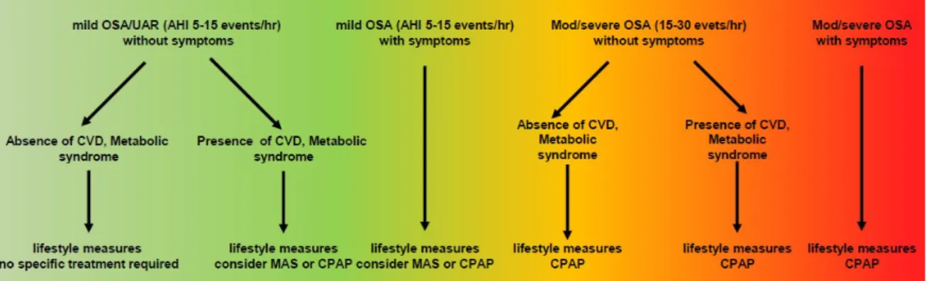
There are many treatments for OSA and their application depends in part on the severity of the disease (Fig 1.4). For patients with mild disease modifying lifestyle (weight loss, stopping smoking, and cardiovascular exercise), increasing total sleeping time, and improving the sleeping environment (e.g. remove digital screens etc. from the bedroom) can help. Additionally, improving diet and reducing stimulants, such as caffeine, before bedtime is advisable. For patients who only have OSA when sleeping in the supine position, positional aids can help. Optimising medical management of co-morbidities is another key part of treatment e.g. pain control, and optimising cardiorespiratory function. In patients with mild to moderate disease, oral mandibular advance devices are often helpful. The most commonly used treatment for OSA is positive airway pressure, which acts as a pneumatic splint blowing air into the upper airway and preventing collapse (Fig 1.5). There are different forms of pressure delivery, including continuous positive airway pressure (CPAP), autotitrating CPAP (auto-CPAP), bilevel ventilation and adaptive servoventilation (ASV). OSA is most effectively treated with CPAP, and this section will summarise the reasons why CPAP is considered the mainstay of treatment for patients with moderate to severe OSA.
20
Figure 1.4 Algorithm for the treatment of mild, moderate, and severe obstructive sleep apnoea (OSA). Upper airway resistance (UAR), cardiovascular disease (CVD).
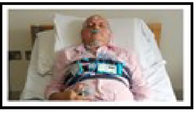
Figure 1.5 A patient using CPAP
The recent National Institute for Health and Care Excellence (NICE) Health Technology Appraisal of the use of CPAP concluded that it was an efficacious and cost-effective treatment for OSA syndrome
21
(McDaid et al., 2009). The benefit of CPAP is measured in most trials as an improvement in sleepiness as measured by the Epworth Sleepiness Scale (ESS; See Chapter 2, Section 2.3.1). The ESS is a subjective measure of sleepiness and treatment with CPAP (versus conservative treatment) reduces sleepiness by approximately 2.7 points (95% confidence interval -3.45 to -1.96). The reduction in sleepiness is proportional to the severity of the disease and therefore patients with severe OSA often feel more relief of symptoms than those with mild disease.
The cost of CPAP treatment is relatively cheap (compared to no treatment) per quality adjusted life year (QALY) gained at <£4,000 (McDaid et al., 2009). However, the majority of the cost is associated with a reduction in the incidence of cardiovascular disease, stroke, and road traffic accidents associated with sleepiness. Thus in patients who are not sleepy, and have a low cardiovascular and/or cerebrovascular risk CPAP may not be cost-effective.
The effectiveness of CPAP therapy is also related to the adherence to treatment. Adherence to CPAP is estimated to range from 46% to 83%, with an average nightly usage of 2.39 hours per night in minimally symptomatic middle-aged OSA patients (Craig et al., 2012) and 2 hours 22 mins in older OSA patients at 12 months (McMillan et al., 2014). This is somewhat lower than the 4 hours per night on 70% of nights which is commonly used as the benchmark for CPAP adherence, and less than the optimal outcomes achieved with a usage of at least 5 hours per night. Factors that impact CPAP compliance are shown in Table 1.1 (adapted from the Oxford Handbook of Medicine chapter submitted for publication). The percentage of patients who continue using their CPAP falls over time, e.g. from 84% at the end of the first year to 68% after 4 years, remaining at this level for a further 3 years. The weighted average for studies that report discontinuation rates over 3 years was estimated to be 3.8% per annum
22
Table 1.1: Predictors of poor CPAP adherence include
Patient characteristics – increased nasal resistance, depression
Disease characteristics – either severe or mild minimally symptomatic disease
Psychological or social – less self-efficacy, poor social support, limited disease or treatment knowledge
Technical – lack of heated humidification and flexible pressure
1.3 COGNITIVE FUNCTION AND OSTRUCTIVE SLEEP APNOEA
There have been many studies of OSA and cognitive function. These have shown an association between OSA and a broad range of neurocognitive impairments (Bucks et al., 2013). A recent study by our group demonstrated deficits in verbal memory (Twigg et al., 2010), while other studies have found deficits in spatial memory (Varga et al., 2014) (Daurat et al., 2008), reaction time (Bedard et al., 1991; Bedard et al., 1993) and numerical memory (Verstraeten et al., 2004). Even in cognitive tests where no impairment has been detected, functional imaging has demonstrated that sleepy OSA patients utilise spare capacity in brain regions not utilised by healthy controls to achieve normal cognitive function (Ayalon et al., 2006). However, despite these data, the impact of OSA on cognitive function remains controversial. Indeed, it has recently been argued that there is only a weak correlation between (subjective) cognitive complaints, and objective cognitive function in patients with OSA (Vaessen et al., 2014). The wide variation in the reported cognitive dysfunction of patients with moderate to severe OSA was the starting point for the studies reported in Chapters 3-5 of this thesis.
1.3.1 Impaired Cognitive function in OSA
A recent meta-analysis determined that deficits in attention/vigilance, delayed long-term visual and verbal memory, visuospatial/constructional abilities and executive dysfunction have been consistently reported in patients with OSA (Twigg et al., 2010; Bucks et al., 2013). The cognitive impairment appears to be associated with the severity of hypoxaemia, while the attention and vigilance dysfunction is more likely to be associated with the degree of sleep fragmentation (Bucks et al., 2013). Working memory, short-term memory, and global cognitive functioning and language ability appear to be largely unaffected by OSA (Bucks et al., 2013).
There are several possible explanations for the wide range of results seen in these studies: The subjective tests used may not be specific enough to detect accurately the cognitive deficits (see
23
Chapter 2, Section 2.4 for a detailed explanation of the cognitive tests used in this thesis), indeed the objective tests used are frequently designed to detect deficits in patients with brain-injuries, and therefore may not be able to capture the milder impairments that occur in OSA (Vaessen et al., 2014). Similarly, one day (or night) of testing provides only a ‘snapshot’ of the patient’s ability. This kind of testing is unable to take account of time-dependent factors, such as fluctuation over time and various circadian influences. Moreover, cognitive domains frequently involve more than one construct. Therefore only carefully deconstructed analyses of the different cognitive test results can accurately assess patient deficits (Olaithe et al., 2014). Finally, the cognitive dysfunction may be a sign of psychological distress, rather than impairment (Pullens et al., 2010; Vaessen et al., 2014). Of note, cognitive reserve and genetic vulnerability (e.g. apolipoprotein e4 genotype) are likely to be factors in the susceptibility to cognitive dysfunction (Rosenzweig et al., 2013c; Olaithe et al., 2014; Rosenzweig
et al., 2014). Some other important factors considered during the design of the studies presented in this thesis were the duration of exposure to the disease, the role of the blood-brain barrier and co-morbidities such as hypertension, metabolic dysfunction and systemic inflammation.
1.3.2
Treatment of Cognitive Function in OSA
Both pharmacological and non-pharmacological treatments for OSA have been shown to improve cognitive function in patient with OSA. Several meta-analyses suggest that CPAP treatment reduces sleepiness and improves mood, and that these changes are associated with improvements in objective cognitive functioning (Giles et al., 2006; Marshall et al., 2006; Kylstra et al., 2013; Vaessen et al., 2014). Although less successful, drugs such as donepezil, phylostigmine, and fluticasone have also been used to try to improve cognitive outcomes in treated OSA patients (Kohler et al., 2009; Mason et al., 2013). Of interest for the studies presented in this thesis, CPAP treatment also appears to reverse some, but not all, neuroanatomical changes. Specifically the study by Canessa et al, 2011 showed that 3 months of CPAP treatment improved cognitive function in several domains and that these improvements were correlated to grey matter volume increases in frontal and hippocampal regions (Canessa et al., 2011). Another recent study showed significant improvements involving memory, attention, and executive functioning that correlated with white matter changes after 12 months of treatment with CPAP (Castronovo et al., 2014). However, in these studies not all areas showed complete reversal of tissue damage, or deficits in cognition, suggesting that initiation of prolonged treatment may be needed as early as possible in the disease process (Kushida et al., 2012; Prilipko et al., 2012; Ferini-Strambi et al., 2013; Yaffe et al., 2014).
24
To investigate the effect of disease duration on cognitive function, studies have been carried out looking into the cognitive performance and effects of treatment in children with OSA (Gozal, 1998; Biggs et al., 2014). In a recent study of children with sleep disordered breathing (SDB), followed-up for four years, treatment led to improvements in several aspects of neurocognition, collectively categorised as performance IQ (Biggs et al., 2014).
Performance IQ represents fluid intelligence and is reflective of incidental learning. It describes the subject’s ability to adapt to new situations (Cattell, 1967). In the study by Biggs et al, improvements were recorded in tasks associated with spatial visualisation, visuo-motor coordination, abstract thought and non-verbal fluid reasoning (Biggs et al., 2014). However, overall improvements in the academic ability or behaviour were less clear. Furthermore, worsening of verbal IQ, which, unlike performance IQ, is more likely to be affected by formal education and learning experiences, was noted in a treated group (Biggs et al., 2014). No conclusive explanation for this finding was provided, and no statistically significant association between the reduction in verbal IQ performance and treatment was demonstrated (Biggs et al., 2014).
Conversely, in the seminal study by Gozal in 1998, younger children with SDB were followed for 12 months after treatment and significant improvements in academic performance were recorded (Gozal, 1998). The different neurodevelopmental ages of children, and different test parameters used may account for the differences in findings between the two groups. Nonetheless, similarities may be emerging from these studies of children and earlier work. Specifically, associations between performance IQ and slow wave activity (SWA) during the non-rapid eye movement (NREM) sleep (Biggs et al., 2012; Biggs et al., 2014).
In healthy adults, sleep progresses through NREM stages N1 to N3 followed by a period of REM sleep occurring approximately 60- 90 minutes into sleep (Vyazovskiy & Delogu, 2014). It has been argued that cognitive improvements in treated OSA patients may reflect increased stability of brain activity during sleep, allowing crucial synaptic repair and maintenance to occur during SWA, and counteracting the toxic effects of OSA mediated by arousal and hypoxia (Neubauer & Fink, 2009; Biggs et al., 2014). This argument is supported by findings showing that sleep presents a crucial period during which the brain can restore cellular homeostasis (Fig 1.6), increase signal to noise ratio, and reinforce neuronal circuitry for subsequent cognitive processing demands (Poe et al., 2010; Abel et al., 2013; Tononi & Cirelli, 2014).
25
Figure 1.6: “Sleep is the price we pay for [neural] plasticity” Tononi & Cirelli 2014. Figure shows neural connections in a fruit fly (left) following sleep, enriched wakefulness, sleep after enriched wakefulness and wake after enriched wakefulness with no sleep. Note the smallest number of synapses occur following sleep, and the most following sleep deprivation. New learning occurs during wakefulness via synaptic potentiation. Neurones strengthen synapses during enriched wakefulness, while interacting with the environment e.g. fear conditioning, cue-reward learning etc. Neurones renormalize synapses in sleep when the brain is off line; synapses have a high energy cost and neural ‘space’ may be reduce if sleep does not occur.
1.3.3
Sleep Deprivation and Cognitive Function in OSA
Sleep alters the molecular signalling pathways that regulate synaptic strength (Fig 1.6), plasticity-related gene expression and protein translation (Abel et al., 2013). Moreover, sleep deprivation can impair neuronal excitability, decrease myelination and lead to cellular oxidative stress and misfolding of cellular proteins (Abel et al., 2013; Picchioni et al., 2014).
The frequent arousals and ensuing fragmented sleep that occur in OSA, have been shown to impact on cognitive function the following day, in a manner similar to that of total sleep deprivation (Daulatzai, 2013). Studies of the effects of sleep deprivation on cognition in the general population suggest comparable cognitive impairments to those seen in OSA (Killgore, 2010). Furthermore, an association has been shown between excessive daytime somnolence and cognitive impairment and an increased risk of cognitive decline and dementia (Yaffe et al., 2014). In a prospective cohort study (The Honolulu-Asia Aging Study), lower nocturnal oxygenation and a reduction in SWA NREM sleep were associated with the development of micro-infarcts and brain atrophy. Conversely, men with longer SWA sleep showed slower cognitive decline (Gelber et al., 2014).
The impact of OSA on selected sleep stages may be particularly important, as each sleep stage is associated with its own functional learning and memory processes (Poe et al., 2010). In OSA patients, stage N2 NREM sleep has been shown to increase, while stages N1, N3 and REM sleep decrease Fig 1.7 (Andreou et al., 2014).
26
Figure 1.7: Overnight sleep patterns (hypnogram), obtained from electroencephalography, illustrating sleep cycles in a young healthy person (Top panel). Note that NREM is the first sleep, and the first REM sleep occurs after approximately 90 minutes; throughout the night there are occasional brief arousals from sleep. The Bottom Panel illustrates sleep cycles in an OSA patient. Note that throughout the night there are frequent, brief arousal from sleep with a reduction in both NREM stage 3 and REM sleep. W: wake; R: REM sleep (marked in blue); 1, 2, 3: NREM stage 1, 2 and 3 sleep.
The consequences of sleep fragmentation in OSA are difficult to assess. In one study of mild OSA, where sleep fragmentation did not significantly reduce the scoring of N3 Sleep, the exponential decay function of SWA was demonstrated as significantly slower in patients than in controls (Ondze et al., 2003). This was due to the more even distribution of SWA throughout the night. These results show that mild sleep fragmentation can alter the dynamics of SWA, without decreasing significantly the amounts of SWA and REM sleep. This means that to fully understand the impact of sleep deprivation in OSA it may be necessary to perform SWA decay analysis (Ondze et al., 2003). In the same study, a decrease of spindle activity was observed in N2 and N3 which was not attributable to an increase of SWA (Ondze et al., 2003; Schonwald et al., 2012). Such a reduction in total spindle density has also been reported in sleep maintenance insomnia, and is likely to be related to sleep fragmentation (Ondze et al., 2003; Schonwald et al., 2012).
27
REM Sleep: Traditionally, obstructive events during NREM sleep were viewed as associated with greater cognitive deficits or impaired quality of life, whilst REM sleep events were shown to be associated with greater sympathetic activity, hypertension and cardiovascular instability in patients with OSA (Mokhlesi et al., 2014) (Mokhlesi & Punjabi, 2012). Recently, a role for fragmented REM sleep in spatial navigational memory problems in OSA patients was suggested (Varga et al., 2014). During this study, patients spent two different nights in the laboratory, during which they performed timed trials, before and after sleep, on one of two unique 3D spatial mazes (Varga et al., 2014). The normal consolidation of sleep was achieved with use of therapeutic CPAP throughout the first night, whereas during the second night CPAP was reduced only during the REM stages. Here, patients showed improvements in maze performance after a night of normal sleep, and those improvements were significantly reduced following a night of REM disruption without changes in psychomotor vigilance. Cognitive improvements were significantly positively correlated with the mean REM run duration across both sleep conditions (Varga et al., 2014).
In some OSA patients, reduction of REM sleep can lead to dissociation of REM traits to other sleep stages, further impacting on critical sleep windows for memory formation and consolidation (Poe et al., 2010). Of particular note, several studies have now demonstrated that, if high homeostatic demands are not fully met during sleep, microsleeps can occur in highly active regions of the brain during the subsequent wake period (Vyazovskiy & Delogu, 2014). This can lead to the concomitant disability of that region (Tononi & Cirelli, 2014; Vyazovskiy & Delogu, 2014). To what extent this takes place in OSA patients, and whether this also contributes to attention/vigilance dysfunction and the higher rate of road traffic accidents seen in OSA patients has yet to be fully understood. Previously reports of retarded SWA decay through the night in even mild OSA patients further supports the notion of non-restorative sleep in OSA (Ondze et al., 2003) .
Sleep Spindles: Intriguingly, in a study of spindle frequency changes in OSA, it has been shown that, unlike healthy controls, OSA patients continued displaying a significant proportion of slow spindles in frontal, central and parietal regions during the night. This may suggest that deregulated spindle formation contributes to cognitive deficits in OSA patients (Schonwald et al., 2012). In another study, sleep architecture of patients with mild OSA showed a high degree of sleep fragmentation resulting in a different time course of SWA and a decreased sleep spindle index when compared to controls (Ondze et al., 2003).
Taken together, these studies further highlight the possible role for OSA brain injury in the
acceleration, or even initiation, of cognitive decline especially in older people (Sforza & Roche, 2012; Daulatzai, 2013; Pan & Kastin, 2014; Yaffe et al., 2014).
28
1.4 ISCHAEMIC CONDITIONING AND NEURO-INFLAMMATION IN OSA
In OSA, repetitive occlusions of the upper airways lead to intermittent hypoxia and recurrent hypoxaemia, typically characterized by short cycles of hypoxia and reoxygenation (Almendros et al., 2014). These cycles can lead to either adaptive or maladaptive processes (Almendros et al., 2014) and the outcome will vary depending on the dynamic interplay between the specific type and amount of reactive oxygen/nitrogen species produced, their duration and frequency, the intracellular localization, and the micro-environmental antioxidant activity (Lavie, 2014). Additional interplay depends on factors such as the genetic makeup, nutrition and other lifestyle related variables, which all affect the redox status (see also (Almendros et al., 2014; Lavie, 2014)). A variety of studies to date suggest that severity of hypoxia, its duration, and cycle frequency, are fundamental determinants of outcomes (Ayalon et al., 2010). For example, it has generally been acknowledged that short, mild, and lower cycle frequency may generate beneficial and adaptive responses in brain, such as ischaemic preconditioning (Almendros et al., 2014). Conversely, chronic moderate-to-severe, and high frequency intermittent hypoxia can induce maladaptive disruption of homeostatic mechanisms, leading to dysfunction and sterile neuroinflammation (Almendros et al., 2014; Lavie, 2014).1.4.1
Ischaemic preconditioning
Ischaemic preconditioning represents a generalized adaptation to ischaemia by a variety of cells (Lavie & Lavie, 2006; Dirnagl et al., 2009). In OSA, induction of ischaemic preconditioning is thought to be due to the activation of several gene programs, including the hypoxia inducible factor-1, vascular endothelial growth factor, erythropoietin, atrial natriuretic peptide and brain derived neurotrophic factor (Brzecka, 2005; Nanduri et al., 2008).Various end-mechanisms and pathways have been shown to play a role, including that of long-term facilitation of diaphragmatic motor output, chemo-reflex activation, vascular remodelling, neo-angiogenesis, productive autophagy, reactive gliosis, various synaptic alterations. Modulation of adult hippocampal neurogenesis has also been suggested (Haddad & Yu, 2009; Aviles-Reyes et al., 2010; Tsai et al., 2011; Papadakis et al., 2013; Tsai et al., 2013; Lavie, 2014).
CPAP treatment of OSA has been shown partially to reverse the damage in the hippocampus, and to ameliorate some of the associated cognitive deficits, possibly also by modulating adult neurogenesis (Canessa et al., 2011). It is proposed that at any given time ongoing maladaptive neuro-inflammatory processes probably exist alongside adaptive mechanisms of increased brain plasticity and ischaemic preconditioning (Ferriero, 2005; Lledo et al., 2006; Dirnagl et al., 2009; Seki, 2011; Rosenzweig et al., 2013a). In a recent study that compared the cognitive performance of patients with high and low
29
hypoxemia after controlling for demographic factors and other aspects of OSA severity, an unexpected advantage of higher hypoxemia on memory was demonstrated in a carefully matched clinical cohort (Hoth et al., 2013).
Another powerful central neuroprotective adaptive mechanism for ischaemic events was demonstrated following the activation of the intrinsic neurons of the cerebellar fastigial nucleus (Reis
et al., 1997). Neurostimulation of these nuclei appears to provide protective reduction in excitability of cortical neurons during the subsequent ischaemic episodes, and to lead to reduced immunoreactivity of cerebral microvessels by down-regulating their production of intracellular cell adhesion molecule (ICAM-1) and consequently preventing the release of pro-inflammatory cytokines (Rosenzweig et al., 2014). The compensatory entraining of the cerebellum by hypertrophic hippocampi may also occur in younger patients with mild OSA (See Chapter 3).
Although there are no direct monosynaptic anatomical connections between hippocampi and cerebellum, their connectivity is thought to be important for the control of movement under states of heightened emotion and novel conditions, and for associative learning. Of note, failed adaptation of cerebellar networks to injury was shown to lead to cognitive deficits and hyperactivity, distractibility, ruminative behaviour, dysphoria and depression in some patients (Rosenzweig et al., 2014). Several studies also suggest that, under certain conditions, intermittent hypoxia can increase immune defences without exacerbating inflammation (Almendros et al., 2014; Lavie, 2014). Moreover, in animals, short-lasting hypoxic exposures mimicking OSA were associated with recruitment of bone-marrow derived pluripotent stem cells, which exhibited up-regulation of stem cell differentiation pathways, particularly involving central nervous development and angiogenesis (Almendros et al., 2014).
1.4.2
Neuro-inflammation
Intermittent hypoxia can lead to maladaptive effects, including neuro-inflammation. Although the exact neurocellular sources for associated processes are still incompletely understood, it is likely that activation of astroglia is important (Dale et al., 2014; Lavie, 2014). In addition, the oligodendrocytes, myelin-producing cells of the CNS, were shown to be selectively sensitive to hypoxia and sleep fragmentation (Rosenzweig et al., 2012; Bellesi et al., 2013). The subsequent loss of buffering functions can ultimately contribute to pathological processes, such as increased glial proliferation and microglial activation (Dale et al., 2014). Astroglial and microglial cells play critical roles in regional blood flow regulation and inflammatory processes in the brain, as well as critical coordination of bioenergetics through lactate transport (Dale et al., 2014). Under normal conditions, microglia in the
30
healthy CNS exhibit a surveillance phenotype that synthesizes and releases neuroprotective growth/trophic factors (Dale et al., 2014). However, severe and prolonged hypoxia can activate microglia toward a toxic, pro-inflammatory phenotype that triggers pathology, including change in hippocampal structure, impaired synaptic plasticity, and cognitive impairment (Dale et al., 2014) (See Chapter 5). Neuro-inflammation has been shown independently to raise the brain's sensitivity to stress, resulting in stress-related neuropsychiatric disorders, such as anxiety and depression (Skaper
et al., 2014).
Dynamic changes in transcription of inflammatory genes were demonstrated following exposure to intermittent hypoxia, with most inflammatory markers increasing over time (Dale et al., 2014). Increased concentrations of prostaglandin E2 in neural tissue have also been demonstrated in hippocampal and cortical regions accompanied by lipid peroxidation of polyunsaturated fatty acids (Dale et al., 2014). Similarly, it was shown that increased carbonylation- and nitrosylation-induced oxidative injury emerges in susceptible brain regions and promotes increased daytime somnolence (Dale et al., 2014; Lavie, 2014). Recently, toll-like receptor 4 (TLR4) expression and activity was demonstrated to be increased in monocytes of patients with OSA (Unnikrishnan et al., 2014). Expression of TLR4 was also demonstrated in the microglia of the cortex and brainstem after chronic intermittent hypoxia where it was postulated to play region specific and differential adaptive or maladaptive role (Unnikrishnan et al., 2014). This finding is of particular interest since TLR4 has also been strongly implicated in several inflammatory and neurodegenerative disorders, including vascular dementia and Alzheimer’s disease (Unnikrishnan et al., 2014). In line with this, in cognitively healthy adults, intermittent hypoxia was correlated with increases in phosphorylated and total tau and amyloidᵝ1-42 concentrations in cerebral spinal fluid, which are key components of Alzheimer’s pathology (Yaffe et al., 2014). Similarly, cerebral amyloidogenesis and tau phosphorylation along with neuronal degeneration and axonal dysfunction were shown in the cortex and brainstems of animals exposed to intermittent hypoxia (see Chapter 5) (Daulatzai, 2013).
Taken together, these findings support the role for neuro-inflammatory processes in cognitive and emotional deficits of OSA patients. They further suggest a close association between hypoxaemia- induced maladaptive processes and dementia.
31
1.5. DESCRIPTION AND STRUCTURE OF THESIS
The primary aim of this thesis is to investigate the effects of OSA on brain structure. The thesis will also investigate whether treatment of OSA with CPAP leads to resolution of any changes in brain structure, possibly mediated by ischaemic preconditioning. Finally the thesis will investigate whether these changes in brain structure are associated with corresponding changes in cognitive function. The general methods used for all the studies in this thesis will be described in Chapter 2. Additional background to the individual studies will be presented in each chapter, with further details of study protocols, data analysis and statistical analyses where necessary.
In Chapter 3 I will describe the reanalysis of brain imaging data gathered previously by the group using more modern techniques. The aim of this study was to investigate the effect of intermittent hypoxia on brain structure in OSA patients. I was interested in brain hypertrophy, suggestive of ischaemic preconditioning, as well as the hypotrophy demonstrated in other studies.
In Chapter 4 I will describe a randomised controlled trial of CPAP in OSA. Brain structure and cognitive function were measured in younger people newly diagnosed with OSA before and after randomisation to CPAP or best supportive care. This aim of this study was to investigate the association of brain structural changes with cognitive dysfunction in OSA and to investigate whether these changes were reversible.
Chapter 5 will describe an analysis of brain structure in older people with OSA before and after randomisation to CPAP. The aim of this study was to investigate whether neuroplasticity still existed in older patients with OSA, or whether any brain structural changes caused by OSA were irreversible. In Chapter 6 the results of the studies presented in this thesis will be discussed, with some suggestions as to how the results, and areas of uncertainty identified during this research, could be developed in the future
32
CHAPTER 2
General Methods
33
2.1 PARTICIPANT AND LABORATORY INFORMATION
OSA patients were recruited from The Royal Brompton (Chapters. 3, 4 and 5), Harefield (Chapter 4) and Charing Cross (Chapter 3) Hospitals and Queen Elizabeth Hospital, Woolwich (Chapter 4). Controls were recruited from the Healthy Volunteers Database in the Academic Department of Sleep and Ventilation and from hospital staff at The Royal Brompton Hospital. Sleep studies were performed at The Royal Brompton as part of routine clinical practice. In addition, research nocturnal polysomnography was carried out (Chapter 4) in the NIHR Respiratory Biomedical Research Unit (BRU) at The Royal Brompton Hospital. MRI scans were performed at both The Royal Brompton Hospital (Chapter 4), and Charring Cross Hospital (Chapter 3 & 5). In the study presented in Chapter 4, I was the first investigator to use the Respiratory BRU Clinical Research Facility for overnight studies and helped to develop the pathway for future overnight studies. The study in Chapter 4 was also the first collaboration between the Respiratory BRU and the Cardiology BRU at The Royal Brompton and the first study to use the Cardiology BRU MRI scanners to perform brain imaging. I set up this collaboration, and arranged the sharing of knowledge and expertise between our previous imaging collaborators and the physicists and radiographers at The Royal Brompton to ensure the correct MRI sequences were used and the images obtained were of appropriate quality. I also set up two new collaborations, to recruit patients from Harefield Hospital and Queen Elizabeth Hospital, Woolwich to Royal Brompton sleep research projects for the first time.
All studies were carried out with Research Ethics Committee approval, and all participants gave written informed consent. Note, the IRAS ethical approval numbers are given in each experimental chapter.
2.2 NOCTURNAL POLYSOMNOGRAPHY
Polysomnography was performed in all OSA patients using the SOMNOscreen system (SOMNOmedics GmBH, Germany). This system allows data to be was transmitted wirelessly in real time to a computer, ensuring that the patient has as comfortable a night’s sleep as possible (Fig 2.1).
34
Figure 2.1: Studies presented in this Thesis used nocturnal polysomnogram to diagnose the severity of the OSA. Note that the patient is able to move freely as the system used is wireless.
2.2.1 Assessment of sleep : EEG, EOG and EMG
Electroencephalogram (EEG), electro-oculogram (EOG) and surface electromyogram (EMG) are used to monitor sleep and wake, using standard sleep scoring criteria (Reschtschaffen, 1968). EEG was measured using 10mm gold cup electrodes (Grass technologies, Rhode Island, USA). The electrodes were sited according to the International ten twenty EEG electrode system. This system uses the nasion and inion as reference points, with EEG electrodes placed at increments of 10 or 20% along transverse and sagittal lines linking these landmarks. Electrodes were placed in the C3 and C4 positions, with a common reference electrode in the Cz position (Fig 2.2).
The electrode positions were marked with a chinagraph pencil and the site was cleaned with an abrasive paste (Nuprep, Weaver and Company, Colorado, USA) and an alcohol soaked swab. This process reduced the electrical impedance between the electrode and the skin. It also helped the attachment of the electrode. The gold cup electrodes were then filled with conducting paste (Ten20
conductive paste, Weaver and Company,
Colorado, USA ) and fixed in place with
adhesive (Collodion, SLE Ltd.,
South Croydon, UK). Reference
electrodes were placed over left and
right mastoid processes (A1 and A2
respectively) and a ground electrode was
placed on the centre of the forehead
35
Figure 2.2. A view of the head showing the electrode positions of the international ten twenty system for recording of sleep using electroencephalography (EEG). (Figures adapted from Malmivo & Plonsey. Bioelectromagnetism: Principles and Applications of Bioelectric and Biomagnetic Fields. New York: Oxford University Press; 1995)
Self-adhesive electrodes were used to measure EOG activity. These were placed 1 cm lateral and superior to the right outer canthus, and 1 cm lateral and inferior to the left outer canthus and referenced to the A1 mastoid electrode. By placing the EOG electrodes asymmetrically conjugate vertical eye movements, which produce opposing deflections in the right and left EOG signals, can be distinguished from electrode artefact or high voltage which produce in-phase deflections in both EOG signals. Self-adhesive electrodes were also used to record EMG activity. The skin was prepared as for the EEG and electrodes were positioned 2 cm posterior to the inferior edge of the mandible, and 2 cm to either side of the midline.
After application of all electrodes, an impedance test was performed to ensure all electrodes had a low resistance for optimal detection of brain electrical activity. An impedance of <10,000 Ohms was considered acceptable, and where this was exceeded, further conducting paste was added to gold cup electrodes, or the electrode was replaced until impedance was satisfactory.
The EEG and EOG signals were digitally sampled at a rate of 128 Hz, whilst the EMG signals were sampled at a rate of 256 Hz (Silber et al., 2007). The signals were processed by differential amplifiers to generate a conventional EEG montage (C3/A2; C4/A1, Right EOG/A1 and Left EOG/A1) for sleep scoring (Reschtschaffen, 1968). Conventional filter settings (High pass filter: 0.2 Hz; low pass filter: 35 Hz) were applied to the differential amplifier output to remove signals outside of the frequencies of interest.
36
Airflow from the nose and mouth was monitored using a ‘nasal pressure’ signal, generated by changes in pressure at the nares. The pressure changes were measured by a nasal cannula (Embla, Colorado, USA) connected to a differential pressure transducer that was referenced to atmospheric pressure. The voltage output of the transducer was proportional to the pressure change across a membrane located within the transducer.
The measurement of nasal pressure provides a semi-quantitative measure of the velocity of air passing through the nostrils (Fig 2.3). However, because the airflow is most commonly non-linear, nasal pressure overestimates apnoeas compared to other sensors (Redline et al., 2007). For example, if the amplitude of the nasal pressure signal falls below 10% (the commonly used criteria to score apnoea) when nasal airflow decreases to 30%. The Bernouilli equation states that the velocity of a gas is proportional to the square root of the gas pressure. Therefore, the nasal pressure signal is equivalent to the square of the airflow; for example, when nasal airflow decreases to 70% of baseline, the nasal pressure will decrease to 49%. It has been suggested that a square root transformation of the nasal pressure signal could be used in this case. However, in practice this is not often used.
The within-breath shape of the nasal pressure airflow signal can also provide information about the occurrence of airflow limitation that does not fulfil the requirements to be scored as apnoeas or hypopnoeas (Fig 2.3).
Figure 2.3: Nasal airflow, respiratory effort and oxygen saturation (SpO2) measured during sleep in a health person (left) and a patient with airflow limitation (right). Notice the flattened inspiratory airflow profile (green) in the patient with flow imitation, compared to the healthy subject.
2.2.3
Measurement of respiratory effort using respiratory inductance
plethysmography
Respiratory effort was measured by respiratory inductance plethysmography. This requires placement of a belt around the upper chest and a further belt around the abdomen, midway between the inferior ribs and the iliac crest (Fig 2.1). The belts were sized according to patient girth and were fastened with
37
Velcro. To ensure the belts did not become displaced during the sleep study they were further secured with tape.
The respiratory inductance plethysmography bands measure changes in current, caused by an alteration in cross sectional area enclosed by a wire. A magnetic field is created by passing alternating current through a thin zigzagging wire which has been sewn into the RIP belt (Sleepsense, SLP Inc., Illinois, USA). This magnetic field is affected by changes in the cross-sectional area enclosed by the wire and this, in turn, alters the frequency of the current. The change in current can be measured, which provides a metric of respiratory and abdominal expansion during inspiration as shown in Fig 2.3.
2.2.4
Measurement of arterial oxygen saturation using pulse oximetry
Finger pulse oximetry (Nonin 8000 Softsensor, Nonin, Minnesota, USA) was used to measure haemoglobin oxygen saturation. This measurement represents the average oxygen saturation of haemoglobin. It relies on the principle that the light absorption characteristics of haemoglobin are altered by the binding of oxygen. One arm of the clip contains LEDs that emit light at red (660nm) and infrared (910nm) wavelengths. Light transmitted through the finger is detected by a photodetector on the other arm of the clip. The absorption of light by blood constituents can be determined from changes in the quantity of transmitted light occurring during fluctuations in arterial volume with each cardiac cycle. The ratio of red and infrared light absorbed during these pulsatile fluctuations is used to calculate the oxygen saturation of haemoglobin. The oxygen saturation is then displayed as a moving time average as shown in Fig 2.3.
2.2.5
Measurement of heart rate using ECG
Heart rate was measured by placing ECG electrodes on the skin below each clavicle and below the lower left ribcage.
2.2.6
Measurement of leg movement using EMG
During sleep phasic limb movements (called Periodic limb movements [PLM]} can disrupt sleep in a similar way to OSA. Therefore, to differentiate between sleep disruption caused by occlusion of the upper airway, and that caused by PLM, EMG of the anterior tibialis muscle is monitored, and phasic
38
bursts of activity are scored. If more than 4 consecutive limb movements are noted, separated by not less than 5 seconds, but not more than 90 seconds the patients is said to have PLM. Limb movements can also occur during an arousal from sleep at the end of an apnoea/hypopnoea. Therefore, if the increase in anterior tibialis EMG activity occurs within 0.5 seconds of a respiratory event it is NOT scored as a limb movement. The PLM index is calculated as the mean number of PLMs per hour of sleep.
2.3 ASSESSMENT OF SLEEPINESS
One of the most common symptoms of OSA is excessive daytime sleepiness. The measurement of sleepiness is difficult. Subjective measurements, such as the commonly used ESS, are influenced by factors such as motivation, and objective measurements are often complex and time consuming. In this thesis, therefore, sleepiness has been measured subjectively using the ESS.
2.3.1
Epworth Sleepiness Scale
The Epworth Sleepiness Scale (Johns, 1991) is a widely used, validated questionnaire that assesses daytime sleepiness. The Epworth scale asks subjects to rank how likely, on average, “in recent times” they are to doze in 8 different situations. Each situation is given a mark out of 3 depending on how likely the subject is to doze in that situation (0 = never 1 = slight chance, 2 = moderate chance, 3 = high chance). The score for each situation is added to give a total out of 24. A total score of 10 or more indicates excessive daytime sleepiness.
Although it was designed to be self-administered, a recent study found that 33% of patients made errors and 23% required assistance when completing the Epworth Sleepiness Scale for the first time. Even among patient who had used the Epworth scale previously, 16% made errors. Furthermore 4% of patients were unable to complete the form because of poor reading skills or because they did not have their glasses with them (Ghiassi et al., 2011). Another criticism is that ESS scores do not correlate (Furuta et al., 1999) or correlate only weakly with objective measures of sleepiness such as the multiple sleep latency test (MSLT).
Despite these criticisms the ESS is widely used around the world. As well as the English language version, it has been translated into and validated in numerous other languages including: Croatian, French, German, Greek, Italian, Japanese, Korean, Mandarin, Norwegian, Portuguese, Serbian, Spanish, Thai and Turkish and a pictorial version has recently been validated (Drakatos et al., 2015). It

![Figure 1.6: “Sleep is the price we pay for [neural] plasticity” Tononi & Cirelli 2014](https://thumb-us.123doks.com/thumbv2/123dok_us/8997445.2797571/25.892.127.608.115.249/figure-sleep-price-pay-neural-plasticity-tononi-cirelli.webp)