The role of auxin transport in the
control of shoot branching
Martin van Rongen
Supervisor: Prof. Ottoline Leyser
Prof. Henrik Jönsson
Department of Plant Sciences
University of Cambridge
This dissertation is submitted for the degree of
Doctor of Philosophy
The role of auxin transport in the control of shoot
branching
Martin van Rongen
Branching is a highly plastic trait, enabling plants to adapt their growth form in response to environmental stimuli. In flowering plants, shoot branching is regulated through the activity of axillary buds, which grow into branches. Several classes of plant hormones have been shown to play pivotal roles in regulating bud outgrowth. Auxin derived from the primary shoot apex and active branches inhibits bud outgrowth, whereas cytokinin promotes it. Strigolactones also inhibit bud outgrowth, by changing properties of the auxin transport network, increasing the competition between buds. This occurs by modulating access to the polar auxin transport stream (PATS) in the main stem. The PATS provides directional, long distance transport of auxin down the stem, involving basal localisation of the auxin transporter PIN-FORMED1 (PIN1). Buds need to export their auxin across the stem towards the PATS in order to activate, but since PIN1 is mainly expressed in narrow files of cells associated with the stem vasculature, PIN1 itself it is unlikely to facilitate this connectivity. This thesis re-examines the role of auxin transport in the stem, showing that, besides the PIN1-mediated PATS, other auxin transport proteins constitute a more widespread and less polar auxin transport stream, allowing auxin exchange between the PATS and surrounding tissues. Disruption of this transport stream is shown to reduce bud-bud communication and to partially rescue the increased branching observed in strigolactone mutants. Furthermore, it is shown that distinct classes of auxin transport proteins within this stream can differentially affect bud outgrowth mediated by BRANCHED1(BRC1). BRC1 is a transcription factor proposed to determine bud activation potential. Taken together, the data presented here provide a more comprehensive understanding of the shoot auxin transport network and its role in shoot branching regulation.
Declaration
This dissertation is the result of my own work and includes nothing which is the outcome of work done in collaboration except as declared in the Preface and specified in the text. It is not substantially the same as any that I have submitted, or, is being currently submitted for a degree or diploma or other qualification at the University of Cambridge or any other University or similar institution except as declared in the Preface and specified in the text. I further state that no substantial part of my dissertation has already been submitted for any such degree, diploma or other qualification at the University of Cambridge or any other University or similar institution except as declared in the Preface or specified in the text. It does not exceed the prescribed word limit of 60,000 words excluding bibliography, figures, tables and appendices.
Martin van Rongen September 2017
Acknowledgements
Many people have supported me in the past years during my Ph.D, for which I am very grateful. Although I cannot thank everybody involved individually, I would like to acknowl-edge some people in particular. Foremost, I would like to thank Ottoline for her continuous support and advice. Her interest in my research and the freedom she has given me to pursue some of my own research interests have made the last few years very enjoyable. Her advice has helped me become a better scientist by taking my ability to think critically and write clearly to a new level. Lastly, her supportive attitude towards combining science and family life gave me the confidence to start a family during my Ph.D, something I would have been very unsure of doing in any other lab. Further thanks also go to Henrik Jönsson and Siobhan Braybrook, whose supervision and support have enabled me to do this research.
All the people in the Leyser lab, past and present, have been very supportive. In particular I would like to thank Tom Bennett for his great support, on more than one occasion helping me find direction when I felt I had lost the plot. A special mention to Genevieve Hines, whose computational modelling and resulting discussions have contributed so much to understanding the sometimes complicated results. I would also like to thank Sally Ward, Maddy Seale, Fabrizio Ticchiarelli, Tanya Waldie, Devin O’Connor and Graeme Mitchison for their help and discussions. Thanks also go to Hugo Tavares, for his help with and discussions on data analysis and statistics.
There has been a lot of technical assistance over the years. First, I would like to thank Ruth Stephens for her technical support in setting up some experiments and looking after plants. Also, Rebecca Butler and Alice Thomas for their help with the nitrate experiments. General support within the Sainsbury Laboratory has been exceptional. I would like to thank Ray Wightman for his microscopy support, the Horticulture staff for their great work on keeping the plant growth facilities going and the Support staff for keeping the lab running so smoothly.
All the experiments were designed and carried out by myself, but I received general technical assistance from Ruth with sowing out experiments and, specifically, setting up the jar assays (Fig. 4.6; 4.10; 5.9). The nitrate experiments were sown out and looked after by Rebecca and Alice (Fig. 5.10). All primary data were collected by myself. The following data,
vi
or part thereof, have been published in Bennett et al. (2016a): Fig. 3.4; 3.8A; 4.2B; 4.5C; 4.8A. Plant lines have been received from various people, as indicated in Appendix A, for which I am grateful.
Lastly, I would like to thank my family. At times the hardest parts, but the ones which also brought the greatest sense of achievement, were mammalian in nature rather than plant-based. For sure, my lovely little girls Lily and Tess have helped me keep things in perspective. Finally, I would like to thank Sarah for her amazing support which enabled me to get to this exciting new starting point. Sarah, we are a far cry from our life as diving instructors in Egypt and I am grateful for every step along the way.
Table of contents
List of figures xi
List of tables xiii
1 Introduction 1
1.1 Plant architecture . . . 1
1.2 Plant hormones in shoot development . . . 3
1.2.1 Auxin . . . 3
1.2.2 Cytokinin . . . 4
1.2.3 Strigolactone . . . 6
1.3 The auxin transport network . . . 7
1.3.1 The mechanism of auxin transport . . . 7
1.3.2 Energising auxin transport through proton ATPase activity . . . 9
1.3.3 Feedback between auxin and proton ATPase regulation . . . 10
1.3.4 Auxin transport in stems . . . 11
1.4 Development and regulation of shoot apices . . . 13
1.4.1 Formation of shoot apices . . . 13
1.4.2 Hormonal control of shoot meristem initiation and activity . . . 14
1.5 Models for shoot branching control . . . 15
1.5.1 The effect of auxin on lateral bud outgrowth . . . 15
1.5.2 The auxin transport canalisation model for shoot branching control 16 1.5.3 The role of strigolactones in auxin transport canalisation . . . 17
1.5.4 The second messenger model for shoot branching control . . . 19
1.5.5 The role ofBRC1in shoot branching . . . 20
1.5.6 Reconciling models for shoot branching control . . . 21
Table of contents viii
2 Materials and methods 23
2.1 Materials and methods . . . 23
2.1.1 Plant lines . . . 23 2.1.2 Growth conditions . . . 23 2.1.3 Growth substrates . . . 23 2.1.4 Seed sterilisation . . . 24 2.1.5 Crosses . . . 24 2.1.6 Hormone solutions . . . 24 2.2 Physiological assays . . . 25
2.2.1 One- and two-node explant setup . . . 25
2.2.2 Jar assays . . . 25
2.2.3 Intact branching assays . . . 27
2.2.4 Decapitation assay . . . 27
2.2.5 Plant height . . . 27
2.2.6 Branch angle . . . 27
2.2.7 Stem diameter . . . 28
2.2.8 Bulk auxin transport assay . . . 28
2.2.9 Auxin pulse assay . . . 28
2.2.10 Auxin uptake assay . . . 28
2.3 Molecular biology . . . 29 2.3.1 DNA extraction . . . 29 2.3.2 PCR . . . 29 2.3.3 Restriction digests . . . 30 2.3.4 Gateway cloning . . . 30 2.3.5 Plasmid isolation . . . 31
2.3.6 Sequencing and analysis . . . 31
2.3.7 Transformation . . . 31
2.3.8 Plant selection . . . 32
2.4 Microscopy . . . 32
2.4.1 Fluorescence microscopy . . . 32
2.4.2 Confocal microscopy . . . 32
2.5 Bioinformatics and primer design . . . 33
2.5.1 Statistics . . . 33
2.5.2 Primer design . . . 33
Table of contents ix
3 The auxin transport network in stems 35
3.1 Introduction . . . 35
3.2 Results . . . 36
3.2.1 The role of PIN proteins in auxin movement in the stem . . . 36
3.2.2 The role of PIN proteins in auxin transport dynamics in the stem . . 39
3.2.3 The role of ABCB proteins in auxin transport in the stem . . . 44
3.2.4 The role of AUX1/LAX proteins in auxin transport in the stem . . . 52
3.3 Summary . . . 54
4 The role of connective auxin transport in the control of shoot branching 55
4.1 Introduction . . . 55
4.2 Results . . . 56
4.2.1 The role of PIN3/PIN4/PIN7 in communication between apices . . 56
4.2.2 The relationship between strigolactones and PIN3/PIN4/PIN7 . . . 60
4.2.3 The role of ABCB1/19 in communication between apices . . . 70
4.2.4 The relationship between strigolactones and ABCB1/19 . . . 72
4.2.5 The relationship between PIN3/PIN4/PIN7 and ABCB19 . . . 75
4.2.6 The role of auxin importers in communication between apices . . . 77
4.3 Summary . . . 81
5 Interplay betweenBRC1and auxin transport in the control of shoot branching 82
5.1 Introduction . . . 82
5.2 Results . . . 84
5.2.1 The effect of PIN3/PIN4/PIN7 on shoot branching inbrc1brc2and
strigolactone mutants . . . 84
5.2.2 The effect of ABCB1, ABCB19 on shoot branching inbrc1brc2and
strigolactone mutants . . . 90
5.2.3 Environmental responses inbrc1brc2mutants lacking PIN3/PIN4/
PIN7 or ABCB19. . . 97
5.2.4 BRC1 protein function . . . 99
5.3 Summary . . . 101
6 The role of proton pumps in auxin transport and shoot branching 102
6.1 Introduction . . . 102
6.2 Results . . . 103
6.2.1 The role of AHA proton ATPases in auxin movement in the stem . . 103
Table of contents x
6.3 Summary . . . 110
7 Discussion 112 7.1 Shoot branching and polar auxin transport . . . 112
7.1.1 Polar auxin transport in the stem . . . 112
7.1.2 Stem auxin transport is multimodal . . . 113
7.1.3 PIN3/PIN4/PIN7 contribute to connective auxin transport . . . 114
7.2 Connective auxin transport allows communication between tissues . . . 114
7.2.1 PIN3/PIN4/PIN7 are required for rapid bud activation . . . 115
7.2.2 PIN3/PIN4/PIN7 mediate bud-bud communication . . . 116
7.3 Additional contributors to connective auxin transport . . . 117
7.3.1 ABCB1 and ABCB19 contribute to stem auxin transport . . . 117
7.3.2 ABCB1 and ABCB19 may contribute to rapid bud activation . . . . 118
7.3.3 ABCB19 mediates bud-bud communication . . . 119
7.3.4 PIN3/PIN4/PIN7 and ABCB19 can act synergistically in shoot branching control . . . 120
7.3.5 Auxin importers contribute to stem auxin transport . . . 121
7.3.6 Auxin importers are unlikely to be regulators of bud activation . . . 123
7.4 Regulation of shoot branching by strigolactone . . . 123
7.4.1 Stem auxin transport in strigolactone mutants . . . 124
7.4.2 PIN3/PIN4/PIN7 contribute to strigolactone-mediated shoot branch-ing control . . . 125
7.4.3 ABCB19 contributes to strigolactone-mediated shoot branching control127 7.5 Providing energy for auxin transport and shoot branching . . . 128
7.5.1 AHA1 contributes to stem auxin transport . . . 129
7.5.2 AHA1 affects shoot branching . . . 130
7.6 Interaction betweenBRC1and connective auxin transport . . . 131
7.6.1 Shoot branching control byBRC1is independent of PIN3/PIN4/PIN7 132 7.6.2 Shoot branching control byBRC1is partially dependent on ABCB19 133 7.7 Conclusions and further directions . . . 133
References 136
Appendix A Plant lines and sources 152
Appendix B Primers and genotyping strategies 155
List of figures
1.1 Arabidopsis plant architecture . . . 2
1.2 The strigolactone signalling pathway in Arabidopsis . . . 8
1.3 Arabidopsis stem anatomy . . . 12
2.1 Chromosome map with commonly used genes . . . 26
3.1 Bulk auxin transport and PIN1 levels inpin3/pin4/pin7mutant stems . . . . 37
3.2 Bulk auxin transport inpin1/pin4/pin7mutant stems . . . 38
3.3 Bulk auxin transport inpin2, pin5, pin6andpin8mutant stems . . . 39
3.4 Movement of an auxin pulse inpin3pin4pin7mutant stems . . . 41
3.5 Movement of an auxin pulse inmax2mutant stems . . . 43
3.6 Movement of an auxin pulse inmax2pin3pin4pin7 mutant stems . . . 45
3.7 Individual auxin pulse traces inmax2pin3pin4pin7mutant stems . . . 46
3.8 Bulk auxin transport inabcb1/abcb19mutant stems . . . 48
3.9 PIN1-GFP levels inabcb1/abcb19stems . . . 49
3.10 Movement of an auxin pulse inabcb19mutant stems . . . 50
3.11 Bulk auxin transport inabcb19/pin3pin4pin7mutant stems . . . 51
3.12 Bulk auxin transport inaux1/laxmutant stems . . . 52
3.13 Auxin uptake and movement inaux1/laxmutant stems . . . 53
4.1 Bud outgrowth dynamics inpin3/pin4/pin7mutants . . . 57
4.2 Bud competition inpin3pin4pin7mutant two-node explants . . . 59
4.3 Bulk auxin transport and PIN1 levels inmaxpin3pin4pin7mutant stems . . 61
4.4 Bud formation and activation dynamics inmaxpin3pin4pin7mutants . . . . 63
4.5 Shoot phenotypes ofmaxpin3pin4pin7mutants . . . 65
4.6 Whole-plant responses ofmaxpin3pin4pin7mutants to GR24 . . . 67
4.7 Bud competition inmaxpin3pin4pin7mutant two-node explants in response to GR24 . . . 69
List of figures xii
4.9 Shoot phenotypes inmaxabcb1andmaxabcb19mutants . . . 73
4.10 Whole-plant and bud competition responses ofabcb19mutants to GR24 . . 74
4.11 Bud activation dynamics and bud formation inabcb19pin3pin4pin7 mutants 76 4.12 Shoot phenotypes inabcb19pin3pin4pin7 mutants . . . 78
4.13 Bud formation and branching responses inaux1/laxmutants . . . 80
5.1 Meristem stages ofbrc1brc2andd14plants after short day to long day growth 85 5.2 Effect of growth conditions on branching in brc1brc2 and d14 mutants lacking PIN3/PIN4/PIN7 . . . 87
5.3 Shoot phenotypes ofbrc1brc2andd14mutants lacking PIN3/PIN4/PIN7 . 88 5.4 Bulk auxin transport inbrc1brc2andd14mutants lacking PIN3/PIN4/PIN7 89 5.5 Effect of growth conditions on branching inbrc1brc2 abcb1/abcb19mutants 91 5.6 Shoot phenotypes ofbrc1brc2plants lacking ABCB1/ABCB19 . . . 92
5.7 Bulk auxin transport inbrc1brc2mutants lacking ABCB1/ABCB19 . . . . 93
5.8 Bud competition in brc1brc2pin3pin4pin7 mutant two-node explants in response to GR24 . . . 95
5.9 Whole-plant responses to GR24 ofbrc1brc2mutants with impaired PIN3/ PIN4/PIN7 or ABCB19 function . . . 96
5.10 Branching response to nitrate limitation and crowding inbrc1brc2abcb19 andbrc1brc2pin3pin4pin7mutants . . . 98
5.11 BRC1 reporter construct and genetic complementation . . . 100
6.1 Bulk auxin transport and PIN1 levels inahastems . . . 104
6.2 Movement of an auxin pulse in theaha1mutant . . . 105
6.3 Auxin uptake inahastems . . . 107
6.4 Bud activation inahamutants . . . 108
6.5 Shoot phenotypes inaha1andaha2mutants . . . 109
6.6 Shoot branching in strigolactone mutants lacking AHA1/AHA2 function . . 111
List of tables
2.1 Plant growth conditions . . . 24
2.2 DNA extraction buffer . . . 29
2.3 PCR conditions . . . 30
A.1 Plant line sources . . . 152
A.2 Plant line descriptions . . . 154
B.1 Genotyping primer sequences . . . 155
B.2 Sequencing primer sequences . . . 157
B.3 Cloning primer sequences . . . 157
B.4 Genotyping strategies . . . 158
Chapter 1
Introduction
This thesis concerns the hormonal control of shoot branching and, specifically, the role of transport of the plant hormone auxin.
1.1
Plant architecture
The sessile nature of plants means that they are unable to escape from unfavourable habitats. Instead they need to adapt to the prevailing environmental conditions. Plants are able to do so by changing their body architecture. The root system can change its architecture in response to soil nutrient availability and increases nutrient foraging when there is a shortage of nutrients. The shoot system has to cope with a range of biotic and abiotic factors, such as herbivory and optimising photosynthetic input.
The ability of plants to adapt their body architecture lies in the indeterminate nature of their development. The general body plan is determined very early in the plant’s life cycle, during embryogenesis. Here the apical-basal axis is formed, with a shoot apical meristem at the apex and a root apical meristem at the base. The shoot apical meristem determines the subsequent growth of all above ground parts of the plant. It does so in a modular manner by forming phytomers. A phytomer is a repeating unit, which consists of a node and its associated leaf or leaves and axillary meristem, and an internode connecting the phytomer to the next node (Fig. 1.1). Axillary meristems have the same growth potential as the primary shoot apical meristem and can give rise to higher order plant structures. Axillary meristems can form axillary buds, which can either remain dormant or continue to grow and form a branch. The number of phytomers formed, and the activity of the axillary buds thus make an important contribution to determining shoot architecture.
The regulation of axillary bud outgrowth is important because it can influence the reproductive success of the plant. Lateral branches provide a new source of growth when the
1.1 Plant architecture 2 shoot apex cauline branch rosette branch internode leaf
Fig. 1.1Arabidopsis plant architecture.
Photograph of a 5-week old Arabidopsis plant, grown under long day conditions. The main architecture of the plant is indicated. Bar = 50 mm.
plant is damaged. Lateral branches also produce flowers, which in turn can determine the number of seeds the plant produces in its life. Furthermore, lateral branches can optimise light capture for photosynthesis. All these factors are strongly affected by environmental conditions, making the axillary bud a crucial integrator for the plant’s growth response to its environment.
The outgrowth of lateral branches is strongly affected by the activity of the primary shoot apex which, when active, is able to prevent lateral branches from growing. This process is called apical dominance and prevents excessive proliferation of lateral branches when the primary shoot is growing. Directing growth at the shoot apex can have adaptive advantages to the plant, since concentrating growth in the vertical axis can optimise light capture for photosynthesis and allows the plant to outcompete neighbouring plants. The mechanism of apical dominance also ensures rapid activation of lateral branches when, for example due to herbivory, the primary shoot is damaged and the degree of apical dominance is reduced.
The inhibitory effect of the primary shoot apex on lateral branch outgrowth would suggest that branches activate in a basal to apical pattern, since the lateral buds closest to the apex would experience the strongest inhibition. Axillary meristems are formed in the rosette leaf axils when they are approximately 17 nodes removed from the primary shoot apex (Stirnberg et al., 1999). This normally only occurs during prolonged vegetative growth, since under standard long day growth conditions the plant transitions to flowering and at this transition the formation of rosette leaves normally stops. During prolonged vegetative growth lateral buds activate in a basal to apical pattern (Grbic and Bleecker, 1996). However, after floral
1.2 Plant hormones in shoot development 3
transition lateral branches activate rapidly along the primary shoot axis in an apical to basal pattern (Hempel and Feldman, 1994; Stirnberg et al., 1999).
Branching patterns are highly influenced by plant hormones, which change their distri-bution and activity throughout development and according to growth conditions. As such, they enable the plant to regulate its growth. The role of several hormones in this process is discussed in the following section.
1.2
Plant hormones in shoot development
Plant hormones are substances that affect plant growth and are able to do so at very low concentrations. Hormones are active throughout the entire life cycle of the plant and can have major effects on the shoot architecture of the plant. Transition from a dormant to an active state in axillary buds is strongly influenced by the activity of plant hormones. Three hormones, auxin, cytokinin and strigolactone, appear to be particularly important in the switch to activation and are the focus of the following discussion.
1.2.1
Auxin
Auxins are a class of plant hormones of which indole-3-acetic acid (IAA) is the most abundant. The two main sources of free IAA are de novo synthesis of IAA and release of IAA from stores of conjugates. Synthesis of IAA in plants can occur via different biosynthetic pathways. In most pathways the aromatic amino acid tryptophan is used as a precursor, but mechanisms independent of tryptophan also exist (reviewed in Zhao, 2010). The main biosynthetic pathway for IAA in Arabidopsis uses the intermediate indole-3-pyruvate (IPA), which is catalysed from tryptophan by TAA1 (TRYPTOPHAN AMINOTRANSFERASE of ARABIDOPSIS) and several other closely related proteins (Stepanova et al., 2008; Tao et al., 2008). Members of the YUCCA protein family then catalyse the conversion of IPA to IAA (Mashiguchi et al., 2011; Stepanova et al., 2011; Won et al., 2011; Zhao, 2012).
Auxin conjugates are formed when amino acids or sugars are added to IAA. Auxin conjugates are thought to be inactive, providing a pool of auxin that can be released into its biologically active form (Bartel and Fink, 1995; Jakubowska and Kowalczyk, 2005; Rampey et al., 2004; Östin et al., 1998). The addition of amino acids to IAA is facilitated by the GH3 (GRETCHEN HAGEN3) family of proteins (Staswick et al., 2005). Sugar addition to IAA is catalysed by UGTs (UDP GLUCOSYL TRANSFERASE) (Jackson et al., 2001; Szerszen et al., 1994). Auxin is also degraded and the oxidised form of IAA, 2-oxindole-3-acetic acid
1.2 Plant hormones in shoot development 4
(oxIAA), is a primary catabolite in Arabidopsis (Peer et al., 2013; Pˇenˇcík et al., 2013; Östin et al., 1998).
Perception of auxin occurs through an Skp1-Cullin-F-box (SCF) E3 ubiquitin ligase complex. In Arabidopsis, ASK1 (ARABIDOPSIS SKIP-LIKE1) and CUL1 (CULLIN1) form the Skp1 and Cullin subunits, respectively. Target specificity is mediated by the F-box protein subunit, of which the TIR1 (TRANSPORT INHIBITOR RESPONSE1) protein is an important member (Dharmasiri et al., 2005; Gray et al., 2001; Kepinski and Leyser, 2005; Ruegger et al., 1998). The SCF complex targets proteins for ubiquitination, which marks them for degradation via the 26S proteasome. The TIR1 protein is related to a family of F-box proteins, called AFBs (AUXIN RESPONSE F-BOX) and collectively these proteins mediate transcriptional auxin responses (Dharmasiri et al., 2005). Upon binding with auxin, TIR1/AFBs interact with Aux/IAA (AUXIN/INDOLE-3-ACETIC ACID) proteins, which act as co-receptors. Aux/IAAs contain the conserved DII domain, which forms a lid-like structure upon binding to auxin and TIR1/AFBs (Tan et al., 2007). Auxin affinity is largely determined by the differential auxin sensing properties which arise from the different TIR1/AFB - Aux/IAA combinations (Calderón Villalobos et al., 2012). Through these interactions, auxin brings the Aux/IAAs to the SCF complex, resulting in their ubiquitination and degradation via the 26S proteasome pathway (Gray et al., 2001; Maraschin F dos et al., 2009). Aux/IAAs act as transcriptional repressors and in the absence of auxin they bind to AUXIN RESPONSE FACTOR (ARF) transcription factors, which prevents the ARFs from activating the transcription of auxin-inducible genes (Guilfoyle and Hagen, 2007; Guilfoyle et al., 1998; Ulmasov et al., 1997).
Transport of auxin occurs predominantly through the activity of specialised transport proteins. The function of these proteins is discussed in Section 1.3.
1.2.2
Cytokinin
Cytokinins are a class of plant hormones that induce cytokinesis, or cell division, in the
presence of auxin. Several natural cytokinins occur in plants, of which trans-zeatin (tZ)
and isopentenyladenine (iP) are most abundant in angiosperms. The initial step of iP and
tZ cytokinin synthesis is mediated through the activity of ADENYLATE
ISOPENTENYL-TRANSFERASE(IPT) genes (reviewed in Kudo et al., 2010), which are expressed throughout the plant (Miyawaki et al., 2004). Active cytokinin is formed by conversion of iP and tZ nucleotides by LONELY GUY (LOG) family members (Kurakawa et al., 2007; Kuroha et al., 2009).
Degradation of cytokinins in Arabidopsis is catalysed by the members of theCYTOKININ
1.2 Plant hormones in shoot development 5
enhanced breakdown of cytokinins and the catabolism of cytokinins contribute to the correct regulation of cytokinin function in development (Werner et al., 2003).
Perception of cytokinin occurs through a two-component phosphorelay system (reviewed in Hwang et al., 2012). The ARABIDOPSIS HIS KINASE (AHK) receptors are membrane localised and autophosphorylate upon binding with cytokinin. The phosphate is relayed to the ARABIDOPSIS HIS PHOSPHOTRANSFER PROTEINS (AHP), which then move from the cytoplasm into the nucleus. Within the nucleus there are two types of ARABIDOP-SIS RESPONSE REGULATORS (ARRs), type A and type B. The type B ARRs act as DNA-binding transcription factors, promoting cytokinin signalling responses. They directly promote the expression of type A ARRs. Type A ARRs are positively regulated by cytokinin and complete a negative feedback loop through their ability to negatively regulate the activity of the type B ARRs, although the mechanism by which they do this is still unclear. Down-stream, CYTOKININ RESPONSE FACTORS (CRFs) can mediate further the cytokinin response (Rashotte et al., 2006).
Transport of cytokinins can take place in multiple ways. Movement of tZ can occur from the root towards the shoot, via the xylem. In the opposite direction iP can move in the phloem (reviewed in Kudo et al., 2010). To date, three different protein families involved in cytokinin
transport have been identified, PUP, ENT and ABCG. The first member of the PURINE
PERMEASE1(PUP) family was shown to be able to transport kinetin and zeatin in adenine-deficient yeast mutants that were complemented with Arabidopsis cDNA libraries (Gillissen et al., 2000). Recently, PUP14 was shown to localise to the plasma membrane in Arabidopsis embryos, removing apoplastic cytokinin and regulating development by inhibiting perception of cytokinin by plasma membrane-localised cytokinin sensors (Zurcher et al., 2016). However, it is unlikely that much plasma membrane perception of cytokinin occurs, because the AHK receptors are predominantly localised on the endoplasmic reticulum (Wulfetange et al., 2011). Another family of putative cytokinin transporters are the EQUILIBRATIVE NUCLEOTIDE TRANSPORTER (ENT) proteins. The ENTs are thought to transport inactive forms of cytokinin (reviewed in Hirose et al., 2008), but are also able to transport many non-cytokinin compounds, suggesting that they might not act specifically in cytokinin transport. Both the PUP and ENT proteins have been shown to affect the uptake of cytokinin. Cytokinin export has been reported for the ABCG14 protein, which belongs to a sub-clade of the ATP-binding
cassette family. Loss-of-function abcg14 mutants show reduced long distance cytokinin
transport of root-derivedtrans-zeatin and use of radiolabelled tZ shows that the protein acts
1.2 Plant hormones in shoot development 6
1.2.3
Strigolactone
Strigolactones are a class of cartenoid-derived hormones that were first discovered as a
germination stimulant for parasitic plants, such asStriga(Cook et al., 1966). Strigolactones
are exuded from roots into the rhizosphere. They promote hyphal branching of arbuscular mycorrhizal (AM) fungi, which form symbioses with most land plants, and enhance the efficiency of AM colonisation (Akiyama et al., 2005; Besserer et al., 2006). They have only been identified recently as important regulators of shoot branching control (Gomez-Roldan et al., 2008; Umehara et al., 2008) and have been shown to influence other plant developmental processes, such as cambial growth (Agusti et al., 2011) and leaf shape (Stirnberg et al., 2002).
Strigolactone synthesis in Arabidopsis occurs in sequential steps catalysed by DWARF27
(D27), MORE AXILLARY GROWTH3 (MAX3), MAX4 and MAX1, where all-trans-β
-carotene is converted into carlactone, a common precursor of diverse strigolactones (Alder et al., 2012). In Arabidopsis, MAX1 converts carlactone into carlactonoic acid, which is then methylated to produce MeCLA (Abe et al., 2014). LATERAL BRANCHING OXIDOREDUCTASE (LBO) further converts MeCLA into an unidentified strigolactone-like compound (Brewer et al., 2016).
Similar to the auxin hormone signalling pathway in Section 1.2.1, strigolactone signalling is based on targeted protein degradation via an SCF complex (Fig. 1.2). The F-box protein determines the specificity of the complex, which in strigolactone signalling is mediated by the MAX2 protein (Stirnberg et al., 2007, 2002). Mutants lacking functional MAX2 are unable to respond to the synthetic strigolactone, GR24, suggesting that MAX2 is involved in
strigolactone signalling (Gomez-Roldan et al., 2008; Umehara et al., 2008). The SCFMAX2
complex leads to polyubiquitination of SUPPRESSOR OF MORE AXILLARY GROWTH2-LIKE (SMXL) proteins SMXL6, SMXL7, SMXL8, which act as growth regulators, and whose activity is suppressed by strigolactone signalling (Soundappan et al., 2015; Wang et al., 2015).
Recent advances have shown that strigolactone perception occurs through the α/β
-hydrolase superfamily protein DWARF14 (D14), which has been identified in different species, such as Arabidopsis, petunia and rice (Hamiaux et al., 2012; Kagiyama et al., 2013; Waters et al., 2012; Zhao et al., 2013). D14 proteins function as strigolactone receptors by cleaving strigolactone, and retain one of the hydrolysis products, leading to a conformational change that allows interaction with MAX2 (de Saint Germain et al., 2016; Yao et al., 2016).
Strigolactone binding enhances the physical interaction of D14 and MAX2, with the latter named D3 in rice, which apparently destabilises D14 (Chevalier et al., 2014; Hamiaux et al., 2012; Zhao et al., 2015). The structure of the D14-D3-ASK1 complex showed that during strigolactone hydrolysis the lid-like structure collapses, reducing the volume of the
1.3 The auxin transport network 7
hydrophobic cavity and triggering strigolactone signalling (Yao et al., 2016). The D3-ASK1 complex apparently stabilises the closed state of D14, which provides an explanation for
the failure to capture D14 in its active signalling state. Thed14-5mutant retains its ability
to hydrolyse strigolactone, but is unable to interact with MAX2/D3 and fails to act as a strigolactone receptor, demonstrating that the enzymatic activity and signalling functions of D14 can be uncoupled (Yao et al., 2016).
Movement of strigolactones in Arabidopsis occurs from the root towards the shoot, but not vice versa, as demonstrated by grafting experiments (Turnbull et al. 2002; Booker et al. 2002). Strigolactones have recently been discovered in the xylem sap from tomato and Arabidopsis, which provides further support for their unidirectional movement (Kohlen et al., 2011). To date, the only characterised strigolactone transporter is the ABC transporter PLEIOTROPIC DRUG RESISTANCE1 (PDR1) in petunia (Kretzschmar et al., 2012). Strigolactone excretion
into the soil in petuniapdr1mutants is strongly compromised, and overexpression ofPDR1
in Arabidopsis confers increased tolerance to high concentrations of GR24 (Kretzschmar et al., 2012). No other strigolactone transporters have been identified to date, and there is no
orthologue ofPDR1in Arabidopsis, so the role of strigolactone transport and any possible
contribution to shoot branching control remains unclear.
1.3
The auxin transport network
Hormonal movement through stems is tightly regulated and plays an important role in the regulation of many plant developmental processes, including the control of shoot branching. A key example of tightly controlled hormone movement is the transport of auxin. Because auxin movement plays a central role in the regulation of shoot branching (see Section 1.5), its transport is explained in more detail below.
1.3.1
The mechanism of auxin transport
Transport of auxin can occur both passively and actively. Auxin is a weak acid and the
pH of the environment determines whether it exists as a negatively charged anion (IAA-)
or as an uncharged proton-associated molecule (IAAH). The pH of the apoplast is acidic (pH ~5.5), and here a relatively large proportion of auxin molecules will exist in their uncharged, protonated state. In this state the auxin molecule can passively cross the plasma membrane. The pH within the cell is higher than in the apoplast, usually around 7, which
results in disassociation and formation of IAA-, which is unable to passively cross the plasma
1.3 The auxin transport network 8 D27 MAX3 MAX4 MAX1 SCFMAX2 D14 SMXL6,7,8 D14 ubub ub ub
all-trans-β-carotene
strigolactone
Fig. 1.2The strigolactone signalling pathway in Arabidopsis.
Strigolactones are synthesised from all-trans-β-carotene, which is converted into strigolactones by
sequential action of D27, MAX3, MAX4 and MAX1. Perception occurs through D14, which cleaves strigolactone and retains one of the hydrolysis products. The resulting conformational change enables interaction with the MAX2 F-box protein. MAX2 forms part of an SCF-complex which enables ubiquitination and targeted protein degradation of SMXL6,7,8. This allows growth responses to strigolactones. Redrawn and adapted from Morffy et al. (2016).
1.3 The auxin transport network 9
of the cell. This mechanism is known as the chemiosmotic model of auxin transport (Raven, 1975; Rubery and Sheldrake, 1974).
The ability of auxin to move passively across the plasma membrane suggests that active import into the cell might not be important. However, there is a family of four closely related auxin importers, which in Arabidopsis are the AUXIN RESISTANT1 (AUX1) and three LIKE-AUXIN RESISTANT1 (LAX1-3) proteins (Bennett et al., 1996; Peret et al., 2012; Swarup et al., 2001). Measurements on auxin influx in protoplasts show that auxin influx mediated by AUX1 is of a comparable magnitude as that of auxin efflux, suggesting active AUX1-mediated import may dominate auxin influx into the cell (Rutschow et al., 2014).
Auxin efflux from cells is mediated by several protein families, of which the PIN-FORMED (PIN) family is of particular importance. The PIN family of proteins in Arabidopsis consists of 8 members. The PIN1 protein plays an important role in the movement of auxin down the stem and the regulation of auxin distribution at the shoot apex (Bennett et al., 1995; Gälweiler et al., 1998; Okada et al., 1991). Three other members, PIN3, PIN4 and PIN7, cluster closely together phylogenetically and share the most similarity with PIN1 (Krecek et al., 2009; Paponov et al., 2005). These four proteins often polarise to particular plasma membranes of cells, giving directionality to the auxin movement across tissues. Other members, such as PIN5, PIN6 and PIN8 appear to affect mainly auxin movement within cells (Dal Bosco et al., 2012; Mravec et al., 2009; Sawchuk et al., 2013).
Auxin transport has also been shown to depend on another class of proteins, the ATP-BINDING CASSETTE (ABC) transporters, sub-class B (Geisler et al., 2005; Noh et al., 2001). These proteins localise predominantly in a non-polar manner, although localisation can be polar (Geisler et al., 2005). Apart from their role in auxin export, some ABCB proteins are able to act as both importers and exporters (Kamimoto et al., 2012; Kubes et al., 2012).
Active auxin export can be blocked through pharmacological inhibitors. Two commonly used inhibitors are 1-N-naphthylphthalamic acid (NPA) and 2,3,5-triiodobenzoic acid (TIBA). How they function exactly remains unclear, but both inhibitors appear to have a rather general effect on protein transport (Geldner et al., 2001).
1.3.2
Energising auxin transport through proton ATPase activity
In order to function, transport proteins need energy. In ABCB proteins, the protein itself is able to generate the required energy. ABC transporters have cytoplasmic ATP-binding domains which are able to bind to ATP and the subsequent hydrolysis of ATP drives the transport. Proteins without such ATP-binding domains, like PIN and AUX1/LAX proteins, rely on the proton motive force to energise their transport. The proton motive force across the plasma membrane consists of a membrane potential and a pH gradient, both of which
1.3 The auxin transport network 10
are generated through the activity of H+-ATPases. The H+-ATPases are proton pumps which
pump positive charges (H+) out of the cell. This creates a membrane potential and the
increase in protons also acidifies the apoplast. The pH of the apoplast is typically between 5 and 6, whereas the pH of the cytoplasm inside the cell is around 7, and this difference results in a pH gradient across the plasma membrane.
There are 11 isoforms of the Arabidopsis H+-ATPase (AHA). These proteins are a
sub-family of the P-type ATPase supersub-family of ion pumps (Axelsen and Palmgren, 2001), which appear to be the only members to specifically catalyse the ATP-dependent efflux of protons across the plasma membrane (Haruta et al., 2010). Two of the genes encoding these proteins, AHA1andAHA2, are expressed at high levels throughout the plant. Relatively moreAHA1
transcript is found in shoots, whereas AHA2transcript is more abundant in roots (Haruta
et al., 2010). They play an important role in plant growth, since mutants carrying mutations in both these genes are embryo-lethal (Haruta et al., 2010).
1.3.3
Feedback between auxin and proton ATPase regulation
The activity of H+-ATPases is at least in part regulated by auxin, since auxin promotes plasma
membrane H+-ATPase activity (Takahashi et al., 2012). This can affect auxin transport and
growth. An increase in plasma membrane H+-ATPase activity increases the proton gradient
across the plasma membrane, which is predicted to enhance the accumulation of auxin within the cell and increase both the influx and efflux of auxin (Steinacher et al., 2012).
Auxin-induced H+-ATPase activity also leads to acidification of the cell wall, which can
loosen the cell wall and result in cell expansion (Hager, 2003). This mechanism underlies the acid growth theory, which predicts that growth can occur through rapid elongation of cells in response to an acidic environment (reviewed in Rayle and Cleland, 1992). The
molecular mechanism underlying auxin-induced H+-ATPase activity has only recently been
uncovered. In etiolated hypocotyls, auxin application is able to phosphorylate a critical Thr-947 residue in the auto-inhibitory domain of AHAs, activating the proton pump (Takahashi et al., 2012). This effect is mediated through the transcriptional upregulation of members
of the SMALL AUXIN UP-RNA (SAUR) gene family, which are primary auxin response
genes. SAUR proteins are highly unstable, but use of stabilised proteins has shown that a cluster of these proteins, including SAUR19, can positively regulate cell expansion (Spartz et al., 2012, 2014, 2017). Stabilised SAUR19 plants exhibit increased plasma membrane
H+-ATPase activity, which results from increased phosphorylation at the critical Thr-947
residue (Spartz et al., 2014). SAUR19 and several other SAUR proteins mediate this effect by physically interacting with the PP2C.D subfamily of protein phosphatases, which negatively
1.3 The auxin transport network 11
et al., 2014). Upon binding to SAURs, PP2C.D phosphatase activity is inhibited, leading to auxin-mediated cell expansion (Spartz et al., 2014, 2017).
Although cell expansion contributes to growth, sustained growth also requires plants to transport their auxin effectively from the shoot towards the root. The vasculature in the stem is important for this, as discussed below.
1.3.4
Auxin transport in stems
Active auxin transport is also strongly associated with the vasculature. The polar auxin transport stream (PATS) in stems makes a major contribution to the transport of auxin from the shoot towards the root. The PATS is associated with the vascular bundles, particularly the xylem parenchyma and the cambium (Goldsmith, 1977) and the composition and number of vascular bundles within the stem is likely to affect auxin transport. The arrangement of tissues within a stem can vary greatly between species. In the majority of higher plants the vascular system is organised in discrete vascular bundles, which have a collection of xylem cells, with phloem cells opposite them. The xylem and phloem are set in the ground tissue, or parenchyma cells, and are separated by the cambium, which consists of meristematic cells that give rise to the xylem and phloem cells. In monocots, the arrangement of the vascular bundles is more or less irregular whereas in dicots the discrete vascular bundles are arranged in a ring. In dicots in the mature stem the cambium can extend between the vascular bundles, forming a complete ring of vascular tissue. This can give rise to extensive xylem and phloem development, which is most obvious in woody plants, such as trees (reviewed in Scarpella and Meijer, 2004).
The stem anatomy in Arabidopsis follows the general arrangement of dicot stems and the different tissues in Arabidopsis stems can be distinguished in cross sections (Fig. 1.3). Following the tissues from the outside towards the centre of the stem at the position of a vascular bundle, there is the epidermis, cortex, phloem, cambium, xylem and xylem parenchyma. Surrounding the vascular bundles, the sclerenchyma cells have very thick cells walls and form a fibrous tissue that provides support to the plant. The inner tissue of the stem is comprised of pith parenchyma cells.
Auxin in the stem moves in a basipetal manner, from the apex towards the root. The bulk of the auxin transport occurs through the PATS, to which the PIN1 protein makes a major contribution (Gälweiler et al., 1998). In the stem, PIN1 is localised predominantly on the basal plasma membrane of xylem parenchyma and cambium cells (Gälweiler et al., 1998). In
accordance with this role, the bulk movement of radiolabelled auxin throughpin1mutants
1.3 The auxin transport network 12 xylem parenchyma xylem xylem parenchyma xylem phloem phloem cambium pith epidermis
Fig. 1.3Arabidopsis stem anatomy.
Cross section through the basal internode an Arabidopsis inflorescence stem, grown under long day growth conditions, at terminal flowering. Cell walls are stained with toluidine blue. The main types of tissues discussed in the text are indicated. Bar = 100 µm.
1.4 Development and regulation of shoot apices 13
transport inmaxmutants, which show high levels of PIN1 in the stem, is increased (Bennett
et al., 2006; Crawford et al., 2010; Shinohara et al., 2013).
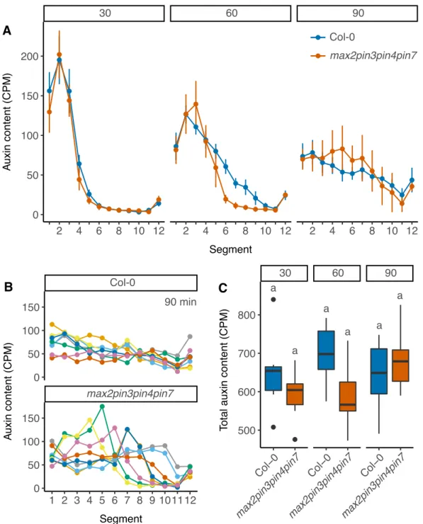
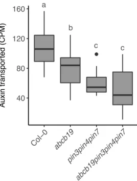
Other PIN proteins also contribute to stem auxin transport (Bennett et al., 2016a; Boot et al., 2016). Furthermore, ABCBs, which are expressed in much broader domains in the stem than PINs (Bennett et al., 2016a) can also contribute to stem auxin transport (Noh et al., 2001). The presence of many different auxin efflux proteins in the stem suggests that stem auxin transport dynamics are likely to be complex. In accordance with this, auxin pulses applied to stems generally show a degree of spreading (e.g. Brewer et al., 2009; Rashotte et al., 2003), which would not be expected if auxin moved simply from shoot to root with linear kinetics. Interestingly, the rate of spreading of a pulse over time can provide information about the underlying transport mechanism (Mitchison, 2015) and auxin pulse dynamics suggest that stem auxin transport is likely comprised of multiple auxin transport regimes with different transport properties (Bennett et al., 2016a; Boot et al., 2016; Mitchison, 2015).
1.4
Development and regulation of shoot apices
The capability of long term growth in lateral branches lies at the axillary meristem. Meris-tematic cells in the axillary meristem give rise to all the tissues that make up the branch and, for branches to grow, these meristematic cells must be formed, maintained and regulated. The regulation of meristems is under tight genetic control. The three hormones discussed above, auxin, cytokinin and strigolactone, all affect axillary meristem activity and their roles are discussed below.
1.4.1
Formation of shoot apices
All aerial parts of the plant derive from the primary shoot apical meristem (SAM). In Arabidopsis, the SAM is a dome of cells with a specific spatial arrangement. The outer L1 layer of cells divides predominantly anticlinally, forming the shoot epidermis. Immediately subtending the L1, the L2 also typically acts as a single layer of cells contributing to sub-epidermal tissues. Further inwards, the L3 divides in both anticlinal and periclinal directions and cells derived from it form core tissues in leaves and stems. Radially, distinct functional zones can be distinguished in the SAM with, at the centre of the dome, a central zone which contains slow dividing meristematic cells, which give rise to the surrounding tissues. Adjacent to this is the peripheral zone, where an increased rate of division amplifies cell
1.4 Development and regulation of shoot apices 14
populations and new organs are initiated. Underlying these two zones is the rib zone, where division and elongation contribute to the stem tissues.
Axillary meristems in Arabidopsis exhibit the same cellular and functional organisation as the primary SAM. The SAM is initiated very early in embryogenesis, whereas axillary meristems form post-embryonically in leaf axils. Axillary meristem formation is under the control of at least two hormones, auxin and cytokinin. Their role is discussed below.
1.4.2
Hormonal control of shoot meristem initiation and activity
Recent analyses have characterised the role of plant hormones in the specification of axillary meristem formation (Wang et al., 2014a,b). Low auxin levels in leaf axils precede the initiation of axillary meristems and artificially elevating auxin concentration in this region disrupts axillary meristem formation (Wang et al., 2014a,b). Axillary meristem formation is also impaired when auxin transport is disrupted. Mutants lacking PIN1 form significantly fewer axillary meristems than wild type plants and a similar effect is seen in plants lacking PINOID, an important regulator of PIN1 activity (Wang et al., 2014a,b). Axillary meristem formation is not only compromised in backgrounds where auxin export is impaired, but also inaux1lax1lax2auxin importer mutants (Wang et al., 2014a).
Axillary meristem formation is also dependent on cytokinin. Cytokinin signalling, perception and biosynthesis are increased during axillary meristem formation (Wang et al., 2014b) and mutants impaired in these processes show decreased levels of axillary meristem formation (Muller et al., 2015; Wang et al., 2014b).
Apart from their roles in meristem formation, auxin and cytokinin also play important roles in the regulation of meristem activity. The initiation of new organs at the SAM coincides with the accumulation of high concentrations of auxin (Benkova et al., 2003; Heisler et al., 2005; Vernoux et al., 2011). These auxin maxima correspond to the polarisation pattern of PIN1 in the SAM. Here, PIN1 localisation in the L1 layer is polar, and directed towards sites of inferred high auxin accumulation (Bayer et al., 2009; Benkova et al., 2003; Heisler et al., 2005; Reinhardt et al., 2003). Using computational models, the phyllotactic patterns at the shoot apex can largely be reproduced using a system where PIN1-mediated auxin accumulation controls patterning (Jönsson et al., 2006; Smith et al., 2006). Mutants lacking
PIN1 provide further evidence. The Arabidopsispin1mutant fails to produce flowers, and
applying pharmacological inhibitors of auxin transport to wild type plants can mimic this effect (Bennett et al., 1995; Okada et al., 1991). Conversely, exogenous application of auxin to such barren meristems can trigger flower production, but only when applied to the peripheral zone of the meristem, suggesting that the role of auxin maxima in organ initiation is highly tissue specific (Reinhardt et al., 2003).
1.5 Models for shoot branching control 15
The formation of auxin maxima at the SAM depends on the correct localisation of auxin
exporters, but it is also influenced by auxin importers. The aux1lax1lax2lax3 quadruple
mutant, lacking all four known auxin import proteins, displays disrupted phyllotactic pattern-ing (Bainbridge et al., 2008). The formation of auxin maxima, as well as PIN polarisation is disrupted in this mutant background and it appears that auxin importers are necessary to stabilise PIN-mediated patterning mechanisms (Bainbridge et al., 2008). Further evidence for this comes from the observation that PIN1 expression in the L1 alone is not sufficient
to restore a normal phyllotactic pattern inpin1aux1lax1mutants, and that in these mutants
PIN1 is ectopically expressed in the L2 (Kierzkowski et al., 2013).
1.5
Models for shoot branching control
The outgrowth of branches is a coordinated interaction between the shoot apices and the stem. It is likely that systemic and local effects are involved in the regulation of bud outgrowth and models which attempt to explain this are discussed below.
1.5.1
The effect of auxin on lateral bud outgrowth
Auxin produced by the shoot apex has an inhibitory effect on the outgrowth of underlying axillary buds. Experiments conducted in the 1930s showed that when the shoot apex is removed through decapitation, axillary buds which were previously inhibited were able to activate. Application of auxin to the decapitated stump was shown to restore bud outgrowth inhibition, demonstrating that auxin is the signal responsible for the inhibition (Thimann and Skoog, 1933). However, the inhibitory effect of auxin is indirect, since auxin itself does not enter the bud (Booker et al., 2003; Prasad et al., 1993). Auxin movement through the stem occurs in a basipetal manner, which precludes upward movement into the bud. Interestingly, this basipetal movement of auxin does not only result in inhibition of buds located basally to the growing shoot, but more apical buds can also be inhibited (Ongaro et al., 2008; Snow, 1929). Together, these observations suggest that auxin inhibits bud outgrowth in an indirect manner.
Two main non-exclusive hypotheses have been proposed to explain the indirect effect of auxin on bud inhibition. One model predominantly takes into account the manner in which auxin transport is dynamically regulated in the context of bud outgrowth. The other model suggests that second messengers relay the inhibitory effects of auxin. Both models are discussed next.
1.5 Models for shoot branching control 16
1.5.2
The auxin transport canalisation model for shoot branching
control
The auxin transport canalisation model for shoot branching control proposes that buds need to efficiently export their auxin in order to activate and that this export occurs through a process called canalisation. The concept of auxin transport canalisation was proposed to explain patterns of vascular strand formation in response to wounding and describes the process where an initial auxin flow from an auxin source towards an auxin sink is upregulated and polarised, and becomes limited to increasingly narrower files of auxin transporting cells, and thus becomes ‘canalised’ (Sachs, 1981). This process relies on a positive feedback between auxin flux and transport. Auxin flux upregulates and polarises its own transport in the direction of the flux (Sachs, 1981). Although the concept of canalisation was proposed prior to the molecular genetic era, observations on PIN auxin efflux proteins have confirmed the positive feedback auxin exerts on its own transport, as well as the importance of auxin transport in vascular patterning (Paciorek et al., 2005; Sauer et al., 2006; Sawchuk et al., 2013; Scarpella et al., 2006). Furthermore, when PIN localisation was traced in experiments comparable to Sachs’, the dynamic behaviour was consistent with the early predictions (Balla et al., 2011; Sauer et al., 2006).
In the context of shoot branching, canalisation is thought to play an important role in enabling buds to establish efficient and sustained auxin export into the stem. Inactive buds are potential sources of auxin which produce and export auxin upon activation (Balla et al., 2011; Li and Bangerth, 1999; Morris, 1977; Thimann and Skoog, 1933). Auxin export from buds is correlated with their growth (Li and Bangerth, 1999; Morris, 1977; Prusinkiewicz et al., 2009). An explanation for this export requirement might be that it allows continued leaf initiation and expansion (Bayer et al., 2009).
The ability of buds to canalise their auxin transport into the stem is predicted to be influenced by the sink strength of the main stem for auxin, relative to the auxin source strength of the bud. Crucial in this process is the initial auxin flux from the bud towards the stem, which, as a result of the positive feedback between auxin flux, and the upregulation and polarisation its auxin transport, is amplified to establish a polar auxin transport stream out of the bud. It is important to note that the initial flux out of the bud depends on relative, not absolute differences between auxin sources and sinks, as well as the strength of the feedback between auxin flux and its upregulation and polarisation (Prusinkiewicz et al., 2009). For example, auxin concentrations at an auxin source may remain constant in absolute terms, but become relatively stronger if the sink strength is reduced. This situation could arise if less auxin is exported into the sink. In the context of the main stem, this would occur following
1.5 Models for shoot branching control 17
decapitation of the primary shoot apex, or when other sources of auxin reduce their auxin export into the sink.
A key determinate of sink strength is the amount of auxin that is present in the main stem. Catabolism and synthesis of auxin in the main stem typically appears to be limited (discussed in Kramer and Ackelsberg, 2015). Therefore the auxin concentration in the main stem is primarily determined by the amount of auxin fed into it and the amount of auxin that is transported away towards the root. Growing shoot apices produce auxin and feed this into the stem. The amount of auxin produced by each active apex is not necessarily constant, so the individual source strength of each active apex can vary. However, each actively growing apex feeds auxin into the stem, thereby affecting the sink strength. The rate at which auxin is transported towards the root also affects the sink strength, since high levels of polar auxin transport away to the root will result in a stronger sink. The resulting relative balance between any given source and sink then determines how readily a bud is able to activate. A bud acting as a strong auxin source, combined with a stem providing a strong sink will enable a bud to canalise its auxin export easily and activate. In contrast, a bud which produces little auxin will find it more difficult to establish an initial auxin flux towards a comparable sink, and would be predicted to remain dormant. Importantly, this system assumes dynamic modulation of the auxin transport network as buds activate. All buds feed into a common sink and as such they compete for access to the polar auxin transport stream in the main stem that constitutes an important part of this sink. As one bud activates, it reduces the stem auxin sink strength by exporting auxin into it. In turn, this makes it harder for other buds to activate. This provides an explanation for the indirect inhibitory effect of auxin in this process, and also explains how buds are able to prevent other buds from activating (Prusinkiewicz et al., 2009).
1.5.3
The role of strigolactones in auxin transport canalisation
The importance of the relative differences between auxin source and sink, as well as the dynamic properties of the auxin transport network, can be used to explain the action of strigolactone on shoot branching. Mutants impaired in strigolactone signalling or biosynthesis show high levels of branching (Booker et al., 2004; Sorefan et al., 2003; Stirnberg et al., 2002; Waters et al., 2012). The high degree of branching in these mutants results at least in part from reduced competition for outgrowth between buds. Competition between buds can be measured by taking young inflorescences bearing two cauline buds and removing the primary shoot apex to release these buds from inhibition (Ongaro et al., 2008). After decapitation, both buds can activate or only one bud grows out and subsequently inhibits the activation of the other bud. Interestingly, the top and the bottom bud are both capable
1.5 Models for shoot branching control 18
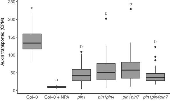
of inhibiting outgrowth (Ongaro et al., 2008). In strigolactone mutants both buds grow out, but exogenous application of strigolactone enhances competition between shoot apices, such that the ability of one bud to inhibit the outgrowth of another bud is enhanced (Crawford et al., 2010). Applying strigolactone to isolated single buds at physiologically meaningful concentrations has no effect on bud outgrowth. However, when an additional auxin source is provided through apical application of auxin to the decapitated stump, the inhibitory effect of apical auxin is enhanced by basal application of strigolactones, a process that is strigolactone signalling dependent (Crawford et al., 2010). Strigolactones are able to enhance bud-bud competition at least in part by affecting the PIN1 accumulation on the basal plasma membrane. In strigolactone mutants, levels of PIN1 on the basal plasma membrane of xylem parenchyma
cells are increased and, consistent with this, max mutants show increased levels of stem
auxin transport (Bennett et al., 2006; Crawford et al., 2010). The branchy phenotype of these mutants is correlated with the increased auxin transport, since applying low levels of NPA, an auxin transport inhibitor, is able to reduce stem auxin transport to wild type levels and results in a subsequent decrease in branching (Bennett et al., 2006; Lazar and Goodman, 2006).
Strigolactones promote PIN1 endocytosis from the plasma membrane and in themaxmutants
this process is impaired, resulting in the observed overaccumulation of PIN1 (Shinohara et al., 2013). PIN1 endocytosis by strigolactones is dependent on strigolactone signalling
through MAX2, since the response is abolished inmax2mutants. Furthermore, it occurs
independent of new protein synthesis, suggesting that strigolactone acts post-transcriptionally on PIN1 (Shinohara et al., 2013).
In the context of auxin transport canalisation, the reduced removal of PIN1 from the plasma membrane in strigolactone mutants may contribute to higher sink strength in the stem, because more basal PIN1 is able to transport auxin towards the root. In the bud, the elevated PIN1 levels may enable the bud to export and canalise its auxin more easily towards the stem, influencing source strength. Perhaps most significantly, reduced removal of PIN1 from the plasma membrane will enhance the positive feedback between auxin flux and the polarisation and upregulation of auxin transport, such that a very small initial flux of auxin from the bud to the stem will be sufficient to drive auxin transport canalisation (Prusinkiewicz et al., 2009). This combined effect allows strigolactone mutants to activate their branches more readily, resulting in increased levels of branching (Bennett et al., 2006; Prusinkiewicz et al., 2009; Shinohara et al., 2013). Thus, strigolactone is able to regulate the levels of shoot branching at least partially through its effect on the auxin transport network.
1.5 Models for shoot branching control 19
1.5.4
The second messenger model for shoot branching control
Another model for auxin-mediated inhibition of shoot branching has been proposed in which auxin acts through one or more second messengers. In this model, auxin in the stem regulates the production of a second messenger that moves directly into the bud to regulate its activity (Sachs and Thimann, 1967; Snow, 1929). Both cytokinin and strigolactone fit well within this model.
In the second messenger model, cytokinin and strigolactone are thought to act antago-nistically, with cytokinin promoting and strigolactone inhibiting bud outgrowth. Consistent with these roles, direct application of cytokinin to inhibited buds is able to activate them, even in the presence of apical auxin (Chatfield et al., 2000; Wickson and Thimann, 1958). Similarly, direct application of strigolactone to buds is able to inhibit outgrowth (Brewer et al., 2009; Gomez-Roldan et al., 2008; Umehara et al., 2008). Furthermore, strigolactone is able to reduce the stimulatory effect of cytokinin on bud outgrowth (Dun et al., 2012).
The indirect effect of auxin on bud outgrowth in this model is proposed to occur via transcriptional regulation of cytokinin and strigolactone biosynthesis. Here, auxin has been shown to act through the AXR1-AFB (AUXIN RESISTANCE PROTEIN1-AUXIN SIGNALLING F-BOX PROTEIN)-dependent auxin signalling pathway (Hayward et al., 2009; Nordstrom et al., 2004). Through this pathway auxin is able to negatively regulate
transcription of members of the IPT gene family which are involved in cytokinin synthesis,
leading to decreased cytokinin production (Nordstrom et al., 2004; Tanaka et al., 2006). Upon decapitation, repression is reduced, and cytokinin produced at the stem-bud node is thought to enter the bud, stimulating bud outgrowth.
Conversely, AXR1-AFB-dependent auxin signalling is likely to positively affect the production of strigolactones, because strigolactone biosynthesis genes are upregulated by auxin (Brewer et al., 2009; Foo et al., 2005; Hayward et al., 2009; Sorefan et al., 2003). In accordance with this, reducing auxin in the main stem through decapitation or NPA treatment decreases transcript levels of these genes (Foo et al., 2005; Hayward et al., 2009). Movement of auxin from the shoot to the root could thus stimulate strigolactone production in the root, which would lead to increased movement of strigolactone from the root to the shoot, where they could modulate bud activity. Strigolactone levels in the shoot are not only determined by synthesis in the root. Grafting experiments have demonstrated that wild type shoots grafted to strigolactone deficient roots can still suppress branching (Booker et al., 2005), suggesting that strigolactones are also synthesised in the shoot at sufficient levels to modulate bud outgrowth. Therefore it is possible that auxin is able to locally upregulate strigolactone production in the shoot, which enables the movement of strigolactone into the bud, where it can inhibit bud outgrowth.
1.5 Models for shoot branching control 20
1.5.5
The role of
BRC1
in shoot branching
A proposed mechanism by which cytokinin and strigolactone are able to inhibit bud
out-growth is via the transcriptional regulation of BRANCHED1 (BRC1). The BRC1 gene
belongs to the TCP family of transcription factors. These transcription factors contain a TCP domain, named after the first characterised members TEOSINTE BRANCHED1 in
maize, CYCLOIDEA in snapdragon and PCF1 and PCF2 in rice. In Arabidopsis,BRC1is
closely related toTEOSINTE BRANCHED1(TB1) in maize (Aguilar-Martinez et al., 2007).
During domestication of maize from its wild relative teosinte, a mutation which confers overexpression of TB1 has been selected, resulting in reduced branching and different flow-ering patterns in the shoot. Loss-of-function mutants in TB1 display increased branching
phenotypes, resembling teosinte (Doebley et al., 1997), suggesting thatTB1negatively
reg-ulates branching. Consistent with this,TB1-like mutants in Arabidopsis, such asbrc1and,
to a lesser extentbrc2, show increased levels of branching (Aguilar-Martinez et al., 2007;
Finlayson, 2007). TB1in maize andBRC genes in Arabidopsis are predominantly expressed
in the axillary buds (Aguilar-Martinez et al., 2007; Doebley et al., 1997), correlating well with a possible function in regulating axillary bud activity. Although the function of BRC1 is not specifically known, other TCP transcription factors include cell cycle regulators genes among their transcriptional targets (Kosugi and Ohashi, 2002; Li et al., 2005; Schommer et al., 2014; Trémousaygue et al., 2003). This has led to the assumption that BRC1 regulates cell cycle progression in buds, although there are many other transcriptional targets for this gene family (reviewed in Li, 2015).
Hormones can regulate the transcript levels ofBRC1. In pea, thePsBRC1orthologue is
positively regulated by cytokinin, and negatively by strigolactone (Braun et al., 2012; Dun et al., 2012), consistent with the stimulatory and inhibitory effects of these hormones on bud
outgrowth. BRC1transcript levels frequently correlate with bud activity, with highBRC1
levels correlating with bud outgrowth inhibition, and vice versa (Aguilar-Martinez et al., 2007; Finlayson, 2007; Seale et al., 2017). However, this correlation can be broken. For
example, strigolactone mutants in maize constitutively express high levels ofTB1, but still
exhibit high levels of branching (Guan et al., 2012). In Arabidopsis,brc1buds can remain
inhibited and buds with high levels ofBRC1transcript can be active, suggesting that the role
ofBRC1is more complex than that of a straightforward bud outgrowth regulator (Seale et al.,
1.5 Models for shoot branching control 21
1.5.6
Reconciling models for shoot branching control
Although the second messenger model is able to account for many shoot branching responses, some data are not easily explained. Application of physiologically meaningful concentrations of strigolactone to single buds has no effect on bud outgrowth (Crawford et al., 2010), which is difficult to explain if strigolactones are direct inhibitors of bud outgrowth. Moreover, the ability of buds to compete for outgrowth is difficult to reconcile with the direct effects predicted by the second messenger model. Following decapitation, the buds on stems with two nodes are delayed in their outgrowth, compared to a bud on stem segments bearing only one node (Crawford et al., 2010). This means that the presence of the more basal bud in the two-node situation is able to affect the outgrowth of the more apical bud, even though it is located further away from the decapitation site (Crawford et al., 2010). Auxin concentration changes at the apical node, with the presumed effects on cytokinin and strigolactone concentration, would not be able to affect bud-bud competition in this manner. Even more difficult to explain with the second messenger model is the effect of basal application of strigolactone on two-node bud outgrowth. Here, strigolactone mostly reduces the growth of only one bud (Crawford et al., 2010), which is not easy to reconcile with a direct effect of strigolactone on bud inhibition.
The auxin transport canalisation model for shoot branching readily explains these obser-vations. Here, the main stem provides a common auxin transport pathway for all the growing shoot apices. Branching patterns arise from the dynamic properties of the network, which changes the ability of shoot apices to export their auxin into the stem - a requirement for activation. In this context strigolactone does not directly regulate bud outgrowth, but instead affects the auxin transport network, in turn affecting the ability of buds to export their auxin by dampening polar auxin transport in the stem, leading to enhanced competition (Crawford et al., 2010; Prusinkiewicz et al., 2009; Shinohara et al., 2013). This dynamic regulation of auxin transport can also account for the striking observation that strigolactones can actually
promote branching in some instances, such as in thetransport inhibitor3(tir3) auxin transport
mutant, which has increased levels of branching associated with reduced levels of auxin transport (Ruegger et al., 1997; Shinohara et al., 2013).
Arguing against the auxin transport canalisation model are recent suggestions that strigo-lactone acts independently of auxin (Brewer et al., 2015). Here, application of the auxin transport inhibitor NPA to pea buds was unable to fully prevent bud outgrowth in both wild type and strigolactone-deficient mutants. Application of strigolactone to NPA-treated buds was able to inhibit bud outgrowth to a greater extent than NPA alone. This suggests that a bud activation mechanism may exist that is not correlated with auxin transport (Brewer
1.6 Aims 22
et al., 2015), but the limited understanding of the mechanism by which NPA works makes interpretation of these results difficult.
It is important to note that the auxin transport canalisation and second messenger models for shoot branching are not mutually exclusive. Both mechanisms could act in parallel, with the contribution of each varying depending on the circumstances.
1.6
Aims
The aim of this thesis is to increase the understanding of the components of the auxin transport network that may underlie the auxin transport canalisation hypothesis for shoot branching control. Specifically, the aim was to answer the following questions:
• How does auxin move down the stem?
– Which auxin exporters contribute to stem auxin transport?
– How is auxin distributed in the stem?
– Do auxin importers affect stem auxin transport?
• To what extent does auxin transport regulate bud-bud communication?
– Which auxin exporters are involved in communication between shoot apices?
– Do auxin exporters other than PIN1 act in the strigolactone signalling pathway?
– Does auxin influx play a role in bud outgrowth regulation?
• What is the relationship between the regulation of shoot branching byBRC1and auxin
transport?
– In what way does the auxin transport network affectBRC1-mediated bud
out-growth?
• Does H+-ATPase activity affect shoot branching?
– Do H+-ATPases affect auxin transport in the stem?
Chapter 2
Materials and methods
2.1
Materials and methods
2.1.1
Plant lines
Arabidopsis thaliana(‘Arabidopsis’ hereafter) plants were all in the Col-0 ecotype. Wild type refers to the Col-0 ecotype. Details of the plant lines used are shown in Appendix A. The physical location on the chromosomes of the most commonly used genes in this study can be found in Fig. 2.1.
2.1.2
Growth conditions
Arabidopsis seeds were stratified on wet filter paper at 4◦C for two to five days prior to
sowing. Plants were grown on Levington’s F2 compost pre-treated with Intercept at 0.02 g/l or Exemptor at 0.03 g/l (Levington Horticulture, Ipswich, UK). Plants were grown in P40
cellular trays (16 cm2per pot) or P24 cellular trays (25 cm2per pot). Plants were grown in
glasshouses or in controlled environment rooms according to the growth conditions and light regimes shown in Table 2.1.
2.1.3
Growth substrates
Arabidopsis thaliana salt (ATS) solution was used for in vitro growth (Wilson et al., 1990). Sucrose was added at 1 % and agar was added at 0.8 % to solidify the media, where stated.
Nitrate experiments were conducted on a mixture of sand and terra green, as described in de Jong et al. (2014). Each pot was supplied with 25 ml ATS with nitrate solution at the start of the experiment. An additional 10 ml ATS with nitrate solution was added weekly after the