R E S E A R C H
Open Access
Complement is activated in progressive
multiple sclerosis cortical grey matter
lesions
Lewis M. Watkins
1, James W. Neal
2, Sam Loveless
3, Iliana Michailidou
4, Valeria Ramaglia
4, Mark I. Rees
1,
Richard Reynolds
5, Neil P. Robertson
3, B. Paul Morgan
2and Owain W. Howell
1,5*Abstract
Background:The symptoms of multiple sclerosis (MS) are caused by damage to myelin and nerve cells in the brain and spinal cord. Inflammation is tightly linked with neurodegeneration, and it is the accumulation of neurodegeneration that underlies increasing neurological disability in progressive MS. Determining pathological mechanisms at play in MS grey matter is therefore a key to our understanding of disease progression.
Methods:We analysed complement expression and activation by immunocytochemistry and in situ hybridisation in frozen or formalin-fixed paraffin-embedded post-mortem tissue blocks from 22 progressive MS cases and made comparisons to inflammatory central nervous system disease and non-neurological disease controls.
Results:Expression of the transcript forC1qAwas noted in neurons and the activation fragment and opsonin C3b-labelled neurons and glia in the MS cortical and deep grey matter. The density of immunostained cells positive for the classical complement pathway protein C1q and the alternative complement pathway activation fragment Bb was significantly increased in cortical grey matter lesions in comparison to control grey matter. The number of cells immunostained for the membrane attack complex was elevated in cortical lesions, indicating complement activation to completion. The numbers of classical (C1-inhibitor) and alternative (factor H) pathway regulator-positive cells were unchanged between MS and controls, whilst complement anaphylatoxin receptor-bearing microglia in the MS cortex were found closely apposed to cortical neurons. Complement immunopositive neurons displayed an altered nuclear morphology, indicative of cell stress/damage, supporting our finding of significant neurodegeneration in cortical grey matter lesions.
Conclusions:Complement is activated in the MS cortical grey matter lesions in areas of elevated numbers of complement receptor-positive microglia and suggests that complement over-activation may contribute to the worsening pathology that underlies the irreversible progression of MS.
Keywords:Complement, Grey matter lesion, Innate immunity, Multiple sclerosis, Neurodegeneration
Background
Multiple sclerosis (MS) is an inflammatory, demyelinat-ing and neurodegenerative disease of young adults. Damage can occur throughout the central nervous sys-tem, and the pathology of the grey matter can be exten-sive [1, 2]. Progresexten-sive MS, marked by increasing
irreversible disability and reduced quality of life, is char-acterised pathologically by extensive cortical demyelin-ation [3]. Magnetic resonance imaging has demonstrated that an increasing number of cortical lesions, and lesions of deep grey matter, are predictive of disease course [4]. Grey matter pathology is seen from the earliest stages, and inflammation is linked to neurodegeneration throughout the disease [5]. There are now concerted efforts to better understand the innate and adaptive immune mechanisms that drive this pathology to identify disease relevant prog-nostic markers and new therapeutic opportunities. * Correspondence:[email protected]
1Institute of Life Sciences, Swansea University School of Medicine, Swansea
SA2 8PP, UK
5Division of Brain Sciences, Imperial College London, London, UK
Full list of author information is available at the end of the article
The complement system is central to innate and adaptive immune responses. Complement is synthe-sised in the brain, and adult human neurons are a major source of parenchymal complement [6], which can also be generated systemically. Complement is im-portant for synaptic pruning during development, complement signalling causes neuronal morphological changes in the adult and complement-labelled neurons are targeted by complement receptor-bearing phago-cytes [7–10]. Complement is activated through the classical, lectin and alternative pathways that generate anaphylatoxins C3a and C5a and opsonins, including C3b [11, 12]. Build-up of complement fragment C3b can lead to C5 convertase formation with proteolysis of the C5 component and subsequent activation of the terminal pathway leading to the formation of the membrane attack complex (MAC). To avoid self-injury, host cells express an array of complement regu-latory proteins (Cregs) that, for example, inhibit C3-cleaving enzymes (factor H), prevent C1q assembly with C1r, s and initiation of the classical pathway (C1-inhibitor) or block assembly of the MAC (clusterin) [13]. Intrathecal and blood-borne levels of comple-ment proteins mirror the MS disease profile [14–17], but we need to know more about the role of comple-ment in pathogenesis.
Evidence for complement activation in MS grey mat-ter is mixed, with some reporting little evidence for complement activation in purely cortical lesions [18]. Others have shown complement to be differentially
expressed [19–21] and the complement recognition
and initiation protein C1q to be associated with degen-erating synapses in the MS hippocampus [22]. As yet, Creg expression in MS grey matter has not been re-ported. We have examined the localisation of comple-ment recognition molecules (C1q), activation products (C3b, Bb, MAC), regulators (factor H, C1 inhibitor, clusterin) and receptors (C3aR, C5aR and complement receptor 3/ CD11b) for the first time in order to better understand the immune mechanisms of MS cortical grey matter pathology relevant to disease progression.
Methods Study cohort
Tissue was provided by the UK Multiple Sclerosis Society Tissue Bank (MSSTB) at Imperial College (MS and non-neurological controls, including cryopreserved samples) and the Oxford Brain Bank, Oxford University (inflamma-tory and non-neurological controls) with appropriate re-search ethics approval (South West Wales REC study approval 13/WA/0292). Of these samples, 22 cases of neu-ropathologically confirmed MS, 15 non-neurological con-trols and eight non-MS neurological concon-trols with glial activation and neuroinflammation (four ischaemic stroke, four viral encephalitis; referred to collectively as“non-MS inflammatory controls”) were used for the main data col-lection (Table 1 and Additional file 1:Table S1). Non-neurological and non-MS inflammatory controls were selected based on the availability of tissue blocks from the pre-determined study regions and gender and age similar to the available MS cohort. In all instances, the fresh brain was cut into 1-cm-thick coronal slabs before either cryo-preserving in cold isopentane on a bed of dry ice or placed in freshly prepared fixative (for formalin-fixed, paraffin-embedded samples). Sections from representative anatom-ically matched brain regions were prepared for each case as follows: (1) superior frontal gyrus, (2) cingulate gyrus, (3) thalamus, and (4) hippocampus and inferior temporal lobe. Cryosections from region-matched blocks of frontal, cingulate and hippocampus and inferior tem-poral lobe were available for the complete MS cohort and, together with an additional eight non-neurological controls (MSSTB), were used to assess complement ter-minal pathway activation (staining for C9neo and clus-terin) and complement receptor staining.
Tissue characterisation
All cases were processed histologically for luxol fast blue/cresyl fast violet and haematoxylin/eosin, and sub-sequent sections were stained with antibodies to myelin-oligodendrocyte-glycoprotein (MOG) and human leukocyte antigen (HLA-D) to determine tissue architecture, cellular infiltration, demyelination and the density of microglia/
Table 1Summary of multiple sclerosis and control groups used for quantitative analysis
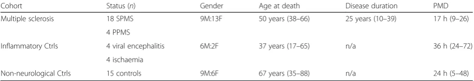
Cohort Status (n) Gender Age at death Disease duration PMD
Multiple sclerosis 18 SPMS 9M:13F 50 years (38–66) 25 years (10–39) 17 h (9–26)
4 PPMS
Inflammatory Ctrls 4 viral encephalitis 6M:2F 37 years (17–65) n/a 36 h (24–72)
4 ischaemia
Non-neurological Ctrls 15 controls 9M:6F 67 years (35–88) n/a 24 h (5–48)
Inflammatory controls are non-MS neurological controls characterised by neuroinflammation and gliosis. Group values showed as median (data range). See (Additional file1: Table S1) for individual case details including cause of death, disease activity at death and sampled blocks
macrophages (Figs. 1 and 2 and Additional file 1: Figure S1). White matter lesions (WML), deep grey matter (thal-amus) and hippocampus lesions of MS were classified as follows: active (demyelinated lesion core confluent with HLA-D+ microglia/macrophages and the presence of early myelin (MOG+ inclusion) degradation products); chronic active (HLA-D+ microglia/macrophages restricted to the lesion edge and the presence of early and late (LFB+ inclu-sion) myelin degradation products) or chronic inactive (pale or no rim of ramified microglia (with late myelin deg-radation products) at the edge). Grey matter lesions (GML) of the frontal, cingulate and temporal gyri were charac-terised based on location within the cortical laminae and were described as subpial (type III), intracortical (type II) and leukocortical involving the subcortical white matter (type I) or as lesions spanning the entire width of cortex from the pia to the leukocortical junction, but without in-volving the white matter (type IV) [23]. Our analysis of le-sions affecting the deep cortical laminae (layers V and VI) included both type I and IV lesion areas. All cortical GMLs used in this study (subpial and those affecting the deep cortical laminae) were characterised as chronic (ac-tive or inac(ac-tive) based on the number and distribution of HLA-D+ microglia/macrophages and the presence of early (chronic active) or late (chronic inactive) myelin deg-radation products [24].
Immunostaining protocols and image capturing
Paraffin wax sections were de-waxed, rehydrated and subjected to heat-induced epitope retrieval as described previously [25]. Following overnight incubation with the primary antibody, sections were incubated with biotinyl-ated secondary antibody and avidin-biotin peroxidase complex with diaminobenzidine as the chromogen (Impact DAB; Vector Laboratories Ltd., Peterborough, Cambridgeshire, UK). Individual antibody details are listed in Additional file 1: Table S2. Snap-frozen, unfixed cryosections were air-dried, fixed in methanol or 4 %
paraformaldehyde and quenched with H2O2 before
immunostaining. Immunofluorescence staining was performed on wax or snap-frozen sections by sequen-tial antibody incubation and detection, followed by diamidino-2-phenylindole (DAPI) counterstaining. In all instances, sections from each MS case for each anti-body were immunostained in the same experimental run to ensure comparability of labelling. All experi-ments included primary antibody-negative controls and irrelevant species-specific antisera as positive con-trols. Sections were viewed on a Leica DRMB bright-field microscope (Leica Microsystems, Milton Keynes, Buckinghamshire, UK), a Zeiss Axio Imager under epi-fluorescence or a Zeiss LSM 710 confocal (Carl Zeiss Ltd., Cambridge, Cambridgeshire, UK).
Quantitative analysis
All quantification and analysis was performed with the researcher blinded to the case identity. The mean num-ber of immunostained cells was calculated for each com-plement protein or cell phenotypic marker of interest from ×100 images (field of view (fov) area = 0.3 mm2) of regions of interest: normal or demyelinated cortical lam-inae I–III (i.e. subpial lesions); normal or demyelinated cortical laminae V–VI (i.e. leukocortical and type IV le-sions); normal or demyelinated subcortical white matter; and for C3b-iC3b only, normal or lesioned thalamus (ventral nucleus) and normal or lesioned CA1 of the ros-tral hippocampus. Positively stained cells were manually tagged in ImageJ (http://imagej.nih.gov/ij/) using the
“multipoint”tool. Layer V NeuN-immunopositive neurons were automatically counted using the “analyse particles” tool following image transformation and thresholding. Changes in nuclear area and shape indicate cell stress/ damage [26, 27]. We investigated the nuclear morphology of Smi32+ pyramidal neurons of layers V–VI co-labelled with anti-C3b-iC3b using high-resolution confocal z-stacks (captured under sequential scanning of the blue, green and red channels, using a plan apochromat ×63/ 1.40 oil immersion objective, image scaling = 0.07 μm/ pixel, optical section = 0.5μm). Images of single (Smi32+, C3b−)- or double (Smi32+/C3b+)-stained cells were imported to ImageJ, and optical sections, where the nu-cleus was sectioned most centrally (visible nucleolus and z-section where nucleus at its widest diameter), were out-lined using the“wand”tool and morphometric parameters calculated for each nucleus using the “shape descriptors” tool. A minimum of twenty co-labelled Smi32/C3b-iC3b+ neurons per case, from eight MS cases, were assessed.
In situ hybridisation
and a rabbit anti-sheep immunoglobulin G/horseradish peroxidase (HRP) (Dako). HRP was visualised using Vector NovaRED (red-brown reaction product) prior to perman-ent mounting (VectaMount, Vector Laboratories).
Data presentation and statistical testing
Data was handled in Excel (Microsoft Office, 2010) and analysed using GraphPad Prism (v6.05, GraphPad Soft-ware, CA, USA). Appropriate multi-group comparisons and correlation analysis were performed following D’Agostino and Pearson normality testing. In all in-stances, case means per region of interest (e.g. GML, WML) were compared and a p< 0.05 was regarded as significant.
Results
Complement is activated in MS cortical and deep grey matter
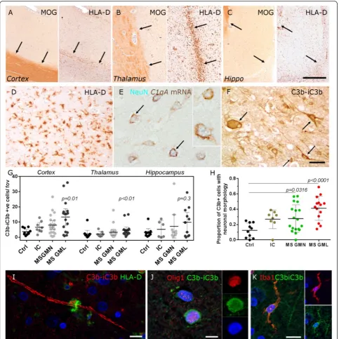
Chronic inflammatory demyelinating lesions of the neo-cortex, thalamus and hippocampus grey matter (GM) were identified by histological and immunohistochemical assessment (Fig. 1a–d) as described in the methods. In
situ hybridisation for complement C1QA transcript
showed that complement C1q is synthesised by neurons of the deep cortical laminae in MS (Fig. 1e). Neurons and glia were immunostained with an antibody to the central complement component C3b (and its initial cleavage product iC3b) (Fig. 1f ). Quantification of C3b immunopositive cells revealed an increased number in MS GMLs (of cortical and deep grey matter—thalamus), in comparison to non-neurological controls (Fig. 1g). There was an increase in the proportion of C3b+ cells with a neuronal morphology out of the total number of C3b+ quantified cells in MS cortical GM (normal appear-ing grey matter (GMN) and GML), in comparison to non-neurological control samples (Fig. 1h). In addition to notable labelling of cells resembling neurons (arrows in Fig. 1f), C3b immunoreactive myelin was present, fre-quently closely apposed with HLA-D+ phagocytes (Fig. 1i); oligodendrocytes (Olig-1+; Fig. 1j) and microglia (Iba-1+; Fig. 1k). The number of C3b immunostained cells was not associated with confounding variables such as age of
death, post-mortem delay or whether death was infection related (Additional file 1: Table S3).
Classical, alternative and terminal pathway activation products are present in MS cortical grey matter lesions
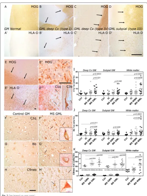
We focussed our attention on describing complement ac-tivation in association with neocortical demyelination and neurodegeneration in progressive MS. Cortical GMLs were described as subpial, leukocortical or spanning the entire cortical ribbon but without affecting the underlying white matter (Fig. 2a–d). The majority of cortical GMLs were chronic inflammatory demyelinating lesions whilst classically active cortical GMLs were infrequently ob-served (Fig. 2e). In a classically active cortical GML (con-fluent with HLA-D+ macrophages containing early myelin degradation products; Fig. 2e′–e′′), complement C1q and complement activation fragment C3b+ cells were noted (Fig. 2e′′′). We stained and quantified the density of C1q, fragment Bb and C9neo immunopositive cells in MS normal-appearing GM and chronic GML areas, in comparison to MS inflammatory controls and non-neurological controls. Cells with a neuronal, oligodendro-cyte and/or astrooligodendro-cyte-like morphology were labelled by antibodies against C1q, fragment Bb and C9neo in grey and white matter areas (Fig. 2f–h; (Additional file 1: Fig-ures S2, S3)). The pattern of cell-associated complement labelling in MS and control brain was similar to that seen in Alzheimer’s disease cortex, which was used as a positive staining control (see Additional file 1: Figure S4). Immu-nostaining revealed a significantly greater number of complement-labelled cells (neurons and glia) in deeper cortical laminae of the MS GMLs (leukocortical and type IV) in comparison to region and neuronal layer-matched controls (Fig. 2i–k). The number of C1q+ and C3b+ cells was elevated in active cortical GMLs (albeit with ann= 2) in comparison to chronic cortical GMLs (n= 18; 12.0 and 31.1 positive cells per field of view compared with 7.7 ± 1.8 and 12.5 ± 2.6 C1q and C3b+ cells in active and chronic GML groups, respectively). The density of C9neo + cells, determined from unfixed, cryopreserved samples from the same cortical regions from the same MS cases, was significantly elevated in deep cortical (leukocortical and type IV) and subpial (type III) GMLs in comparison (See figure on previous page.)
to non-inflammatory controls. WMLs generally displayed two- to tenfold more complement immunopositive cells in comparison to grey matter areas in the same tissue block. The density of C1q-, Bb- and C9neo-positive cells in MS WMLs was increased in comparison to normal-appearing and control white matter (Fig. 2i–k, Additional file 1: Figure S3).
The expression of key complement classical, alternative and terminal pathway regulators in cortical grey matter lesions is similar in MS and controls
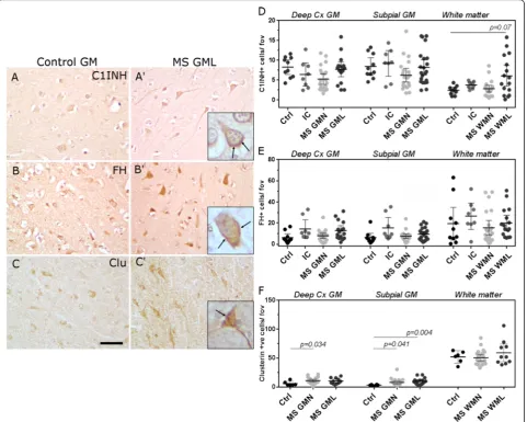
We next determined the numerical density of cells immu-nolabelled for the classical pathway Creg, C1-inhibitor (C1INH), the alternative pathway regulator factor H (FH)
and the terminal pathway regulator clusterin (Fig. 3). In agreement with our description of complement activation fragment staining, Creg staining was seen on and within cells with a neuronal and/or glial morphology in cortical grey matter areas (Fig. 3a–c, Additional file 1: Figures S2, S3). The number of C1INH and FH immunolabelled cells was unchanged between MS and control (Fig. 3d, e), whilst the density of clusterin immunostained cells was increased in MS GMN regions in comparison to non-neurological controls (Fig. 3f, note that cryosections of in-flammatory controls were not available). These data imply that classical and alternative pathways are active despite unchanged expression of key Cregs in lesions of the dee-per cortical laminae.
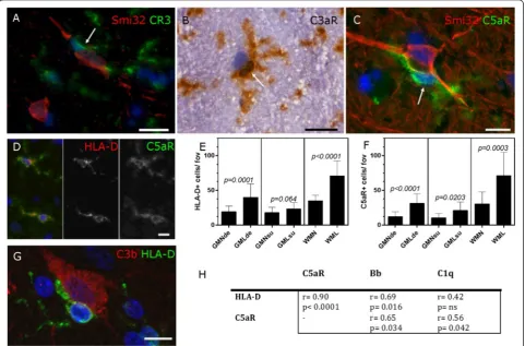
Microglia are complement anaphylatoxin receptor positive and are increased in density in cortical grey matter lesions
We immunostained cryopreserved MS tissue from our complete MS cohort to reveal the presence of com-plement receptor 3+ (CR3/CD11b), C3aR+ and C5aR + cells with a microglial morphology in close contact with cortical neurons (Fig. 4a–c). C5aR+ microglia were activated (HLA-D+, Fig. 4d) and were signifi-cantly increased in number in GMLs and WMLs, in comparison to normal appearing tissues in the same blocks from the same cases (Fig. 4e, f ). HLA-D+ microglia were seen tightly associated with C3b+ cor-tical neurons and the density of activated microglia (C5aR+ and HLA-D+ cells) correlated with the num-ber of classical (C1q+) and alternative (fragment Bb+) cells in GMLs affecting the deep cortical laminae (Fig. 4h).
Complement is associated with morphological and immunophenotypic markers of stress and
neurodegeneration in MS cortical grey matter lesions
C3b+ neurons of the deeper cortical laminae were iden-tified by their co-expression of non-phosphorylated neu-rofilament protein (Smi32+, Fig. 5a, b). Complement activation fragment C3b immunostaining visualised by confocal microscopy was noted on the surface and in the cytoplasm of cortical neurons (Fig. 5a), whilst some of these cells had an altered nuclear morphology (Fig. 5b). The proportion of cortical neurons (Smi32+) that were complement C3b+ was increased in MS GML (Fig. 5c). C3b+ neurons in MS cortex showed signs of nuclear stress as defined by morphometric analysis of nuclear area and shape (aspect ratio; Fig. 5d, e) [27]. Quantitative evidence of neuronal dysmorphia was sup-ported by the presence of C3b+ neurons expressing hypo-phosphorylated neurofilaments (Smi34+), neuronal
nuclei expressing the pro-apoptotic kinase and neur-onal stress/damage marker protein kinase R (pPKR) [29]—that were more numerous in MS GM in compari-son to control GM (Fig. 5g, g′), and the presence of
C3b+/cleaved caspase3+ or C3b+/TUNEL reaction+ cortical neurons (Fig. 5h, i). Within the same regions of interest, there was a significant reduction in the
num-ber of NeuN+ neurons in normal (p= 0.013) and
lesioned cortical layer V MS GM (p= 0.002) in com-parison to region matched non-neurological controls (Fig. 5j–j′′).
Discussion
Progressive MS is associated with a widespread and chronic activation of the central immune response con-fined behind a relatively intact blood-brain barrier [2, 30]. Our quantitative analysis demonstrates that comple-ment classical, alternative and terminal pathways are ac-tivated and we show for the first time that complement expression is notable in and on large neurons in MS cor-tical grey matter lesions. Our data suggest that a dysreg-ulation of complement activation and control occurs in the MS brain, and an increase in complement anaphyla-toxin receptor-positive microglia may serve to sustain the neuroinflammatory response that drives myelin and neuronal pathology in the progressive phase.
Complement is activated in MS cortical grey matter lesions
In the cortical grey matter, C1q expression was seen in and on neurons, neurites and glia. The pattern of stain-ing suggested biosynthesis as well as a deposition of
complement, which is supported byC1q mRNA
expres-sion in NeuN+ neurons, shown here and by others [22]. Evidence for alternative complement pathway activation in our study is seen in the elevated number of activation fragment Bb+ cells. Therefore, localisation of the opso-nin C3b (and its breakdown product iC3b) in and on neurons in GMLs of the cortex, thalamus and hippo-campus may be a consequence of classical and/or alter-native pathway activation.
Proteolysis of C3 yields C3a, a soluble anaphylatoxin that activates both protective and damaging immune re-sponses against neurons through C3aR engagement. Membrane bound C3b activates CR3+ microglia to trig-ger activation that can be detrimental to cell integrity, whilst recent evidence suggests that C3a and C3b can be generated intracellularly [31]. Accumulation of C3b can lead to formation of the C5 convertase and release of C5a, which is damaging to neurons via C5aR activation [32, 33]. We show that the number of C5aR+ microglia is increased in chronic cortical lesions. C5b formation and the subsequent recruitment of C6-9 lead to the for-mation of MAC. MAC deposition is seen in acute and chronic MS WMLs [19, 34, 35] but hitherto has not been demonstrated for cortical GMLs. The MAC may be directly cytopathic (there is a significant loss of cor-tical neurons in these same lesions of interest, Fig. 5), but even sublethal attack can trigger the production of pro-inflammatory cytokines and reactive oxygen species, stimulate binding of damage-associated molecular pat-terns [36], confer protection [37] or mediate NLRP3 inflammasome-induced IL1βsynthesis [38].
Our quantitative findings are in agreement with quali-tative reports describing C1q, C3d and C4d immunore-activity in some cortical GMLs [18–21], and quantitative measures of elevated C1q and C3d labelling in the hippocampus [22], in a cell-associated pattern of immu-nostaining similar to that seen by us in the cortical grey matter. In agreement with previous publications [18, 19], we noted that the pattern of complement immunoreac-tivity (recognition molecules and activation fragments) was most striking in cortical layers V–VI near the white matter border; however, we report that the density of complement-labelled cells was not related to the pres-ence of underlying WMLs (Additional file 1: Figure S5), confirming that a major part of the complement re-sponse is generated locally by cells of the cortical grey matter.
Complement regulator expression in MS cortical grey matter lesions does not increase with activation—evidence for dysregulation?
Uncontrolled activation of complement is detrimental to the host and results in progressive cell and tissue injury in the chronically inflamed organ [13]. Each complement activation pathway is regulated at multiple strategic points to prevent uncontrolled activation. We did not detect a significant difference in the numerical density of cells immunolabelled for C1INH or FH between MS cortical grey matter and control samples. The demon-stration of unchanged number of complement regulator-positive cells despite on-going complement activation suggests that the drive to complement activation over-whelms regulation. We suggest that such a disparity be-tween markers of activation and regulation of the classical and alternative complement pathways manifests as the robust and widespread expression of fragments of complement activation, leading to the unchecked gener-ation of opsonin and soluble anaphylatoxin products that may exacerbate the pathology in the progressive MS brain. It is for these reasons that complement markers could be valuable prognostic indicators of a more severe disease [15, 17].
Microglial activation and neuronal injury and loss
and deposition of products of complement activation, in the presence of increased numbers of complement re-ceptor positive microglia, could be contributory. These findings suggest that in this environment of significant neuronal loss, a substantial proportion of the remaining large (Smi32+) neurons are under inflammatory stress and appear dysmorphic, which would render them dys-functional. Complement may drive neuronal damage through C3b(iC3b)-CR3 activation of phagocytes in a process of primary phagocytic damage [40] or through the activation of local glia through anaphylatoxin recep-tor engagement that can cause dendritic damage and neuronal toxicity [10, 41, 42]. Complement synthesised by neurons could be a physiological response to stress, which may aid synaptic pruning by locally activated microglia or engage neuronal complement receptors resulting in al-tered neuritic and synaptic presentation [9, 22].
Conclusions
The presence of complement activation products and anaphylatoxin receptor positive activated microglia sug-gest that neurons in the MS cortical grey matter are sub-ject to a sustained innate immune attack that may contribute to their dysfunction and death. Our work supports efforts to investigate the utility of complement as a potential biomarker or therapeutic target for pro-gressive MS.
Additional file
Additional file 1: Table S1.Demographic and pathological details for cases used in this study. All cases were retrospectively confirmed as MS, non-inflammatory or inflammatory (viral encephalitis or ischaemia) following detailed analysis of patient health records and full neuropathological work up. Region matched sections (FFPE or cryosections) were available for all cases from the frontal cortex (sup. Frontal gyrus), cingulate cortex and hippocampus and temporal (inf.) gyrus. Tissue sections from inflammatory control cases were selected based on region-matching and not on the presence of pathology in the particular block. Examples of myelin and inflammatory cell staining are shown in Additional file:Figure S1. Abbreviations: PMD- post-mortem delay from death to tissue processing (hrs); Disease activity- neuropathologically defined disease activity based on the presence (Active) or absence (Stable) of active inflammatory lesions; Disease duration is calculated from retrospectively determined time of dis-ease onset; Tissue- available formalin-fixed paraffin embedded (FFPE) or cryopreserved tissue blocks; Quantified lesions- number of GM or WM lesion regions of interest analysed in this quantitative study.Table S2.Primary antibodies used in this study, retrieval conditions and commercial source. Citrate- 100mM sodium citrate, 10mM citric acid, pH 6.0; TRIS/ EDTA- 10mM Tris, 1mM EDTA, pH 8.5.Table S3.Complement cell-associated expression was not related to age of death, post-mortem delay or whether death was infection (inflammatory) or non-infection related. Linear regression and Pearson’s correlation analysis (Age of death and PMD (post mortem delay) versus mean C3b+ cell count per case) or Mann-Whitney test of mean C3b + cell count between those cases with an inflammatory-related or non-inflammatory disease noted at death.Figure S1.Non-neurological controls and inflammatory neurological controls used in this study. Control cortical grey and subcortical white matter was characterised by a uniform pattern of anti-MOG staining (A- D) and small numbers of ramified HLA-D+ microglia (E- H). Inflammatory controls were selected on the basis of available region-matched blocks and frequently displayed an unremarkable pathology.
Occasional areas of microglial activation were noted in some sections of cortical and subcortical tissue (I- P; ischaemic encephalitis inflammatory control), whilst perivascular cuffs of activated microglia and macrophages were sometimes seen in cases of viral encephalitis (Q- X). Scale bars= W (and all low power images) 100µm; X (and all high power images) 20µm.
Figure S2.Complement recognition fragment, activation products and
regulator expression in MS, MS inflammatory controls (IC) and non-neurological control (Ctrl) grey matter. 400x magnification images of C1q (A-C), C1-inhibitor (D-F), Bb (G-I), factor H (J-L), C3b-iC3b (M-O), MAC (P, Q) and clusterin (R, S) in deeper cortical laminae of control (Ctrl; left column & P, R), MS (central column & Q, S) and non-MS inflammatory controls (right column only) showing the cell-specific and often robust pattern of anti-complement immunostaining seen in MS (and to a lesser extent, non-MS inflammatory controls) cortical grey matter in comparison to non-diseased controls. Scale bar= 20µm.Figure S3.Complement recognition fragment, activation product and regulator expression in subcortical white matter samples. 400x magnification images of C1q (A-C), C1-inhibitor (D-F), Bb (G-I), factor H (J-L), C3b-iC3b (M-O), MAC (P, Q) and clusterin (R, S) in sub-cortical white matter of non-neurological control (Ctrl), MS and non-MS inflammatory controls (IC), showing the cell-specific and often robust pattern of anti-complement immunostaining seen in MS white matter in comparison to non-neurological controls. Scale bar= 20µm.Figure S4. Examples of complement immunolabelling in Alzheimer’s disease (AD). Two AD cases (age of death, 85 and 96yrs; gender, both female; post-mortem delay, 30 and 19hrs, respectively), showed complement specific immunolabelling with anti-C1q, C3b-iC3b and C9neo of cells resembling neurons and glia (similar to that seen in MS and controls), as well as diffuse labelling of amyloid plaques (for anti-C1q, C3b and C9neo antibodies). Cellular immunolabelling of C1INH and clusterin resembles that seen in control, MS and inflammatory control cases. C5aR+ glia were also seen in the AD deep grey matter. All images captured at 200x magnification.Figure S5.Complement activation in cortical grey matter is not related to underlying white matter inflammation. Grey matter lesions of the deeper cortical laminae are characterised as type I (involving both the white and grey matter) or type IV (involving the entire vertical extent of the cortex but without affecting the underlying white matter). Our analysis revealed there to be no difference between the density of C1q (A,B and D) and C3b-labelled cells (C) in type I grey matter lesions (that are associated with white matter lesions) and type IV lesions. Scatter graphs of type I versus type IV cell-specific counts for C1q and C3b quantitative immunostaining compared by Mann-Whitney test. (PDF 1849 kb)
Abbreviations
C1INH, C1 inhibitor; Clu, clusterin; Creg, complement regulator; Ctrl, control cohort; FH, factor H; GML, grey matter lesion; GMN, normal appearing grey matter; IC, non-MS inflammatory controls; MAC, membrane attack complex; MOG, myelin-oligodendrocyte glycoprotein; WML, white matter lesion
Acknowledgements
We thank Mr Ryan Harley and Ms Rhian Evans for their excellent technical assistance. We would like to thank Dr Djordje Gveric and the UK MS Society Tissue Bank (funded by the UK MS Society grant 007/14) and Drs Carolyn Sloan and Marie Hamard at the Oxford Brain Bank (supported by the Medical Research Council, Brains for Dementia Research, Alzheimer Society and Alzheimer Research UK, Autistica UK and the NIHR Oxford Biomedical Research Centre).
Funding
This work was funded through the UK Multiple Sclerosis Society (grant number 993), the British Neuropathological Society and the St. David’s Medical Foundation.
Availability of data and materials
The datasets supporting the conclusions of this article are included within the article (and its Additional file 1).
Authors’contributions
interpretation, and literature search. All authors were involved in writing the paper and had final approval of the submitted version.
Competing interests
VR is a co-inventor of a patent that describe the use of inhibitors of the ter-minal complement pathway for therapeutic purposes; she is a co-founder of Regenesance BV and holds IP and equity. NPR has received research support, travel awards and honoraria from Biogen, Novartis, Sanofi, Genzyme and Bayer (for work unrelated to this study). BPM is a Consultant to GlaxoSmithK-line. The other authors have no conflict of interest to declare.
Consent for publication Not applicable.
Ethics approval and consent to participate
The study of human post-mortem tissue at Swansea Institute of Life Sciences was approved by the South West Wales Research Ethics Committee (study approval number 13/WA/0292).
Author details
1Institute of Life Sciences, Swansea University School of Medicine, Swansea
SA2 8PP, UK.2Institute of Infection and Immunity, Cardiff University, Cardiff, UK.3Institute of Psychological Medicine and Clinical Neuroscience, Cardiff University, Cardiff, UK.4Department of Genome Analysis, Academic Medical Centre, Amsterdam, The Netherlands.5Division of Brain Sciences, Imperial College London, London, UK.
Received: 18 March 2016 Accepted: 3 June 2016
References
1. Reynolds R, Roncaroli F, Nicholas R, Radotra B, Gveric D, Howell O. The neuropathological basis of clinical progression in multiple sclerosis. Acta Neuropathol. 2011;122:155–70.
2. Lassmann H, Brück W, Lucchinetti CF. The immunopathology of multiple sclerosis: an overview. Brain Pathol. 2007;17:210–8.
3. Kutzelnigg A, Lucchinetti CF, Stadelmann C, Brück W, Rauschka H, Bergmann M, et al. Cortical demyelination and diffuse white matter injury in multiple sclerosis. Brain. 2005;128:2705–12.
4. Calabrese M, Romualdi C, Poretto V, Favaretto A, Morra A, Rinaldi F, et al. The changing clinical course of multiple sclerosis: a matter of gray matter. Ann Neurol. 2013;74:76–83.
5. Frischer JM, Bramow S, Dal-Bianco A, Lucchinetti CF, Rauschka H, Schmidbauer M, et al. The relation between inflammation and neurodegeneration in multiple sclerosis brains. Brain. 2009;132:1175–89. 6. Shen Y, Li R, McGeer EG, McGeer PL. Neuronal expression of mRNAs for
complement proteins of the classical pathway in Alzheimer brain. Brain Res. 1997;769:391–5.
7. Fraser DA, Laust AK, Nelson EL, Tenner AJ. C1q differentially modulates phagocytosis and cytokine responses during ingestion of apoptotic cells by human monocytes, macrophages, and dendritic cells. J Immunol. 2009;183: 6175–85.
8. Fraser DA, Pisalyaput K, Tenner AJ. C1q enhances microglial clearance of apoptotic neurons and neuronal blebs, and modulates subsequent inflammatory cytokine production. J Neurochem. 2010;112:733–43. 9. Stevens B, Allen NJ, Vazquez LE, Howell GR, Christopherson KS, Nouri N,
et al. The classical complement cascade mediates CNS synapse elimination. Cell. 2007;131:1164–78.
10. Lian H, Yang L, Lu H, Lian H, Yang L, Cole A, et al. NF k B-activated astroglial release of complement C3 compromises neuronal morphology and function associated with Alzheimer’s disease. Neuron. 2015;85:101–15. 11. Walport MJ. Complement. First of two parts. N Engl J Med. 2001;344:1058–66. 12. Ricklin D, Lambris JD. Complement in immune and inflammatory disorders:
pathophysiological mechanisms. J Immunol. 2013;190:3831–8.
13. Zipfel PF, Skerka C. Complement regulators and inhibitory proteins. Nat Rev Immunol. 2009;9:729–40.
14. Sellebjerg F, Jaliashvili I, Christiansen M, Garred P. Intrathecal activation of the complement system and disability in multiple sclerosis. J Neurol Sci. 1998;157:168–74.
15. Ingram G, Hakobyan S, Hirst CL, Harris CL, Pickersgill TP, Cossburn MD, et al. Complement regulator factor H as a serum biomarker of multiple sclerosis disease state. Brain. 2010;133:1602–11.
16. Ingram G, Hakobyan S, Hirst CL, Harris CL, Loveless S, Mitchell JP, et al. Systemic complement profiling in multiple sclerosis as a biomarker of disease state. Mult Scler. 2012;18:1401–11.
17. Aeinehband S, Lindblom RPF, Al Nimer F, Vijayaraghavan S, Sandholm K, Khademi M, et al. Complement component C3 and butyrylcholinesterase activity are associated with neurodegeneration and clinical disability in multiple sclerosis. PLoS One. 2015;10:e0122048.
18. Brink BP, Veerhuis R, Breij ECW, van der Valk P, Dijkstra CD, Bö L. The pathology of multiple sclerosis is location-dependent: no significant complement activation is detected in purely cortical lesions. J Neuropathol Exp Neurol. 2005;64:147–55.
19. Gay F, Drye TJ, Dick GWA, Esiri MM. The application of multifactorial cluster analysis in the staging of plaques in early multiple sclerosis. Identification and characterization of the primary demyelinating lesion. Brain. 1997;120:1461–83. 20. Schwab C, McGeer PL. Complement activated C4d immunoreactive
oligodendrocytes delineate small cortical plaques in multiple sclerosis. Exp Neurol. 2002;174:81–8.
21. Ingram G, Loveless S, Howell OW, Hakobyan S, Dancey B, Harris CL, et al. Complement activation in multiple sclerosis plaques: an
immunohistochemical analysis. Acta Neuropathol Commun. 2014;2:53. 22. Michailidou I, Willems JGP, Kooi E-J, van Eden C, Gold SM, Geurts JJG, et al.
Complement C1q-C3-associated synaptic changes in multiple sclerosis hippocampus. Ann Neurol. 2015;77:1007–26.
23. Bø L, Vedeler CA, Nyland HI, Trapp BD, Mørk SJ. Subpial demyelination in the cerebral cortex of multiple sclerosis patients. J Neuropathol Exp Neurol. 2003;62:723–32.
24. Peterson JW, Bo L, Mork S, Chang A, Trapp BD, Bö L, et al. Transected neurites, apoptotic neurons, and reduced inflammation in cortical multiple sclerosis lesions. Ann Neurol. 2001;50:389–400.
25. Howell OW, Schulz-Trieglaff EK, Carassiti D, Gentleman SM, Nicholas R, Roncaroli F, et al. Extensive grey matter pathology in the cerebellum in multiple sclerosis is linked to inflammation in the subarachnoid space. Neuropathol Appl Neurobiol. 2015;41:798–813.
26. Ayala YM, Misteli T, Baralle FE. TDP-43 regulates retinoblastoma protein phosphorylation through the repression of cyclin-dependent kinase 6 expression. Proc Natl Acad Sci U S A. 2008;105:3785–9.
27. Eidet JR, Pasovic L, Maria R, Jackson CJ, Utheim TP. Objective assessment of changes in nuclear morphology and cell distribution following induction of apoptosis. Diagn Pathol. 2014;9:92.
28. Budde BS, Namavar Y, Barth PG, Poll-The BT, Nürnberg G, Becker C, et al. tRNA splicing endonuclease mutations cause pontocerebellar hypoplasia. Nat Genet. 2008;40:1113–8.
29. Bose A, Mouton-Liger F, Paquet C, Mazot P, Vigny M, Gray F, et al. Modulation of tau phosphorylation by the kinase PKR: implications in Alzheimer’s disease. Brain Pathol. 2011;21:189–200.
30. Leech S, Kirk J, Plumb J, McQuaid S. Persistent endothelial abnormalities and blood-brain barrier leak in primary and secondary progressive multiple sclerosis. Neuropathol Appl Neurobiol. 2007;33:86–98.
31. Liszewski MK, Kolev M, Le Friec G, Leung M, Bertram PG, Fara AF, et al. Intracellular complement activation sustains T cell homeostasis and mediates effector differentiation. Immunity. 2013;39:1143–57. 32. Pavlovski D, Thundyil J, Monk PN, Wetsel RA, Taylor SM, Woodruff TM.
Generation of complement component C5a by ischemic neurons promotes neuronal apoptosis. FASEB J. 2012;26:3680–90.
33. Woodruff TM, Crane JW, Proctor LM, Buller KM, Shek AB, de Vos K, et al. Therapeutic activity of C5a receptor antagonists in a rat model of neurodegeneration. FASEB J. 2006;20:1407–17.
34. Compston DAS, Morgan BP, Cambell AK, Wilkins P, Cole G, Thomas ND, et al. Immunocytochemical localization of the terminal complement complex in multiple sclerosis. Neuropathol Appl Neurobiol. 1989;15:307–16. 35. Storch MK, Piddlesden S, Haltia M, Iivanainen M, Morgan P, Lassmann H.
Multiple sclerosis: in situ evidence for demyelination. Ann Neurol. 1998;43: 465–71.
36. Li YP, Mold C, Du Clos TW. Sublytic complement attack exposes C-reactive protein binding sites on cell membranes. J Immunol. 1994;152:2995–3005. 37. Cole DS, Hughes TR, Gasque P, Morgan BP. Complement regulator loss on
38. Triantafilou K, Hughes TR, Triantafilou M, Morgan BP. The complement membrane attack complex triggers intracellular Ca2+ fluxes leading to NLRP3 inflammasome activation. J Cell Sci. 2013;126:2903–13. 39. Mahad DH, Trapp BD, Lassmann H. Progressive multiple sclerosis 1:
pathological mechanisms in progressive multiple sclerosis. Lancet Neurol. 2015;14:183–93.
40. Brown GC, Neher JJ. Microglial phagocytosis of live neurons. Nat Rev Neurosci. 2014;15:209–16.
41. Gasque P, Singhrao SK, Neal JW, Wang P, Sayah S, Fontaine M, et al. The receptor for complement anaphylatoxin C3a is expressed by myeloid cells and nonmyeloid cells in inflamed human central nervous system: analysis in multiple sclerosis and bacterial meningitis. J Immunol. 1998;160:3543–54. 42. Boos L, Campbell IL, Ames R, Wetsel RA, Barnum SR. Deletion of the
complement anaphylatoxin C3a receptor attenuates, whereas ectopic expression of C3a in the brain exacerbates, experimental autoimmune encephalomyelitis. J Immunol. 2004;173:4708–14.
• We accept pre-submission inquiries
• Our selector tool helps you to find the most relevant journal • We provide round the clock customer support
• Convenient online submission • Thorough peer review
• Inclusion in PubMed and all major indexing services • Maximum visibility for your research
Submit your manuscript at www.biomedcentral.com/submit