Open Access
Primary research
Influence of
RAR
α
gene on
MDR1
expression and P-glycoprotein
function in human leukemic cells
Tatjana P Stromskaya, Ekaterina Y Rybalkina, Tatjana N Zabotina,
Alexander A Shishkin and Alla A Stavrovskaya*
Address: Institute of Carcinogenesis, N.N. Blokhin Russian Cancer Research Centre of the Russian Academy of Medical Sciences, Kashirskoye sh 24, Moscow 115478, Russia
Email: Tatjana P Stromskaya - [email protected]; Ekaterina Y Rybalkina - [email protected]; Tatjana N Zabotina - [email protected]; Alexander A Shishkin - [email protected]; Alla A Stavrovskaya* - [email protected]
* Corresponding author
Abstract
Background: Multidrug resistance (MDR) phenotype of malignant cells is the major problem in the chemotherapy of neoplasia. The treatment of leukemia with retinoids is aimed on the induction of leukemic cells differentiation. However the interconnections between retinoid regulated differentiation of leukemic cells and regulation of MDR remains unclear.
Methods: Four lines of cultured leukemic cells of diverse types of differentiation were infected with RARα gene and stable transfectants were isolated. We investigated the differentiation of these cells as well as the expression of RARα and MDR1 genes and P-glycoprotein (Pgp, MDR protein) functional activity in these cells.
Results: All RARα transfected sublines demonstrated the increase in the quantity of RARα mRNA. All these sublines became more differentiated. Intrinsic activity of MDR1 gene (but not Pgp functional activity) was increased in one of the transfectants. All-trans-retinoic acid (ATRA) induced Pgp activity in two of three infectants to a larger extent than in parental cells.
Conclusion: The data show that RARα regulates MDR1/ Pgp activity in human leukemic cells, in the first place, Pgp activity induced by ATRA. These results show that RARα overexpression in leukemic cells could result in MDR.
Background
Multidrug resistance (MDR) phenotype of malignant cells is the major problem in the chemotherapy of neoplasia. P-glycoprotein (Pgp) activity is recognised to be one of the major mechanisms responsible for MDR. Pgp transports many structurally diverse compounds across the cell membrane and confers the MDR phenotype in tumor cells [1]. A number of signaling pathways participate in the regulation of MDR1 gene expression and the activity
of its product, Pgp [2]. Some of these signaling pathways could participate in coordinated regulation of MDR1/Pgp activity, cell proliferation and cell differentiation. It was shown that retinoic acid (RA) can modulate MDR1 gene expression [3-5]. Retinoids are known to be involved into the regulation of the cell growth, differentiation and apoptosis. In the last decade retinoids became implicated into the treatment of leukemia and some solid tumors [6]. This approach changed the focus of the haematological
Published: 24 May 2005
Cancer Cell International 2005, 5:15 doi:10.1186/1475-2867-5-15
Received: 22 October 2004 Accepted: 24 May 2005
This article is available from: http://www.cancerci.com/content/5/1/15
© 2005 Stromskaya et al; licensee BioMed Central Ltd.
diseases treatment from the cytotoxicity of the anti-cancer drugs to the reversal of arrested maturation of leukemic cells. Retinoids act via two families of receptors (RARs – RARα, RARβ, RARγ) and RXRs (RXRα, RXRβ, RXRγ). There is the evidence that RARα is the crucial receptor mediating the biological effects during retinoid signaling in some cells [7]. Cell differentiation caused by the stable overex-pression of receptor RARα was shown to result in consti-tutive over expression of MDR1 gene in some cultured cells of solid tumors [4]. However the interconnections between RA/RARα regulated differentiation of leukemic cells and regulation of MDR1/Pgp activity remains unclear. In some leukemic cells RA did not influence
MDR1 and/or Pgp activity, while in the others it either augmented or reduced MDR1/Pgp expression [5,8]. The aim of this study is to investigate if effects of all-trans -retin-oic acid (ATRA) on MDR1/Pgp activity in leukemic cells are connected with RARα expression and with the leuke-mic cell differentiation. We isolated sublines of cultured leukemic cells characterized by the stable RARα
overex-pression and investigated the constitutive and ATRA induced MDR1/Pgp activity in these cells. Our data show that various RARα transformed leukemic cell lines
acquired more differentiated phenotype. Constitutive level of MDR1 gene expression increased in one of RARα
overexpressing cell sublines. RARα overexpression did not influence Pgp functional activity while Pgp activity induced by ATRA was elevated in all infectants studied. This shows that the main effect of RARα in the cells stud-ied is its influence on the induced functional activity of Pgp.
Methods
Cell lines and culture
Lines of cultured leukemic cells used in the study: H9 cells (acute human T-cell leukemia) [9], KG-1 cell line (cells of acute myelogenous leukemia) [10], K562 cell line (cells obtained from the patient in blast crisis of chronic mye-loid leukemia) [11], NB4 (acute promyelocytic leukemia) [12].
Cells were grown in RPMI-1640 medium supplemented with 10% fetal calf serum (Gibco, USA), 2 mM L-glutamine, 50 µg/ml gentamycin at 37°C in a fully humidified atmosphere of 95% air and 5% CO2. All the derived cell lines described in this paper were obtained by retroviral infection and selection with the appropriate antibiotic. ATRA (all-trans-retinoic acid, Sigma, USA) was added to the culture medium at seeding or 24 hours after seeding (see Legends to Figures).
Expression vector and retrovilal infection
The PA317/LRARSN retroviral vector-producing cell line was used. All the procedure was described earlier [4]. In brief, the vector used contains a cDNA fragment
harbour-ing the complete codharbour-ing sequence of the RARα gene
driven by the Moloney murine leukemia virus long-termi-nal repeat as well as the SV40 early promoter-driving neo-mycin phosphotransferase gene (neo) as a selectable marker [13]. The cells (4 × 10⁵ per 25-cm2 flask) were
seeded 24 h before infection. Conditioned medium from a retrovirus-producing cell line was filtered through a 0.45-µm membrane (Millipore, USA), diluted 1:1 with medium, containing 1% serum and 8 µg /ml Polybrene and added to the cells for 24 h at 37°C, 5% CO2. Further selection were carried out by culturing cells in medium supplemented with 400 µg/ml G418 (Gibco, USA) for at least 21 days. The medium was changed twice a week. The pool of G418-resistant cells was resuspended in culture medium and progressively expanded.
Assay of cell growth, apoptosis and differentiation Cells were seeded into 24-well plates (1 × 10⁴ cell per
well) and the cell number was counted at days 1, 3, 5 and 8 after seeding. The apoptosis in the populations of the parental and RARαinfected cell lines was performed using the standard procedure [14]. Cells were collected 24 h after seeding, washed with PBS, and fixed in 70% ethanol overnight at 4°C. Fixed cells were suspended in citric buffer and stained with propidium iodide (5 mcg/ml) in PBS for 1 hour at 4°C. DNA content was subsequently measured by FACScan (Becton Dickinson, USA).
The immunophenotype of the cells was evaluated as pre-viously described [15]. Surface expression of the following antigens was determined: CD3, CD5, CD7, CD8, CD11b, CD13, CD15, CD33, CD34, HAE3 and HAE9. In brief, cells were incubated with phycoerythrin-labelled mono-clonal mouse antibodies for 20 min at 4°C (Becton, Dick-inson), washed with RPMI 1640 medium and analyzed with a flow cytometer (Becton Dickinson).
Analysis of rhodamine 123 (Rh123) efflux by the cells The technique used in the study was described in [16]. Cells were loaded with 5 µg/ml Rh123 (Sigma) for 10 min at 37°C, washed twice with cold PBS, pH 7.2, and incu-bated for 30 min in dye-free medium at 37°C. After the completion of incubation, cell were washed twice with cold PBS. Cell fluorescence was measured on a flow cytometer FACScan (Becton Dickinson, USA). Each meas-urement counted 5000 events. Non-viable cells were gated out of the analysis on the basis of side scatter.
RNA isolation and reverse transcriptase polymerase chain
reaction (RT-PCR) analysis of RARα and MDR1 genes
expression
1.8% agarose gels. The samples with clearly visualized 18S and 28S RNA bands were used for further procedures. First-strand cDNA was synthesized using reverse tran-scriptase M-MuLV (MBI Fermentas, Russia) with 4 µg RNA as a template, 2.5 ng random hexamers, 0.25 mM of each deoxynucleotide triphosphate (SibEnzyme, Russia), dithi-othreitol, 4 Units of RNAase inhibitor (MBI Fermentas, Russia) and 100 Units of M-MuLV RT. The reaction was performed at 42°C for 50 min, and 1/60 volume of reac-tion mixture was used for amplificareac-tion. PCR was done in a total volume of 25 µl using the thermocycler "Tercyc" (DNA-technology, Russia). The PCR mixture consisted of (NH4)2SO4-containing PCR buffer ("MBI Fermentas"), 0.160 mM dNTPs mix ("MBI Fermentas"), 2 mM MgCl2, 20 pmoles of each specific primer and 0.8 Unit of Taq-polymerase ("MBI Fermentas"). PCR was done as follows: 94°C for 2 seconds, Tm (different for each gene) for 10 seconds, 72°C for 5 seconds. Semi-quantitative PCR anal-ysis of RARa and MDR1 genes expression were performed using oligomers amplifying a 333 bp and 167 bp prod-ucts, respectively. Specific gene primers used for RT-PCR are given in Table 1. The amounts of template cDNAs were normalized by PCR amplification of β2-microglobulin cDNA (internal control). The optimal numbers of PCR cycles were 24 for the b2-microglobulin, 26 for RARα-spe-cific product, 33 for MDR1 (for all cells lines except KG1 and KG1/RAR, for these cells the numbers of PCR cycles
MDR1-specific product were 26). These numbers of cycles yielded clearly detectable PCR products within an expo-nential range. PCR products were amplified in separate tubes, resolved by electrophoresis in 2% agarose gel, stained with ethidium bromide and visualized in UV light.
Results
Influence of RARα gene overexpression on cell
differentiation, proliferation and spontaneous apoptosis
RARα gene was introduced into the cultured leukemic cells of diverse types of differentiation as described in Methods. The sublines of H9, KG-1, K562 and NB4 cells characterized by the capability to grow in the medium
supplemented with G418 were isolated (H9/RAR, KG-1/ RAR, K562/RAR and NB4/RAR). Semi-quantitative RT-PCR revealed more pronounced expression of RARα
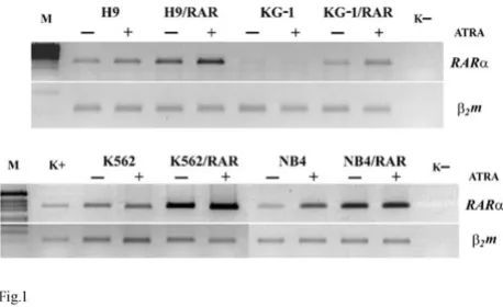
mRNA in all transfected cell lines in comparison with the wild type cells (Fig. 1). ATRA (5 µM applied for 48 h) increased RARαmRNA in some RARαtransformed cells (H9/RAR, KG-1/RAR, K562/RAR) to a greater extent than in parental cells (Fig. 1).
The investigation of the differentiation status of these cells shows that all RARα transfected sublines differ from the parental cell populations (Fig. 2). RARα transfected H9 culture contains more cell variants expressing CD5 and CD8 antigens than parental cell line (Fig. 2A). Thus the number of cells with antigens of later lymphoid differen-tiation markers increased in RARα overexpressing H9 cells. There is phenotypic evidence of granulocytic differ-entiation in KG-1/RAR cell subline as indicated by a reduction in CD13 expression and the increase in the expression of CD11b antigen in comparison with parental cells (Fig. 2B). In KG-1/RAR cell population the portion of CD34 cells decreased and the portion of CD33 cells increased (Fig. 2B). This also testifies to increased differ-entiation of these RARα overexpressing cells. In K562/ RAR population the number of the cells of erythroid dif-ferentiation (expressing HAE9 and HAE3 antigens) is larger than in K562 population (Fig. 2C). Hemoglobin synthesis is increased in K562/RAR culture more than 5-fold in comparison with parental cells (not shown). In Table 1: Specific gene primers used for RT-PCR
Gene Product size
Primer Sequence
RARα 333 bp F 5'-GTCTTTGCCTTCGCCAACCAG-3' R 5'-GCCCTCTGAGTTCTCCAACA-3' MDR1 127 bp F 5'-CCCATCATTGCAATAGCAGG-3' R 5'-GTTCAAACTTCTGCTCCTGA-3'
β2m 114 bp F 5'-ACCCCCACTGAAAAAGATGA-3' R 5'-ATCTTCAAACCTCCATGATG-3'
Expression of RARαmRNA in parental and RARαtransfected cell lines. RT PCR
Figure 1
Expression of RARαmRNA in parental and RARα transfected cell lines. RT PCRM. k- – water. ATRA (5
NB4/RAR population the number of the cells of myeloge-nous differentiation (expressing CD11b and CD15 anti-gens) is larger than in NB4 population (Fig. 2D).
The percentage of cells undergoing spontaneous apopto-sis increased 2-3-fold in all RARαtransfected cell popula-tions (Fig. 3). This could be connected with more differentiated phenotype of RARα transformed cells. It seems that in the population of H9/RAR cells the increased number of apoptotic cells could be at least in part connected with increased expression of CD95 (Fas/ APO1): in this RARαtransformed subline CD95 increased almost 10-fold in comparison with parental cell popula-tion (from 2,6% in H9 to 21,4% in H9/RAR culture). However in KG-1 and K562 cell populations the number of CD95 expressing cell did not increase after RARα trans-formation.
As Fig. 4 shows, RARα transfected KG-1, K562 and NB4 cells proliferated more slowly than parental cells. How-ever H9/RAR cells did not demonstrate slower prolifera-tion rate. Thus, more differentiated status of the RARα
transformed cell populations was not necessary connected with the decrease in the proliferation rates. All RARα
transformed cells seem to be more sensitive than wild type cells to inhibitory action of ATRA on cell proliferation (Fig. 5).
Comparison of antigen expression by the parental and RARα
infected cell lines
Figure 2
Comparison of antigen expression by the parental and RARαinfected cell lines. Cells were incubated for 30 min at 4°C in the presence of an appropriate monoclonal antibody. After three washes with PBS, cells were incubated for 30 min at 4°C with goat antimouse IgG labeled with phy-coerythrin and then analyzed in flow cytometer (Becton Dickinson).
Spontaneous apoptosis in the populations of the parental and
RARαinfected cell lines
Figure 3
Spontaneous apoptosis in the populations of the parental and RARαinfected cell lines. Propidium iodide flow cytometry detection of dead cells was performed using the standard procedure. Cells were collected 24 h after seeding, washed with PBS, and fixed in 70% ethanol overnight at 4°C. Fixed cells were suspended in citric buffer and stained with 5 mcg/ml propidium iodide in PBS for 1 hour at 4°C. DNA content was subsequently measured by FACScan (Bec-ton Dickinson, USA). All cells with sub-G0 DNA content
were regarded as dead cells. This figure is representative of 3 separate experiments.
0 10 20 30 40 50
H9
H9/RAR
KG-1
KG-1/RAR
K562
K562/RAR
NB4
NB4/RAR
Proliferation rates of parental and RARαinfected cells
Figure 4
Proliferation rates of parental and RARαinfected cells. Cells were seeded into 24-well plates (1 × 10⁴ cell per
well) and the cell number was counted at days 1,3,5,8 after seeding. This figure is representative of 3 separate
experiments.
0 10 20 30 40 50 60
1 6 8 10 days
cell number x 10000
H9 H9/RAR
0 20 40 60 80 100
1 6 8 10 days
cell number x 10000
KG1 KG1/RAR
0 10 20 30 40 50
1 6 8 days
cell number x 10000
NB4 NB4/RAR
0 10 20 30 40 50
1 6 8 10 days
cell number x 10000
K562 K562/RAR
Influence of retinoic acid (ATRA, 5 µM) on the proliferation of parental and RARαtransfected cells
Figure 5
Influence of retinoic acid (ATRA, 5 µM) on the prolif-eration of parental and RARαtransfected cells. Cells were seeded into 24-well plates (1 × 10⁴ cell per well), ATRA
was added at seeding and the cell number was counted at days 1, 3, 5 and 8 after seeding. This figure is representative of 3 separate experiments.
A
ATRA ATRA
0 10 20 30 40 50
H9H9/RAR
cel
l number
x 10000
B
ATRA ATRA
0 5 10 15 20 25
KG-1KG-1/RAR
cel
l numb
er
x 100
00
C
ATRA ATRA
0 10 20 30 40
K562K562/RAR
cell numbe
r x 10000
D
ATRA ATRA
0 20 40 60 80
NB4NB4/RAR
ce
ll numbe
r x
Influence of RARα overexpression on MDR1 gene activity We studied intrinsic and ATRA induced expression of
MDR1 gene in all cell lines by semi-quantitative RT-PCR technique. The basal levels of MDR1 mRNA varied in dif-ferent wild type cells: in H9 and NB4 cells constitutive
MDR1 gene expression was not revealed, in K562 wild type cells some MDR1 mRNA was found, in KG-1 cells the quantity of MDR1 mRNA was large (Fig. 6). It is notewor-thy that the optimal number of PCR cycles were 33 for
MDR1-specific product in all cells while for studies of KG1 and KG1/RAR cells we used 26 PCR cycles. In RARα trans-fected H9 cells the constitutive expression of MDR1 gene slightly increased, while in KG-1/RAR and NB4/RAR cells the constitutive MDR1 mRNA quantity was not elevated in comparison with the wild type cells, it seems to be even slightly decreased in K562/RAR (Fig. 6). Thus the altera-tions of the basal level of MDR1 expression in RARα trans-formed cells seem to vary in different cell lines.
ATRA (5 µM applied for 48 h) increased MDR1 gene expression in all examined cell lines either in parental or
RARαtransfected cells (Fig. 6). In H9/RAR cells effect of ATRA on MDR1 expression was significantly greater in comparison with parental cells. In other ATRA treated
RARα transformed cell sublines MDR1 expression was undistinguishable from ATRA treated parental cells (Fig 6).
Intrinsic and retinoic acid induced expression of MDR1 gene in parental and RARαtransfected cells
Figure 6
Intrinsic and retinoic acid induced expression of
MDR1 gene in parental and RARαtransfected cells. k-
– water. ATRA (5 µM) was added to cell cultures 24 h after seeding for 48 h. Then the cells were collected and proc-essed as specified in "Methods" (RNA isolation and reverse transcriptase polymerase chain reaction (RT-PCR) analysis of MDR1 gene expression). The optimal numbers of PCR cycles were 33 for MDR1-specific product for all cells lines except KG1 and KG1/RAR, for these cells the numbers of PCR cycles for MDR1-specific product were 26. This figure is rep-resentative of 2 separate experiments.
Evaluation of Rh123 efflux from the parental and cells RARα
transfected cells
Figure 7
Influence of RARα gene transformation on Rh123efflux by the cells
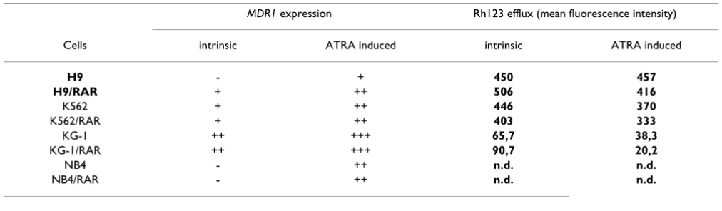
The retention of Rh123 by the cells is considered as a test for Pgp functional activity [16,17]. Rh123 efflux from the cells was increased in K562/RAR cells in comparison with the parental cell population (Fig. 7B, Table 2). In H9 and KG-1 RARαtransformed cells there were alterations in the Rh123 retention (Fig. 7A and 7C): in the populations of H9/RAR and KG-1/RAR cultures the fraction of more dull cells decreased in comparison with parental cultures (mean fluorescence intensity of the cell sublines studied are given in the table 2). This shows that Pgp activity was not elevated in these RARαtransformed cell populations and suggests that there is some decrease in Pgp functional activity.
There was increase in the portion of Rh123 dull cells after ATRA treatment both in K562 and K562/RAR cell popula-tions (mean fluorescence intensity of both populapopula-tions decreased approximately on 17%) (Fig. 8C, D, Table 2). ATRA induced Rh123 efflux from H9/RAR cells, while in H9 parental population this drug had no effect (Fig. 8A, B, Table 2). In KG-1/RAR cells ATRA induced very prominent increase in the number of Rh123 dull cells (more than 70% decrease of mean fluorescence intensity), while in the parental cell population ATRA decreased mean fluo-rescence intensity to a lesser extent (Fig. 8E, F, Table 2).
Discussion
The treatment of leukemia with retinoids is aimed on the induction of leukemic cells differentiation. The question is: are there interconnections between RA/RARα regulated differentiation of leukemic cells and MDR1/Pgp activity? In this study we have isolated more differentiated variants of the cultured leukemic cells by the introduction into the cells of RARα gene encoding one of RA receptors. All
RARαtransformed leukemic cell populations were charac-terized by the higher RARαgene expression in
compari-son with the parental cells. All RARα transformed leukemic cell populations became more differentiated. This was demonstrated by the studies of the differentiation markers, by the increase in the number of cells dying by spontaneous apoptosis and by the decrease of the proliferation rates of most RARα transfected cell sublines. Thus, RARαoverexpression could result in the increase of the differentiation of various leukemic cell populations.
We compared MDR1 gene expression and Pgp functional activity tested by Rh123 retention in parental and RARα
transformed cells. The results are summarized in the Table 2. Increased constitutive (uninduced) expression of
MDR1 gene was found in one of four cell lines after RARα
transformation (H9/RAR, Table 2, Fig. 6). In the previous experiments with melanoma and hepatoblastoma human cells we have shown that constitutive expression of MDR1
gene was increased after RARαtransfection in both RARα
transformed cell sublines [4]. Thus interconnections between regulation of the basal MDR1 and RARα activi-ties could exist both in the cells of solid tumors and in the leukemic cells. Our data suggest, that in the cell popula-tions of solid tumors RARα overexpression could be accompanied by constitutive MDR1 over-expression more often than in the cells of hematopoietic malignancies.
Our study did not reveal the occurrence of the functional Pgp in leukemic cells studied after RARαtransformation. In H9/RAR cells elevation of the constitutive MDR1
expression did not lead to the increase in Rh123 efflux (Fig. 7A, Table 2). Some studies also have described dis-crepancies between Pgp (protein) or MDR1 mRNA expres-sion and Pgp function in leukemic cells [18,19]. These discrepancies could occur for a variety of reasons. Anyway, our data show that increase in the differentiation of leukemic cell populations induced by RARα overexpres-sion did not result in the elevation of constitutive Pgp
Table 2: Influence of RARαtransformation on the intrinsic and induced expression of MDR1 gene
MDR1 expression Rh123 efflux (mean fluorescence intensity)
Cells intrinsic ATRA induced intrinsic ATRA induced
H9 - + 450 457
H9/RAR + ++ 506 416
K562 + ++ 446 370
K562/RAR + ++ 403 333
KG-1 ++ +++ 65,7 38,3
KG-1/RAR ++ +++ 90,7 20,2
NB4 - ++ n.d. n.d.
functional activity. In our previous study we found that
RARαoverexpression did not change Pgp functional activ-ity in two RARα transformed sublines of human cells (melanoma and hepatoblastoma) but did change it in the rat cells [4]. It seems that exogeneous RARαin the cells of human malignancies does not influence basal Pgp func-tional activity.
In KG-1/RAR characterized by the increased differentiaion (Table 1) we had not found increase in the constitutive
MDR1 expression and Pgp functional activity decreased (Fig. 7C, Table 2). It is known that blood stem cells and early progenitors expressing CD34 antigen also express high levels of functionally active Pgp [20]. Maturation of these cells is accompanied by the decrease in Pgp expres-sion and even more rapid decrease in Pgp functional
activ-Influence of retinoic acid (ATRA) on Rh123 efflux from the parental and cells RARαtransfected cells
Figure 8
Influence of retinoic acid (ATRA) on Rh123 efflux from the parental and cells RARαtransfected cells. ATRA (5
Publish with BioMed Central and every scientist can read your work free of charge
"BioMed Central will be the most significant development for disseminating the results of biomedical researc h in our lifetime."
Sir Paul Nurse, Cancer Research UK
Your research papers will be:
available free of charge to the entire biomedical community
peer reviewed and published immediately upon acceptance
cited in PubMed and archived on PubMed Central
yours — you keep the copyright
Submit your manuscript here:
http://www.biomedcentral.com/info/publishing_adv.asp
BioMedcentral
ity [21]. It may be suggested that alterations of Pgp function in KG-1/RAR are connected with the differentia-tion of these cells.
The situation with Pgp functional activity induced by ATRA in the cells studied differs from the situation with constitutive activity of this protein. In all three RARα
transfected cells ATRA had induced Pgp fuctional activity (Fig. 8. Table 2). Moreover, in two RARαtransformed sub-lines (H9/RAR and KG-1/RAR) ATRA activated Pgp, while in the parental cells it had either no effect (H9) or acti-vated Pgp to a lesser extent (KG-1) (Table 2). These data suggest that RARα participate in the control of induced, but not in constitutive Pgp functional activity in leukemic cells.
The regulation of MDR1 gene transcription and Pgp func-tional activities are the complex processes [1,2]. The stud-ies of these processes are underway. Our data show that
RARα gene overexpression could influence the induced Pgp functional activity in leukemic cells, i.e. could participate in the occurrence of multidrug resistance in the populations of these malignant cells. It seems that this influence could depend on the cell context.
Acknowledgements
This work was supported by grants 04-04-48613a and 02-04-48200 from the Russian Foundation for Basic Research.
References
1. Ambudkar SV, Dey S, Hrycyna CA, Ramachandra M, Pastan I, Gottes-man MM: Biochemical, cellular, and pharmacological aspects
of the multidrug transporter. Annu Rev Pharmacol Toxicol 1999,
39:361-398.
2. Scotto KW: Transcriptional regulation of ABC drug
transporters. Oncogene 2003, 22:7496-511.
3. Bates SE, Mickley LA, Chen YN, Richert N, Rudick J, Biedler JL, Fojo
AT: Expression of a drug resistance gene in human
neurob-lastoma cell lines: modulation by retinoic acid-induced
differentiation. Mol Cell Biol 1989, 9:4337-4344.
4. Stromskaya TP, Rybalkina EY, Shtil AA, Zabotina TN, Filippova NA, Stavrovskaya AA: Influence of exogenous RARα gene on MDR1 expression and P-glycoprotein function in human and rodent
cell lines. Br J Cancer 1998, 77:1718-1725.
5. Tokura Y, Shikami M, Miwa H, Watarai M, Sugamura K, Wakabayashi M, Satoh A, Imamura A, Mihara H, Katoh Y, Kita K, Nitta M: Aug-mented expression of P-gp/multi-drug resistance gene by
all-trans retinoic acid in monocytic leukemic cells. Leuk Res 2002,
26:29-36.
6. Altucci L, Gronemeyer H: The promise of retinoids to fight
against cancer. Nature Rev Cancer 2001, 1:181-193.
7. Schneider SM, Offterdinger M, Huber H, Grunt TW: Activation of
retinoic acid receptor α is sufficient for full induction of
retin-oic responses in SK-BR-3 and T47D human breast cancer
cells. Cancer Res 2000, 60:5479-5487.
8. Zhou DC, Marie JP, Maisonneuve L, Faussat-Suberville AM, Zittoun R:
Effect of differentiating agents on modulation of MDR1 gene expression in multidrug-resistant hematopoietic HL60/DNR
cell line. Exp Hematol 1993, 21:779-784.
9. Popovic M, Sarngadharan MG, Read E, Gallo RC: Detection, isola-tion and continuous producisola-tion of cytopathic retroviruses
(HTLV III) from patients with AIDS and at risk for AIDS.
Sci-ence 1984, 224:497-500.
10. Koeffler HP, Golde DW: Acute myelogenous leukemia: a
human cell line responsive to colony-stimulating activity.
Sci-ence 1978, 200:1153-1154.
11. Lozzio BB, Lozzio CB: Properties and usefulness of the original
K-562 human myelogenous leukemia cell line. Leuk Res 1979,
3:363-370.
12. Lanotte M, Martin-Thouvenin V, Najman S., Balerini P, Valensi F, Berger R: NB4, a maturation inducible cell line with t(15;17) marker isolated from human acute promyelocytic leukemia
(M3). Blood 1991, 77:1080-1086.
13. Collins SJ, Robertson KA, Mueller LeM: Retinoic acid-induced granulocytic differentiation of HL-60 is mediated directly
through the retinoic acid receptor (RARα). Mol Cell Biol 1990,
10:2154-2163.
14. Ohta H, Yatomi Y, Sweeney EA, Hakomory S, Igarashi Y: A possible role of sphingosine in induction of apoptosis by tumor
necro-sis factor-alpha in human neutrophils. FEBS Lett 1994,
355:267-270.
15. Turkina AG, Baryshnikov Ju A, Sedjakhina NP, Folomeshkina SV, Sokolova MA, Khoroshko ND, Stavrovskaya AA: Studies of P-glyc-oprotein in chronic myeloid leukemia patients: expression,
activity and correlations with CD34 antigen. Bri J Haematol
1996, 92:88-96.
16. Egudina SV, Stromskaya TP, Frolova EA, Stavrovskaya AA: Early steps of P-glycoprotein expression in cell cultures studied
with vital fluorochrome. FEBS Letters 1993, 329:63-66.
17. Feller N, Kuiper CM, Lankelma J, Ruhdal JK, Scheper RJ, Pinedo HM, Broxterman HJ: Functional detection of MDR/P170 and MRP/ P190 mediated multidrug resistance in tumour cells by flow
cytometry. Br J Cancer 1995, 72:543-549.
18. Marie J-P, Zhou DC, Gurbuxani S, Legrand O, Zittoun R:
MDR1/P-glycoprotein in haematological neoplasms. Eur J Cancer
1996:1034-1038.
19. Bailly JD, Muller C, Jaffre'zou JP, Demur C, Gassar G, Bordier C, Lau-rent G: Lack of correlation between expression and function
of Pgp in acute myeloid leukemia. Leukemia 1995, 9:799-807.
20. Chaudhary PM, Mechetner EB, Roninson IB.: Expression and activ-ity of the multidrug resistance P-glycoprotein in human
peripheral blood lymphocytes. Blood 1992, 80:2735-2739.
21. List AF: Role of multidrug resistance and its pharmacological
modulation in acute myeloid leukemia. Leukemia 1996,