ABSTRACT
Background and Objectives: Vitamin Dis an essential secosteroid that plays a crucial role in the homeostasis of a few mineral elements, particularly calcium. Since vitamin D deficiency and thyroid diseases are two important global health problems, we aimed to investigate a possible relationship of vitamin D and calcium levels with hypothyroidism in an Iranian population.
Methods: This case-control study was conducted on 175 subjects with hypothyroidism (75 males and 100 females) and 175 euthyroid controls (85 males and 90 females) who were referred to a laboratory in Gorgan, Iran. Serum levels of 25-hydroxyvitamin D, calcium, thyroid-stimulating hormone (TSH), free triiodothyronine (free T3) and thyroxine (total T4) were measured in all participants.
Results: Vitamin D and calcium were significantly lower in patients with hypothyroidism (P<0.0001). Free T3 and calcium levels differed significantly among hypothyroid patients based on their vitamin D status (P<0.0001), but vitamin D levels were within sufficient range in all groups. Moreover, there was a positive correlation between free T3 with vitamin D (r= 0.337, P<0.0001) and calcium (r= 0.361, P<0.0001) levels. Conclusions: Our results suggest that there may be a relationship between decreased vitamin D levels and thyroid function parameters.
Keywords: Vitamin D Deficiency, Hypocalcemia, Hypothyroidism, Thyrotropin, Thyroxine.
Mojtaba Zare Ebrahimabad(MSc) Department of Biochemistry and Biophysics, Metabolic Disorders Research Center, Faculty of Medicine, Golestan University of Medical Sciences, Gorgan, Iran
Hanieh Teymoori (MSc)
Department of Biochemistry and Biophysics, Metabolic Disorders Research Center, Faculty of Medicine, Golestan University of Medical Sciences, Gorgan, Iran
Hamid Reza Joshaghani (PhD)
Professor of Clinical Biochemistry, Laboratory Sciences Research Center (LSRC), Golestan University of Medical Sciences, Gorgan, Iran
Corresponding author: Hamid Reza Joshaghani
Tel: +98-1732436108
E-mail: [email protected]
Address: Laboratory Sciences Research Center (LSRC), Golestan University of Medical Sciences, Gorgan, Iran
Vitamin D Status and its Relationship with Thyroid
Function Parameters in Patients with Hypothyroidism
This paper should be cited as: Zare Ebrahimabad M, Joshaghani HR [Vitamin D Status and its Relationship with Thyroid Function Parameters in Patients with Hypothyroidism]. mljgoums. 2019; 13(5): 8-12
Received: 30 Jun 2018
Revised: 27 Aug 2019
Accepted: 05 Oct 2019
This work is licensed under a Creative
Commons Attribution 4.0 License.
MATERIALS AND METHODS
This case-control study that was carried out between April and October 2015 on people with suspected hypothyroidism who were referred to the Kavosh laboratory (Gorgan, Iran). Based on results of thyroid tests, 175 subjects (75 men and 100 women) with hypothyroidism (TSH> 5 mIU/L and T4< 4.5 μg/dL) were enrolled in the study. In addition, 175 euthyroid (0.5<TSH<5 mIU/L and 5<T4<12 μg/dL) individuals (85 men and 90 women) were enrolled as a control group. The two groups were matched in terms of age. Informed written consent was taken from all subjects. The subjects had no history of thyroid disease or interfering chronic disorders and were not taking any medication or supplement that could alter thyroid hormones or vitamin D/calcium levels.
Serum 25-hydroxyvitamin D levels were measured by the Chemiluminescence immunoassay (CLIA analyzer Maglumi, China). A serum vitamin D level of ≥ 30 ng/ml was considered normal, while values lower than 20 ng/ml and in the 21-29 ng/ml range indicated vitamin D deficiency and vitamin D insufficiency, respectively (1, 15). Serum levels of TSH, T4 and free T3 were measured by Chemiluminescence immunoassay (Siemens Inc, Germany). Serum calcium levels were evaluated by a photometric method using an automated biochemistry analyzer (Mindray Medical International Limited, China). The Shapiro–Wilk test was used to evaluate normal distribution of data in both groups.
Comparison of variables between the two groups was made using the independent samples t-test and the Mann-Whitney U test. Pearson's bivariate correlation coefficient was performed to investigate the possible relationship of serum vitamin D and calcium levels with TSH, T4 and free T3 values. All statistical analyses were performed in SPSS software (version 16) and at a significance level of 0.05.
RESULTS
The mean age was 44.21±8.49 years in hypothyroid subjects and 43.19±8.54 years in euthyroid subjects (P=0.61). Serum level of 25-hydroxy vitamin D and calcium was significantly lower in hypothyroid patients than in euthyroid subjects (P<0.0001) (Table 1).
INTRODUCTION
Vitamin D is a fat-soluble compound with a steroid-like structure that plays a key role in maintaining phosphorus and calcium homeostasis as well as bone health. Approximately one billion people suffer from vitamin D deficiency or insufficiency worldwide (1). This vitamin is normally produced in response to ultraviolet radiation exposure and is converted to its active form (calcitriol) in the liver and kidney (2).
Nowadays, investigating the role of vitamin D and treatment procedures for vitamin D deficiency has attracted much attention. Previous clinical and animal studies have revealed that vitamin D deficiency may be involved in numerous skeletal disorders such as rickets and osteoporosis, as well as non-skeletal disorders, such as immune system disorders (3). The rising prevalence of vitamin D deficiency has become a major public health issue, but neither international nor governmental health organizations have come up with effective countermeasures (2). Vitamin D deficiency could lead to development of various endocrine disorders such as type 1 and type 2 diabetes, adrenal insufficiency and polycystic ovary syndrome (4-6). Moreover, it may be involved in the pathogenesis of some autoimmune disorders such as rheumatoid arthritis, systemic lupus erythematosus, inflammatory bowel disease and multiple sclerosis. It has been proposed that vitamin D supplementation could prevent the onset and/or progression of autoimmune diseases (7-9). The association of vitamin D levels with thyroid disorders such as Hashimoto thyroiditis and autoimmune thyroid disease has been well established, but few studies have investigated the relationship of hypothyroidism and vitamin D level (10-12). Clinical manifestations of hypothyroidism include dry skin, cold intolerance and severe fatigue along with distinct laboratory findings in serum thyroid-stimulating hormone (TSH) levels greater than 5 mU/L and serum T4 levels less than 4.5 μg/dL (13). Both vitamin D and thyroid hormones bind to a similar type of receptors, such as intracellular steroid hormone receptors, which may play important roles in the expression of various genes in metabolic pathways (14). In the present study, we aimed to assess vitamin D and serum calcium levels in patients with hypothyroidism and euthyroid subjects.
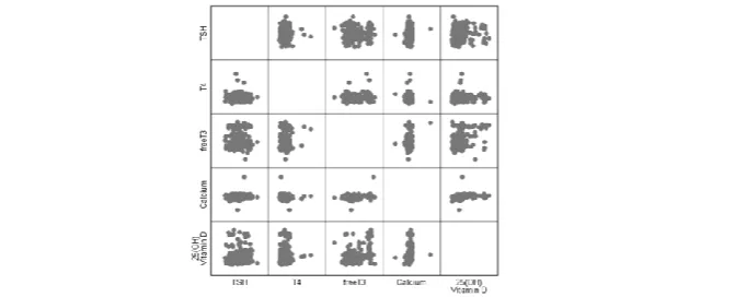
There were positive and significant correlations between free T3 and serum 25-hydroxyvitamin D (r=0.337, P<0.0001) and calcium (r=0.361, P<0.0001) levels among hypothyroid patients. However, hydroxyvitamin D and calcium levels had no significant association with TSH and T4 levels (Figure 1). In euthyroid subjects, there was a positive correlation between serum 25-hydroxyvitamin D and free T3 levels (r=0.168, P=0.026).
insufficiency, but found no significant correlation between overt or subclinical hyperthyroidism and vitamin D insufficiency (10, 11, 23). Studies on the possible association of vitamin D level and development of hypothyroidism have been limited and controversial (24, 25). In the present study, vitamin D, calcium and free T3 levels differed significantly between hypothyroid men and women but not among euthyroid individuals. Results of the studies on variation of serum 25-hydroxyvitamin D levels between men and women have been contradictory (26-28). In Muslim countries, vitamin D level is generally lower in women than in men, which is mainly due to wearing Based on the vitamin D levels, the subjects
were divided into vitamin D sufficiency, insufficiency and deficiency groups. Only free T3 was different among the hypothyroid patients (Table 2). Serum TSH in hypothyroid individuals did not differ significantly between men and women (P=0.91, Cl= -0.66 - 0.74), while serum levels of 25-hydroxyvitamin D, calcium and free T3 differed significantly between men and women (P<0.0001 for all parameters).
DISCUSSION
The role of vitamin D in bone and minerals homeostasis has been well documented (16, 17). Recent studies have demonstrated the association between vitamin D deficiency and various disorders including cardiovascular disease, cancer, infection, obesity and osteoporosis (18-21). Vitamin D levels vary depending on the amount of vitamin D produced in the skin and dietary intake of vitamin D (2, 22). In the present study, vitamin D level differed significantly between the hypothyroid and euthyroid subjects. Numerous studies have addressed the role of vitamin D in the onset and progression of thyroid disorders. Tamer et al. reported a significant association between Hashimoto’s disease and vitamin D
Parameters Hypothyroid Euthyroid P-value
25-hydroxyvitamin D (ng/ml)
46.87±29.7 78.17±44.58 <0.0001
Calcium (mg/dL) 8.74±1.21 9.11±0.47 <0.0001
TSH (mIU/L) 8.72±2.32 1.95±1.1 <0.0001
free T3 (pg/ml) 1.06±0.45 1.14±0.36 0.006
Total T4 (µg/dL) 2.57±1.61 7.72±1.78 <0.0001
Table 1- Laboratory findings of the subjects in the two study groups
Parameters Vitamin D deficiency Vitamin D insufficiency
Vitamin D sufficiency P-value
TSH (mIU/L) 8.79 ± 2.80 8.62 ± 2.26 8.73 ± 2.26 0.98
Serum T4 (µg/dL) 2.51 ± 0.91 2.49 ± 0.87 2.60 ± 1.26 0.98
Free T3 (pg/mL) 0.99 ± 0.49 0.76 ± 0.19 1.15 ± 0.47 <0.0001
Calcium 8.29 ± 0.71 8.28 ± 0.54 8.93 ± 1.35 <0.0001
Data are presented as mean ± standard deviation
Table 2. Comparison of serum TSH, T4 and free T3 levels in hypothyroid patients based on vitamin D levels
Figure 1- Correlation analysis for laboratory parameters in hypothyroid individuals
(≥30 ng/ml). Since the present study was conducted during spring and summer, it is likely that increased sun exposure might have a compensatory effect on vitamin D level of hypothyroid subjects.
CONCLUSION
Our findings indicated a significant positive correlation between serum free T3, vitamin D and calcium levels, suggesting that vitamin D might alter T3 secretion. In addition, vitamin D level was significantly lower among hypothyroid patients compared to euthyroid individuals, but the mean level of vitamin D in hypothyroid patients was still within the sufficient range. This suggests that reduced secretion of thyroid hormones might have a negative effect on the absorption or activation of vitamin D. However, a certain mechanism has not yet been suggested or approved. Given our findings, screening for vitamin D and calcium deficiency as well as vitamin D supplementation are strongly recommended for patients with hypothyroidism.
ACKNOWLEDGMENTS
This article was derived from a research project funded by the Deputy of Research and Technology of Golestan University of Medical Sciences, Iran.
CONFLICT OF INTEREST
The authors declare that there is no conflict of interest.
6. Sari F, Ozdem S, Sari R. Serum 25-Hydroxyvitamin D (3) Levels In Type 2 Diabetic Patients With Normo-, Micro-, And Macroalbuminuria. Acta Endocrinol
(Buchar). 2016; 12(3): 303-308. doi:
10.4183/aeb.2016.303.
7. Cigerli O, Parildar H, Dogruk Unal A, Tarcin O, Kut A, Eroglu H, et al. Vitamin Deficiency and Insulin Resistance in Nondiabetic Obese Patients. Acta
Endocrinol (Buchar). 2016; 12(3): 319-327.doi:
10.4183/aeb.2016.319.
8. Borges MC, Martini LA, Rogero MM. Current perspectives on vitamin D, immune system, and chronic diseases. Nutrition. 2011; 27(4): 399-404. doi: 10.1016/j.nut.2010.07.022.
9. Amital H1, Szekanecz Z, Szücs G, Dankó K, Nagy E, Csépány T, et al. Serum concentrations of 25-OH vitamin D in patients with systemic lupus erythematosus (SLE) are inversely related to disease activity: is it time to routinely supplement patients with SLE with vitamin D?
Ann Rheum Dis. 2010; 69(6): 1155-7. doi:
10.1136/ard.2009.120329 . hijab and social-religious behaviors. However,
in a large-scale study in Iran, Heshmat et al. found no difference in vitamin D status between men and women (29).
We observed that only free T3 differed significantly between vitamin D deficient, insufficient and sufficient individuals. Moreover, free T3 was significantly correlated with vitamin D and calcium levels. Vitamin D and triiodothyronine receptors are both members of DNA-binding transcription factors family and can be activated by a low nanomolar concentration of lipophilic molecules as small as cholesterol(30). Several in vivo studies have reported a positive interaction between triiodothyronine and vitamin D in bone, calcium and vitamin D metabolism. In a study by Colston and Cleeve, treatment of vitamin D deficient rats with T3 improved intestinal absorption of vitamin D through alkaline phosphatase stimulation (31, 32). On the other hand, TSH, T4 and T3 secretion can be influenced by sex hormones, genetic Susceptibility and even environmental factors, which might be also involved in the relationship of vitamin D status with TSH, T4 and free T3 levels. Two possible mechanisms for the decreased vitamin D levels in patients with hypothyroidism are poor intestinal vitamin D absorption and improper vitamin D activation (32-34). Although vitamin D level was significantly lower among hypothyroid patients compared to euthyroid individuals, the mean level of vitamin D in hypothyroid patients was still within the sufficient range REFERENCES
1. Holick MF, Binkley NC, Bischoff-Ferrari HA, Gordon CM, Hanley DA, Heaney RP, et al. Evaluation, treatment, and prevention of vitamin D deficiency: an Endocrine Society clinical practice guideline. The Journal of Clinical Endocrinology & Metabolism. 2011; 96(7): 1911-30.
2. Chung M, Balk EM, Brendel M, Ip S, Lau J, Lee J, et al. Vitamin D and calcium: a systematic review of health outcomes. Evid Rep Technol Assess (Full Rep). 2009; 183: 1-420.
3. Stockigt Jim. Clinical Strategies in the Testing of Thyroid Function. 2011.
4. Basit S. Vitamin D in health and disease: a literature review. British journal of biomedical science. 2013; 70(4): 161-72.
5. Lazúrová I, Figurová J, Dravecká I. Vitamin D and Polycystic Ovary Syndrome. Vnitrni lekarstvi. 2016; 62(Suppl 3): 87-91.
23. Muscogiuri G, Tirabassi G, Bizzaro G, Orio F, Paschou SA, Vryonidou A, et al. Vitamin D and thyroid disease: to D or not to D? Eur J Clin Nutr. 2015; 69(3): 291-6. doi: 10.1038/ejcn.2014.265.
24. Vivek S, Sunita S, Yogesh Kumar R. Vitamin D Induced Hypothyroidism. Indian Journal of Public Health Research & Development. 2016; 7(3): 77. DOI: 10.5958/0976-5506.2016.00133.9.
25. Tau C, Garabedian M, Farriaux JP, Czernichow P, Pomarede R, Balsan S. Hypercalcemia in infants with congenital hypothyroidism and its relation to vitamin D and thyroid hormones. J Pediatr. 1986; 109(5): 808-14. 26. Bolland MJ1, Grey AB, Ames RW, Horne AM, Mason BH, Wattie DJ, et al. Age‐, gender‐, and weight‐ related effects on levels of 25‐hydroxyvitamin D are not mediated by vitamin D binding protein. Clinical endocrinology. 2007; 67(2): 259-64.
27. Mackawy AM, Al-Ayed BM, Al-Rashidi BM.
Vitamin d deficiency and its association with thyroid disease. Int J Health Sci (Qassim). 2013; 7(3): 267-75. 28. Elsammak MY, Al-Wossaibi AA, Al-Howeish A, Alsaeed J. High prevalence of vitamin D deficiency in the sunny Eastern region of Saudi Arabia: a hospital-based study/Prévalence élevée de carence en vitamine D dans la région ensoleillée de l'est de l'Arabie saoudite: une étude en milieu hospitalier. Eastern Mediterranean Health Journal. 2011; 17(4): 317-22.
29. Heshmat R, Mohammad K, Majdzadeh SR, Forouzanfar MH, Bahrami A, Ranjbar GH. Vitamin D deficiency in Iran: A multi-center study among different urban areas. Iran J Public Health. 2008; 37(suppl): 72-8. 30. Carlberg C, Campbell MJ. Vitamin D receptor signaling mechanisms: Integrated actions of a well-defined transcription factor. Steroids. 2013; 78(2): 127-36. doi: 10.1016/j.steroids.2012.10.019.
31. Schiller C, Gruber R, Ho Gm, Redlich K, Gober Hj, Katzgraber F, et al. Interaction of Triiodothyronine With 1α,25-Dihydroxyvitamin D3 on Interleukin-6-Dependent Osteoclast-like Cell Formation in Mouse Bone Marrow Cell Cultures. Bone. 1998; 22(4): 341-6.
32. Colston K, Cleeve HJ. Effect of triiodothyronine on intestinal and kidney isoenzymes of alkaline phosphatase and on vitamin D metabolism in adult female rats. Comp Biochem Physiol B. 1986; 83(3): 681-4.
33. Bikle DD. Vitamin D metabolism, mechanism of action, and clinical applications. Chem Biol. 2014; 21(3): 319-29. doi: 10.1016/j.chembiol.2013.12.016 . 34. Cross HS, Pölzleitner D, Peterlik M. Intestinal phosphate and calcium absorption: joint regulation by thyroid hormones and 1, 25-dihydroxyvitamin D3. Acta endocrinologica. 1986; 113(1): 96-103.
10. Ascherio A, Munger KL, White R, Köchert K, Simon KC, Polman CH, et al. Vitamin D as an early predictor of multiple sclerosis activity and progression. JAMA
Neurol. 2014; 71(3): 306-14. doi:
10.1001/jamaneurol.2013.5993.
11. Yasuda T, Okamoto Y, Hamada N, Miyashita K, Takahara M, Sakamoto F, et al. Serum vitamin D levels are decreased and associated with thyroid volume in female patients with newly onset Graves’ disease.
Endocrine. 2012; 42(3): 739-41.
12. Tamer G1, Arik S, Tamer I, Coksert D. Relative vitamin D insufficiency in Hashimoto's thyroiditis.
Thyroid. 2011; 21(8): 891-6. doi:
10.1089/thy.2009.0200.
13. Dağlar G, Kiliç MÖ, Çelik C, Yüksel C, Terzioğlu SG, Özden S, et al. Is there a relatIonshIp between vItamIn D status anD hypocalcemIa after total thyroIDectomy? Acta Endocrinol (Buchar). 2016; 12(3): 291-296. doi: 10.4183/aeb.2016.291.
14. Schräder M, Müller KM, Carlberg C. Specificity and flexibility of vitamin D signaling. Modulation of the activation of natural vitamin D response elements by thyroid hormone. J Biol Chem. 1994; 269(8): 5501-4. 15. Heaney RP, Dowell MS, Hale CA, Bendich A.
Calcium absorption varies within the reference range for serum 25-hydroxyvitamin D. J Am Coll Nutr. 2003; 22(2): 142-6.
16. Bikle Daniel D. Vitamin D and bone. Handbook of nutrition and diet in therapy of bone diseases: Wageningen Academic Publishers. 2016; 2063-8. 17. Bouillon R, Suda T. Vitamin D: calcium and bone homeostasis during evolution. Bonekey Rep. 2014; 3: 480. doi: 10.1038/bonekey.2013.214 .
18. Norman Pe, Powell Jt. Vitamin D and cardiovascular disease. Circ Res. 2014; 114(2): 379-93. doi: 10.1161/CIRCRESAHA.113.301241.
19. Pludowski P, Holick MF, Pilz S, Wagner CL, Hollis
BW, Grant WB, et al. Vitamin D effects on
musculoskeletal health, immunity, autoimmunity, cardiovascular disease, cancer, fertility, pregnancy, dementia and mortality—a review of recent evidence.
Autoimmunity reviews. 2013; 12(10): 976-89.
20. Pourshahidi LK. Vitamin D and obesity: current perspectives and future directions. Proceedings of the Nutrition Society. 2015; 74(2): 115-24.
21. Man SC, Chiriac M, Militaru MS, Trifa AP, Goia-Socol M, Georgescu CE. Association of Col1a1 Sp1 And Fok-I Vdr Genetic Polymorphisms In Young Male Idiopathic Osteoporosis. Acta Endocrinologica (1841-0987). 2017; 13(2): 224-227.
22. Tang BM, Eslick GD, Nowson C, Smith C, Bensoussan A. Use of calcium or calcium in combination with vitamin D supplementation to prevent fractures and bone loss in people aged 50 years and older: a meta-analysis. The Lancet. 2007; 370(9588): 657-66 .