R E S E A R C H A R T I C L E
Open Access
The eagle jugular syndrome
Paolo Zamboni
1, Alba Scerrati
2*, Erica Menegatti
1, Roberto Galeotti
3, Marcello Lapparelli
2, Luca Traina
4,
Mirko Tessari
1, Andrea Ciorba
5, Pasquale De Bonis
2and Stefano Pelucchi
5Abstract
Background:The elongation of the styloid process is historically associated with two variants of the Eagle syndrome. The classic one, mainly characterized by pain and dysphagia, and the carotid variant characterized by pain and sometimes by cerebral ischemia. We observed a further variant characterized by a styloid elongation coursing adjacent to the transverse process of C1, causing significant compression of the internal jugular vein. Methods:We reviewed all the cases of Eagle syndrome, including the jugular variant, admitted in our Hospital in the last six years.
We compared symptomatology, associated comorbidities and imaging. Data were statistically analyzed.
Results:Overall 23 patients were admitted to the Hospital for symptomatic elongation of the styloid process, 11 male and 12 females. The jugular variant of the Eagle syndrome is clinically delineated by significant differences, as compared to the classic variant and carotid variants. Headache was the more prominent symptom (p< .009) as well as a documented peri-mesencephalic hemorrhage was the more significant comorbidity (p< .0003). The group classic-carotid variant was characterized by ipsilateral pain respect to the jugular variant (p < .0003). CT angiography with venous phase extended to the neck veins and imaging reconstruction is highly recommended as imaging technique, complemented by color-Doppler ultrasound.
Conclusions:The elongation of the styloid process may have different paths which creates compression on the surrounding anatomical structures. There may be a possible association of jugular impingement by an elongated styloid process with symptoms.
Trial registration:Protocol n°45–2013.
Keywords:Eagle syndrome, Jugular compression, Elongated styloid process, Perimesencephalic subarachnoid haemorrhage
Background
In 1937, Eagle proposed two syndromes associated with an elongated styloid process, respectively a classic syn-drome and a carotid artery synsyn-drome. The former seems facilitated by scar tissue developed after a tonsillectomy which stretches the nerve endings in this area, mainly causing pain, dysphagia and otalgia [1–7]. The latter is thought a consequence of an elongated styloid process impinging upon the carotid artery and associated nerve endings, causing pain and sometimes facilitating vascular complications and cerebral ischemia [8–11]. In both syn-dromes, the length of the styloid process was thought to be the major causal factor. In addition, it has also been
observed a variant of the Eagle syndrome, where an elongated styloid process is coursing adjacent to the transverse process of C1 [12–14]. Often, in this anatom-ical situation, a significant compression of the internal jugular vein (IJV) can be observed. However, few anec-dotal reports are available regarding symptoms and clin-ical presentation of such jugular vein bone nutcracker [12–14]. In some articles the symptoms seem similar to the classic syndrome, whereas others report unsuspected symptoms and complications such as intracranial hyper-tension and cerebral hemorrhage [13–16].
Despite the heterogeneity of the symptomatology of the Eagle clinical picture [16], we hypothesized that the jugular variant could be characterized by a more specific clinical picture linked with the venous obstruction.
© The Author(s). 2019Open AccessThis article is distributed under the terms of the Creative Commons Attribution 4.0 International License (http://creativecommons.org/licenses/by/4.0/), which permits unrestricted use, distribution, and reproduction in any medium, provided you give appropriate credit to the original author(s) and the source, provide a link to the Creative Commons license, and indicate if changes were made. The Creative Commons Public Domain Dedication waiver (http://creativecommons.org/publicdomain/zero/1.0/) applies to the data made available in this article, unless otherwise stated.
* Correspondence:[email protected]
2Neurosurgery Unit, Ferrara University Hospital, Ferrara, Italy
Aim of the present study is to compare the jugular vari-ant of the Eagle syndrome with the other more common presentations, from a clinical, imaging, and hemodynamic point of view.
Methods
An extensive review of cases of symptomatic elongated styloid process who were admitted and eventually treated in our University Hospital in the last 6 years was performed.
Institutional review board approval was obtained for this retrospective analysis. A detailed medical history, clinical examination, 3D head CT scan with contrast were under-taken in all patients. In addition, cases with carotid or jugular involvement underwent a hemodynamic study by the means of color Doppler ultrasound (CDU).
Patients affected by classic, symptomatic Eagle’s syn-drome and surgical treated at the ENT Department of the University Hospital of Ferrara have been selected for this study. Physical examination included careful palpa-tion of the tonsillar fossa, lateral pharyngeal wall, chew-ing muscles and above mandibular angle in an attempt to determine the patient’s discomfort. The diagnostic workup included an imaging assessment (orthopantomo-graphy, neck CT scans or neck MRI scans). In addition, we reviewed patients with symptomatic carotid variant of the Eagle’s syndrome who were investigated and treated at the vascular surgery unit, by the means of CDU and 3D angio CT scan.
Finally, patients with radiological and/or CDU evi-dence of an elongated styloid process of extracranial jugular or carotid compression above the carotid bifur-cation were respectively collected from the Neurosurgery unit and Vascular Diseases Center.
Patients clinical history was collected, they underwent a CT angio, angio-MRI, CDU of extracranial vessels and a clinical-neurological evaluation. All CTAs were reviewed and eventually reconstructed by the means of proper softwares by three Authors. Surgery was pro-posed to patients with medically untreatable neuro-logical symptoms. Indeed, the prolonged and sustained intracranial hypertension due to a reduced cerebral ven-ous outflow and not manageable by medical therapy (such as acetazolamid) can cause unbearable headache, papilledema and progressive cognitive impairment.
DUS assessment of brain haemodynamics
The doppler ultra sound (DUS) protocol, previously de-scribed [17, 18] is aimed to assess respectively the brain inflow, the brain outflow, and the venous collateral flow index. The DUS assessment of the cerebral inflow con-sists in sampling the common carotid artery, the exter-nal and the interexter-nal carotid artery and the vertebral artery (VA). Parameters of time average velocity (TAV),
cross sectional area (CSA), and flow rate were assessed according to reporting standard. For the assessment of the outflow the internal jugular veins (IJVs) were inso-nated at classic J3 level (upper part of the neck just below the jugular foramen), J2 (at the level of the mid thyroid gland), and J1 level (outlet with the subclavian vein), as well as the vertebral vein (VV) at V2 level.
Statistical analysis
Data are given as mean and standard deviation. The as-sociation between the presence of an elongated styloid process and symptoms and/or cerebral comorbidities in the classic variant group in comparison with the jugular variant group were analyzed by the means of two-sided Fisher exact test. P value < 0.05 were considered significant.
Results
Overall, patients assessed in our University Hospital in the last 6 years for Eagle syndrome were 23 They were 11 males and 12 females. They were subdivided into 3 groups respectively 14 patients with the classic, 1 patient with the carotid, and 8 patients with the jugular variants. The only patient with the carotid variant was a 48 y.o. female, who was admitted for carotid dissection compli-cated by thrombosis. She spontaneously recovered symp-toms from cerebral ischemia. She was excluded from subsequent comparative evaluations.
Clinical symptomatology and associated neurological comorbidities are given in Table 1. The jugular variant of the Eagle syndrome is clinically delineated by signifi-cant differences, as compared to the classic and carotid variant. Headache was the more prominent symptom (p< .009) as well as a documented peri-mesencephalic hemorrhage was the more significant comorbidity (p< .0003). The group classic variant was characterized by ipsilateral pain respect to the jugular variant (p < .0003).
On the Eagle jugular group one patient underwent surgery (see illustrative case) and 7 patients were man-aged conservatively.
14 patients in this series belong to the classic pattern of Eagle Syndrome. The cohort of patients consists of 6 males and 8 females, (aged between 28 and 68 years, mean 48 ± 9 years). All patients were treated surgically under general anesthesia by cervicotomy and received antibiotic prophylaxis. They were usually discharged in one-two days after surgery (informed consent was col-lected from each patient prior to surgery, according to the Italian laws); no patient presented perioperative and postoperative complications.
Neurosurgery department, 5 male 3 female, mean age 64 ± range 50–87 yo.
The degree of vein stenosis at CTA were > 90% in all cases (Fig.1) at C1 level. The corresponding DUS assess-ment was characterized by flow no-Doppler detectable at J3 level, with increased collateral flow index (> 30% of the inflow). The time-averaged maximum blood flow velocity (TAV) in the contralateral not affected J3 jugu-lar veins ranged between 24 and 38 cm/sec.
Symptoms reported in Table 1 were in part treatable by a conservative approach, especially headache and pain. Numbness and dizziness are still persisting but self-considered not invalidating by the patients.
Follow-up is in course. Until now, we proposed surgery to a pa-tient because of the invalidating symptoms which were not controlled by medical treatment, especially headache and facial edema. We are following up the others pa-tients in order to check symptoms deterioration and eventually offering surgery.
Illustrative case
One patient with the jugular syndrome presented with a 2 years history of dysesthesia, pain in the right side of the face, soft tissue swelling (especially in the morning) and nuchal and orbital headache resistant to drug treat-ment. CT angio showed a right jugular compression due
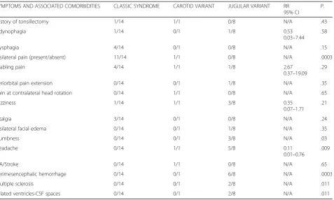
Table 1Patients population demographics and clinical presentation of symptomatic Eagle syndrome subdivided according to the respective clinical variant
SYMPTOMS AND ASSOCIATED COMORBIDITIES CLASSIC SYNDROME CAROTID VARIANT JUGULAR VARIANT RR 95% CI
P.
History of tonsillectomy 1/14 1/1 0/8 N/A .43
Odynophagia 1/14 0/1 1/8 0.53
0.03–7.44 .58
Dysphagia 4/14 0/1 0/8 N/A .15
Ipsilateral pain (present/absent) 11/14 1/1 0/8 N/A .0003
Stabling pain 4/14 1/1 1/8 2.67
0.37–19.09 .29
Periorbital pain extension 0/14 0/1 1/8 N/A .35
Pain at contralateral head rotation 0/14 1/1 0/8 N/A .65
Dizziness 1/14 1/1 3/8 0.35
0.07–1.71 .21
Otalgia 3/14 0/1 0/8 N/A .24
Ipsilateral facial edema 0/14 0/1 1/8 N/A .35
Numbness 0/14 0/1 3/8 N/A .03
Headache 0/14 1/1 5/8 0.11
0.01–0.76
.009
TIA/Stroke 0/14 1/1 0/8 N/A .65
Perimesencephalic hemorrhage 0/14 0/1 6/8 N/A .0003
Multiple sclerosis 0/14 0/1 2/8 N/A .011
Dilated ventricles-CSF spaces 0/14 0/1 2/8 N/A .011
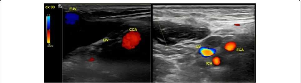
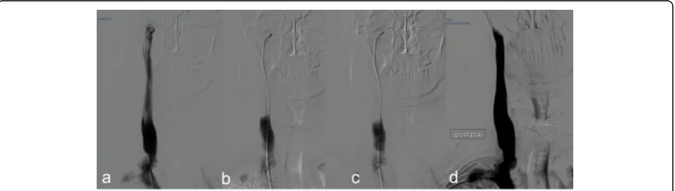
to an elongated styloid process (Fig. 1) and a CDU showed compromised cerebral outflow on the right jugular vein, collateral vein circulation on the face and neck and no evidence of intraluminal causes of occlusion (Fig. 2). Because of the persistent symptoms he under-went surgery consisting of a right lateral transcervical approach to the infratemporal fossa and the complete styloidectomy (Fig.3). The postoperative course was un-eventful. A post operative CT angio (Fig.3c) and a CDU failed to demonstrate an expected increase in diameter of the right internal jugular vein, following the surgical decompression. CDU showed flow re-appearance at J3 level, although the flow rate was lower respect to normal values [19]. Facial oedema was slightly reduced but headache did not. In order to obtain a complete restor-ing of the flow, patients underwent balloon angioplasty either at the level of the J3 residual stenosis or at J1 level, with a satisfactory result [20] (Fig.4). Symptomatic relief was obtained and patient was stable at a 6 months follow-up.
Discussion
The etiopathogenetic mechanism of the elongation of the styloid process has not been ascertained so far; de-velopment abnormalities and/or bone homeostasis al-terations have been proposed in order to explain the occurrence of ossification or elongation of the stylo-hyoid process [1]. Symptoms of the classic Eagle syn-drome (Fig.5) have been associated mainly to an intermittent compressive neuropathy involving differ-ent branches of cranial nerves, which is often exacer-bated by swallowing and yawning [1–7]. These symptoms are caused by a mass effect on cranial nerves eliciting a neuropathic pain which poorly re-sponds to medications. Nowadays the literature tends to support that surgical treatment results in a more definitive treatment and long lasting symptomatic
relief [21]. Other symptoms reported in literature are sense of hypopharyngeal foreign body, odynophagia, dysphagia in addition to cervico-facial pain with differ-ent irradiations. Cases of Horner’s syndrome are usu-ally explained with the compression of the nerve fibers of sympathetic nervous system [1]. Apart from the classic pattern, also a ‘carotid’ variant has been de-scribed, where peculiar symptoms such as arterial dis-section, obstruction, transient ischemic attack and cerebral ischemia were found associated [8–11]. It is more difficult to explain cases of Eagle syndrome asso-ciated with migraine and headache, which could be more consistently related to the external compression of internal jugular vein (Fig.1).
Both at surgery and CTA the classic group did not show any involvement of the jugular vein, and this may explain us the lack of headache in our classic group (Table1).
Clinically relevant extrinsic compression on the extra-cranial internal jugular vein (IJV) due to an elongated styloid process has been rarely described in literature. A study by Jayaraman et al.12enrolled 108 patients whose CT angios were investigated for the incidence of internal jugular vein compression by extrinsic structures in the upper neck. They found 24.1% right side compressions and 30.6% left side compressions. They concluded that jugular vein compression is an anatomic variant and un-likely to be pathologic in nature. However, they did not investigate about the reasons why these CT angios were performed or patients’clinical history [12].
We collected 8 cases of Eagle jugular syndrome. In our study the symptomatology is readily different respect the to the classic or the carotid syndrome, where ipsilateral pain was the more significant symptom (Table1). Head-ache, numbness and dizziness are usually chronic in the jugular group and may be related to the impaired cere-bral venous outflow. Subarachnoid hemorrhage usually presents with an acute headache (described as the worst
headache ever) often associated to vomiting and altered consciousness. In few studies the compression of the IJV was related to pseudotumor cerebri [13,14] or to head-aches [13]. In many others, the jugular variant was sig-nificantly associated with persistent headache, and among comorbidities, to the peri-mesencephalic sub-arachnoid hemorrhage (p-SAH), both possibly related to cerebral venous hypertension [15,16,22,23]. p-SAH is a particular kind of subarachnoid hemorrhage not associ-ated to an evident vascular malformation, like aneurysms or arteriovenous malformations and with an usually good prognosis. The blood is mainly located in the cere-bral basal cisterns and the cause is still debated. Among the hypothesized causes, a hypertensive venous bleeding (like coming from a particular variant of Basal vein of Rosenthal or an occluded venous sinus) is the most reli-able [22,23].
The relationship between the upper internal jugular vein bone nutcracker and the p-SAH could be a novel and intriguing finding. This variety of the subarachnoid hemorrhage is thought to be caused mainly by intracra-nial venous anomalies, but there are no reports in
literature about its relationship with extracranial venous anomalies [22,23]. In our series imaging at the intracra-nial level completely excluded the presence of any kind of vascular anomalies. However we hypothesized a jugu-lar compression with a transient related venous hyper-tension could be a predisposing factor able to facilitate a p-SAH. On the basis of our study, if p-SAH should occur, we suggest extending also at the extracranial level the investigation of the brain vasculature, focusing par-ticularly to the extracranial veins (Fig.1). We also know that the compression of the upper segments of the in-ternal jugular vein can induce chronic intracranial hypertension [24, 25], which was not found in our sur-vey, yet.
CDU investigation allowed us to assess the venous haemodynamics in terms of flow velocity and flow rate of the internal jugular vein, especially just below the nut-cracker, in the upper part of the neck. (Fig. 2) In the more severe cases the internal jugular vein appeared an empty vein, with no Doppler detectable flow in the lumen, even in favor of gravity with the head of the pa-tients in upright position. Out of the initial assessment, Fig. 3a) intraoperative picture of illustrative case, showing the relationship between the elongated styloid process (°) and the internal jugular vein (*);b) the elongation of the removed styloid process;c) the post-operative CTA showing the obtained decompression of the jugular vein due to the styloid process removal, in the absence of the restoration of its flow. This lead us to perform the following jugular PTA
CDU permits also to perform a precise and non-invasive follow-up of patients conservatively treated, in order to monitor any further flow worsening.
Alternatively, jugular outflow in case of external com-pression has been also extensively studied by the means of catheter venography protocol [26]. These authors, by the means of the same protocol, found out that balloon-angioplasty is not an effective treatment in case of exter-nal compression of IJV [27]. CT-angio in our patient clearly demonstrated an external compression by the styloid process. Considering that one of the main advan-tages of venography is the possibility to couple diagnos-tic and endovascular treatment (not indicated in our case, for the above-mentioned reasons) we decided to use CDU to measure the flow and open surgery to treat the obstruction.
In our experience, the treatment is usually conservative, especially when symptoms are not invalidating and appar-ently controlled by medical treatment. Only one patient underwent surgery and endovascular balloon angioplasty, with intriguing results (see illustrative case). In this case both headache and facial edema did not respond to con-servative treatment. Indeed, Higgins et al. [15] stated that in their group of patients with idiopathic intracranial hypertension and impairment in venous outflow, most of those who benefited only did so after subsequent jugular stenting reinforced the surgical decompression. Moreover, Zhou et al. [28] reported in the same field their positive experience with exclusive jugular stenting in a group of 15 patients with IJV stenosis, but without bone compression.
Someone may argue that a potential indication could the prevention of recurrent episodes of cerebral hemorrhage. However, this is a rare eventuality follow-ing p-SAH (1/1220 as reported by Mensfollow-ing et al. [22]). The major limitation of our study is to be a retrospect-ive analysis. Indeed, for the jugular variant we retro-spectively enrolled 8 patients which are not enough to imply causation. Moreover we did not use a classifica-tion of the internal jugular vein phlebograms.
However, our findings are intriguing and warrant fur-ther investigations on the basis of a prospective multi-center epidemiologic data collection.
Conclusions
Our results suggest a possible association of jugular im-pingement by an elongated styloid process with symp-toms. In case of an elongated styloid process, venous narrowing should be always investigated, especially in patients presenting with sign and symptoms of intracra-nial hypertension and may represent in perspective a field of interdisciplinary interest.
Abbreviations
CDU:Color doppler ultrasound; CSA: Cross sectional area; CT: Computer tomography; CTA: Computer tomography angiography; DUS: Doppler ultrasound; ENT: Ear nose and throat; IJV: Internal jugular vein; MRI: Magnetic resonance imaging; p-SAH: Peri-mesencephalic subarachnoid hemorrhage; TAV: Time-averaged maximum blood flow velocity; VA: Vertebral artery; VV: Vertebral vein
Acknowledgements
Not Applicable.
Authors’contributions
P.Z., S.P. and P.D.B. conceived of the presented idea and treated conservatively/surgically the patients. A.S., A.C. and E.M. designed the work, performed the clinical analysis and were major contributor in writing the manuscript. R.G. and M.T. verified the acquired data and the analytical methods. M.L. and L.T. analyzed and interpreted the patient data regarding the carotid and jugular disease and gave their contribution in substantively revising the manuscript. All authors have approved the submitted version (and any substantially modified version that involves the author’s contribution to the study). All authors have agreed both to be personally accountable for the author’s own contributions and to ensure that questions related to the accuracy or integrity of any part of the work, even ones in which the author was not personally involved, are appropriately investigated, resolved, and the resolution documented in the literature.
Funding
This research did not receive any specific grant from funding agencies in the public, commercial, or not-for-profit sectors.
Availability of data and materials
The datasets used and/or analyzed during the current study are available from the corresponding author on reasonable request.
Ethics approval and consent to participate
This study was approved by the ethic committee of the University Hospital of Ferrara (PROTOCOL N 45/2013). Written informed consent from all participants of the study was obtained.
Consent for publication
All participants gave written informed consent for their personal and clinical data publication along with written consent for publication for any identifying images in this study, according to the Italian laws.
Competing interests
The authors declare that they have no competing interests.
Author details
1Vascular Diseases Center, Ferrara University Hospital, Ferrara, Italy. 2Neurosurgery Unit, Ferrara University Hospital, Ferrara, Italy.3Interventional
Radiology Unit, Ferrara University Hospital, Ferrara, Italy.4Vascular Surgery
Unit, Ferrara University Hospital, Ferrara, Italy.5ENT Unit, Ferrara University Hospital, Ferrara, Italy.
Received: 15 March 2019 Accepted: 15 December 2019
References
1. Fusco DJ, Asteraki S, Spetzler RF. Eagle's syndrome: embryology, anatomy, and clinical management. Acta Neurochir. 2012;154(7):1119–26. 2. Babad MS. Eagle's syndrome caused by traumatic fracture of a mineralized
stylohyoid ligament--literature review and a case report. Cranio. 1995;13(3): 188–92.
3. Pierrakou ED. Eagle's syndrome. Review of the literature and a case report. Ann Dent. 1990;49(1):30–3.
4. Han MK, Kim DW, Yang JY. Non surgical treatment of Eagle's syndrome - a case report. Korean J Pain. 2013;26(2):169–72.
5. Politi M, Toro C, Tenani G. A rare cause for cervical pain: Eagle's syndrome. Int J Dent. 2009;2009:781297.
6. Sun CK, Mercuri V, Cook MJ. Eagle syndrome: an unusual cause of head and neck pain. Arch Neurol. 2006;63(2):294–5.
7. Ciorba A, Savini S, Morolli F, Malago M, Bianchini C, Pelucchi S, Pastore A. Orofacial pain and eagle syndrome: cues from a clinical series. Minerva Stomatol. 2014;63(10):361–7.
8. Farhat HI, Elhammady MS, Ziayee H, Aziz-Sultan MA, Heros RC. Eagle syndrome as a cause of transient ischemic attacks. J Neurosurg. 2009;110(1):90–3. 9. Varelas PN, Sinson G, Rand S, Book D. Clipping the Eagle's wings: treatment
of an unusual cause of transient cerebral ischemia. Neurol. 2005;64(2):393–4. 10. Todo T, Alexander M, Stokol C, Lyden P, Braunstein G, Gewertz B. Eagle
syndrome revisited: cerebrovascular complications. Ann Vasc Surg. 2012; 26(5):729.e1–5.
11. Subedi R, Dean R, Baronos S, Dhamoon A: Carotid artery dissection: a rare complication of Eagle syndrome. BMJ Case Rep 2017, 2017:https://doi.org/ 10.1136/bcr-2016-218184.
12. Jayaraman MV, Boxerman JL, Davis LM, Haas RA, Rogg JM. Incidence of extrinsic compression of the internal jugular vein in unselected patients undergoing CT angiography. AJNR Am J Neuroradiol. 2012;33(7):1247–50. 13. Ho S, Luginbuhl A, Finden S, Curry JM, Cognetti DM. Styloid/C1 transverse
process juxtaposition as a cause of Eagle's syndrome. Head Neck. 2015; 37(11):E153–6.
14. Dashti SR, Nakaji P, Hu YC, Frei DF, Abla AA, Yao T, Fiorella D. Styloidogenic jugular venous compression syndrome: diagnosis and treatment: case report. Neurosurgery. 2012;70(3):E795–9.
15. Higgins JN, Garnett MR, Pickard JD, Axon PR. An evaluation of
Styloidectomy as an adjunct or alternative to jugular stenting in idiopathic intracranial hypertension and disturbances of cranial venous outflow. J Neurol Surg B Skull Base. 2017;78(2):158–63.
16. Mendelsohn AH, Berke GS, Chhetri DK. Heterogeneity in the clinical presentation of Eagle's syndrome. Otolaryngol Head Neck Surg. 2006;134(3):389–93. 17. Zamboni P, Menegatti E, Cittanti C, Sisini F, Gianesini S, Salvi F, Mascoli F.
Fixing the jugular flow reduces ventricle volume and improves brain perfusion. J Vasc Surg Venous Lymphat Disord. 2016;4(4):434–45. 18. Zamboni P, Sisini F, Menegatti E, Taibi A, Malagoni AM, Morovic S,
Gambaccini M: An ultrasound model to calculate the brain blood outflow through collateral vessels: a pilot study. BMC Neurol 2013, 13:81–2377-13-81. 19. Zamboni P, Menegatti E, Pomidori L, Morovic S, Taibi A, Malagoni AM, Cogo
AL, Gambaccini M. Does thoracic pump influence the cerebral venous return? J Appl Physiol (1985). 2012;112(5):904–10.
20. Zamboni P, Tesio L, Galimberti S, Massacesi L, Salvi F, D'Alessandro R, Cenni P, Galeotti R, Papini D, D'Amico R, Simi S, Valsecchi MG, Filippini G. Brave dreams research group: efficacy and safety of Extracranial vein angioplasty in multiple sclerosis: a randomized clinical trial. JAMA Neurol. 2018;75(1):35–43. 21. Mortellaro C, Biancucci P, Picciolo G, Vercellino V. Eagle's syndrome:
importance of a corrected diagnosis and adequate surgical treatment. J Craniofac Surg. 2002;13(6):755–8.
22. Mensing LA, Vergouwen MDI, Laban KG, Ruigrok YM, Velthuis BK, Algra A, Rinkel GJE. Perimesencephalic hemorrhage: a review of epidemiology, risk factors, presumed cause, clinical course, and outcome. Stroke. 2018;49(6):1363–70. 23. Fu FW, Rao J, Zheng YY, Song L, Chen W, Zhou QH, Yang JG, Ke JQ, Zheng
GQ. Perimesencephalic nonaneurysmal subarachnoid hemorrhage caused by transverse sinus thrombosis: a case report and review of literature. Med (Baltimore). 2017;96(33):e7374.
24. Sangra MS, Teasdale E, Siddiqui MA, Lindsay KW: Perimesencephalic nonaneurysmal subarachnoid hemorrhage caused by jugular venous occlusion: case report. Neurosurgery 2008, 63(6):E1202–E1203; discussion E1203. 25. De Bonis P, Menegatti E, Cavallo MA, Sisini F, Trapella G, Scerrati A, Zamboni
P. JEDI (jugular entrapment, dilated ventricles, intracranial hypertension) syndrome: a new clinical entity? A case report. Acta Neurochir. 2019;161(7): 1367–70.
26. Veroux P, Giaquinta A, Perricone D, Lupo L, Gentile F, Virgilio C, Carbonaro A, De Pasquale C, Veroux M. Internal jugular veins out flow in patients with multiple sclerosis:a catheter venography study. J Vasc Interv Radiol. 2013; 24(12):1790–7.
27. Giaquinta A, Beggs CB, Veroux M, De Marco E, Sanzone A, Virgilio C, Veroux P. Factors influencing the hemodynamic response to balloon angioplasty in the treatment of outflow anomalies of internal jugular veins. J Vasc Surg Venous Lymphat Disord. 2017;5(6):777–88.
28. Zhou D, Meng R, Zhang X, Guo L, Li S, Wu W, Duan J, Song H, Ding Y, Ji X. Intracranial hypertension induced by internal jugular vein stenosis can be resolved by stenting. Eur J Neurol. 2018;25(2):365–e13.
Publisher’s Note