Available Online at www.ijpret.com 168
INTERNATIONAL JOURNAL OF PURE AND
APPLIED RESEARCH IN ENGINEERING AND
TECHNOLOGY
A PATH FOR HORIZING YOUR INNOVATIVE WORK
LUNG NODULE DETECTION USING PATCHED BASED CONTEXT ANALYSIS
METHOD WITH SUPPORT VECTOR MACHINE: A REVIEW
MAYURI P. BAMNOTE, PROF. R.S. MANGRULKAR, PROF. A. D. GOTMARE
1. PG Student, Dept. of Computer Science &Engg., B.D.C.O.E, Sewagram-442001
2. Head and Associate Professor, Dept. of Computer Science &Engg., B.D.C.O.E, Sewagram-442001. 3. Professor, Dept. of Computer Science &Engg., B.D.C.O.E, Sewagram-442001
Accepted Date: 15/03/2016; Published Date: 01/05/2016
\
Abstract: Prediction and diagnosis of cancer is very important task in healthcare industry. Cancer is of the leading cause of death globally so it is very essential to detect and predict it at very initial stage. Lung nodule, which leads to lung cancer is detected by various medical imaging techniques which includes X-ray, Computerized Tomography (CT). Classification as well as Detection of nodules is important task because the nodules are commonly attached to the blood vessels. Many studies have shown that early prediction is the best way to cure this disease. The classification based upon the four types of lung nodules which is well-circumscribed, vascularized, juxta-pleural and pleural-tail.
Keywords: Lung Cancer, Computerized Tomography, Classification, Detection.
Corresponding Author: MISS. MAYURI P. BAMNOTE
Access Online On:
www.ijpret.com
How to Cite This Article:
Mayuri P. Bamnote, IJPRET, 2016; Volume 4 (9): 168-173
Available Online at www.ijpret.com 169
INTRODUCTION
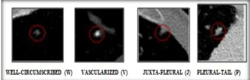
Lung nodules are masses which is small in size present in the human lung, and are spherical in shape; however, they can be pulled by surrounding anatomical structures, such as vessels and adjacent pleura [1]. Figure 1 shows the four types of lung nodules. Lung nodules can be divided into four types:
1. Well-circumscribed (W): nodule located centrally in the lung but without any connection to vasculature.
Figure 1: Transaxial Ct images with four types of Lung Nodule
2. Vascularized (V): location of this nodule is centrally in the lung but closely connected to neighboring vessels.
3. Juxta-Pleural (J): here the large portion of the nodule connected to the pleural surface.
4. Pleural- tail (P): location of nodule near the pleural surface connected by a thin tail.
Available Online at www.ijpret.com 170
LITERATURE REVIEW
Sr. No.
References
Evaluation Approach
1.
Fan Zhang, Yang Song, Weidong Cai, Min-zhao Lee,yun Zhou,Heng Huang, Shimin Shan.
Three main stages were explained :Adaptive patch-based division, con- centric multilevel partition, HOG feature set is designed.
2.
R.Harini Karthika ,
P.Thirugnanam
It describes the Multilevel Pattern Recognition And Semantic Of LDA Analysis
3.
Amal Farag,Shireen Elhabian, James Graham et.al.
Breifly discussed the modeling of the Lung Nodules for Detection in LDCT Scans
4.
Ravivarman.R, Sasirekha.N Survey on Lung Nodule Classifications
5. William J. Kostis, Anthony Reeves,
David F. Yankelevitz,
Three-Dimensional Segmentation and
Growth-Rate Estimation of Small
Pulmonary Nodules in Helical CT Images
6.
Yang Song,
Weidong Cai, Yun Zhou, David Dagan Feng,
Feature-Based Image Patch
Approximation for Lung Tissue
Available Online at www.ijpret.com 171 7.
Prashant Naresh , DR. Rajashree shettar
Image processing, as well as classification techniques were discussed
8.
Stefano Diciotti, Giulia Picozzi,
Massimo Falchini, Mario
Mascalchi, Natale Villari, Guido Valli
3-D Segmentation Algorithm were
discussed
METHODS FOR CLASSIFICATIONS
1. Support Vector Machine (SVM)
SVM introduced by Cortes and is generally used for classification purpose.
The fundamental idea of SVM can be described as follows.
Initially, the inputs are formulated as feature vectors. 2 Then, by using the kernel function, these feature vectors are
Mapped into a feature space. 3 Finally, a division is computed in the feature space to separate the classes of training vectors.
Classifiers having own set of advantages and disadvantages as well as it‘s own area of importance’s. None of algorithms can satisfies all criteria. The following points show the characteristics of the support vector machine.
1. Speed of learning is good
2. Speed of class is excellent
3. Tolerance to missing value is good
4. Irrelevant to attribute is excellent
Available Online at www.ijpret.com 172 6. Highly interdependent to attribute is very good
7. Tolerance to noise is good
8. Attempt for incremental learning is good
2. Probabilistic Latent Semantic Analysis (PLSA)
It is a statistical method for the analysis of two-mode and co-occurrence data. It will overcome the problem of sparse representation in data classification. It is useful to differentiate the overlapping contextual structures shared among various types of lung nodules i.e. well-circumscribed, vascularized, juxta-pleural and pleural-tail.
PROPOSED METHOD
In the proposed system, LDCT lung image dataset were taken as input. Image preprocessing operation carried using the Weiner filter. The process of image patches division in which the segmented limb part, left lung part and right lung part patches were formed. Patches were formed according to the local anatomical structures respectively.
Figure 2: Block Diagram of proposed system
CONCLUSION
Available Online at www.ijpret.com 173
REFERENCES:
1. Fan Zhang, Yang Song, Weidong Cai, Member, Min-Zhao Lee, Yun Zhou, Heng Huang, Shimin Shan, Michael J Fulham, and Dagan D. Feng, “Lung Nodule Classification With Multilevel Patch-Based Context Analysis”, IEEE Transactions On Biomedical Engineering, Vol. 61, No. 4, April 2014.
2. R.Harini Karthika, P.Thirugnanam,” Multilevel Pattern Recognition and Semantic Of LDA Analysis”, Journal of Electronics and Computer Sciecne, Issn- 3967-0867, Vol 2 Issue 3 March 2015.
3. Amal Farag, Shireen Elhabian, James Graham, Aly Farag,Salwa Elshazly, Robert Falk, Hani Mahdi, Hossam Abdelmunim,Sahar Al Ghaafary,” Modeling of the Lung Nodules for Detection in LDCT Scans”,32nd Annual International Conference of the IEEE EMBS Buenos Aires, Argentina, August 31 - September 4, 2010
4. Ravivarman.R,sasirekha.N2 ,”Survey On Lung Nodule Classifications”, International Journal
Of Research In Engineering And Technology ,issn: 2319-1163 | Pissn: 2321-7308,volume: 04
Issue: 01, Jan-2015
5. William J. Kostis, Anthony P. Reeves, David F. Yankelevitz, and Claudia I. Henschke, ” Three-Dimensional Segmentation and Growth-Rate Estimation of Small Pulmonary Nodules in Helical CT Images”, IEEE Transactions On Medical Imaging, Vol. 22, No. 10, October 2003
6. Yang Song, Weidong Cai, Yun Zhou, David Dagan Feng,” Feature-Based Image Patch Approximation for Lung Tissue Classification”, IEEE Transactions On Medical Imaging, Vol. 32, No. 4, April 2013
7. Prashant Naresh, Dr. Rajashree shettar,” Image Processing and Classification Techniques for Early Detection of Lung Cancer for Preventive Health Care: A Survey”, Int. J. of Recent Trends in
Engineering & Technology, Vol. 11, June 2014.