EFFECTS OF EXTERNAL MAGNETIC FIELD ON BLOOD -
A THEORETICAL PERSPECTIVE
ABDULMOIZ MOHAMMED
Department of Basic Sciences, Preparatory Year, University of Ha'il, P.O Box # 2440, Ha'il, Saudi Arabia
Email - [email protected]
1. INTRODUCTION:
Bio-magnetic fluid dynamics (BDF) is not a new area in the field of fluid mechanics, which deals with understanding of dynamics of a selected fluid in the presence of an external field. A fluid that is extracted from a creature or living organism tends to be affected by the magnetic field that can be termed as bio-magnetic fluid. Various fluids are found in a living organism such as blood, urine, mucus and so on, but the most important among these fluids is blood, which acts as a magnetic liquid. This characteristic of blood as magnetic liquid, also sometimes referred to as magnetic fluid, is due to complex reactions and interactions among hemoglobin, cell membrane, and intercellular proteins. A unique form of iron oxide present is red blood cells are called as oxygenated state of hemoglobin, and this unique feature helps in the prediction of behavior of blood in the presence of an external magnetic field [1]. In spite of the immense interest in the understanding of the effects of the external magnetic field, there exists a shortfall of its implication in practical fields such as medicine, engineering, and industries.
Initial research finding that account for diamagnetic susceptibility of oxy-hemoglobin and paramagnetic susceptibility of deoxyhemoglobin were attributed to Pauling and Coryell [2]. These findings were later carried out by Higashi et. al., where they performed various experiments with strong magnetic field of strength 4T to 8T, and explained that the constituent of blood such as erythrocytes tend to orient themselves parallel to the applied magnetic field direction [3].
Under the applied magnetic field strength of 3T and 4T, the red blood corpuscles and platelets tend to orient [4]. Fibrinogen which is a type of plasma protein is observed to polymerized and deflect with the 4T of applied magnetic field [5]. Nakona et. al., explored that when samples of blood cells subjected to magnetic field tends to orient which can be attributed to the magnetic torque. Further, they explained that the magnitude of torque ranges from small to very large depending on the parallel or perpendicular direction of the magnetic field concerning heme planes in the unit blood cells. As a result of this, the surrounding fluid plasma will interact with the applied magnetic force, causing an increase in the viscosity of the blood [6].
The plasma solution in the blood considered a linear Newtonian model of viscosity, while the whole blood is termed as a non-Newtonian fluid [7-10]. The effects of the external magnetic field on blood, increase its apparent viscosity and decreases the flow rate [5]. It is essential to quantify the dynamics of biofluid, especially venous blood flow or flow of blood through arterial branches either in effect of or absence of external magnetic field. Many researchers have attempted to discuss the theoretical implications of external magnetic field effects on blood. This work is an attempt to correlate the experimental results with that of theoretical assumptions.
2. THEORETICAL FORMULATION:
On the basis of Stokes’ principle in its modified form, and the rationales involved in the fluid dynamics; Haik et. al., put forth a theoretical model which describes the biofluid dynamics by inducing the magnetization force in the fluid and minimizing the Lorentz force. This model assumed the biofluid flow to be incompressible, Newtonian, laminar and
Abstract: The effects of applied magnetic field on biological samples such as muscles, tissue, urine, and blood
extracted from living organism or from humans was an arguable topic during the last century. This paper presents a broader scope of mathematical presentation of the effects of external magnetic field on biological tissue, particularly blood is selected as a prime subject for this study. The mathematical presentation involves the magnetization force as well as the Lorentz force. The physical properties of blood find a wide variety of applications in understanding many natural phenomena particularly in the field of medicine, and magnetic properties such as magnetic susceptibilities of blood in oxygenated state under the external magnetic influences opened a way to discover its importance. The paper presents a theoretical approach to understand the effects of external magnetic field on blood.
𝑪𝒐𝒏𝒕𝒊𝒏𝒖𝒊𝒕𝒚 𝑬𝒒𝒖𝒂𝒕𝒊𝒐𝒏: ∇ ∙ V = 0 (i)
𝑳𝒊𝒏𝒆𝒂𝒓 𝑴𝒐𝒎𝒆𝒏𝒕𝒖𝒎 𝑬𝒒𝒖𝒂𝒕𝒊𝒐𝒏:
𝜌 (𝐷𝑉
𝐷𝑡) = −∇𝑃 + 𝜌𝐹 + 𝜂∇
2𝑉 + 𝜇
𝑜(𝑀 ∙ ∇)𝐻 + 𝜉(∇2𝑉 + 2∇ × 𝜔)
(ii)
𝑨𝒏𝒈𝒖𝒍𝒂𝒓 𝑴𝒐𝒎𝒆𝒏𝒕𝒖𝒎 𝑬𝒒𝒖𝒂𝒕𝒊𝒐𝒏:
𝜌𝐼 (𝐷𝜔
𝐷𝑡) = 𝜇𝑜𝑀 × 𝐻 + 2𝜁(∇ × 𝑉 − 2𝜔) + 𝜌𝐹 + 𝜂
′∇2𝜔 (iii)
In comparison to the Navier-Stokes equation for Newtonian fluids, equation (ii) contains two additional terms which are described as:
𝜇𝑜(𝑀 ∙ ∇)𝐻 𝜉(∇2𝑉 + 2∇ × 𝜔)
This term represents the magnetic force as a result of polarization, which is dependent on the presence of the magnetic gradient. In the absence of the magnetic gradient, this force vanishes.
This additional term represents the antisymmetric stress tensor, and it’s directionally dependent on fluid-particle rotation and rotation of the flow field.
Moreover, biological fluid - blood is temperature-dependent and possesses high static electrical conductivity. The electrical conductivity of blood is dependent on its flow rate. Thus, both magnetization caused by the orientation of erythrocytes in the presence of magnetic field, and Lorentz force caused by electric current generating from the moving ions in the plasma, must be taken into account. An importance model considered by Tzirtzilakis is of importance, which considered biofluid as incompressible, laminar flow of a homogeneous Newtonian and electrically conducting [1]. This model accounted for both the magnetization force as well as the Lorentz force. Blood when exposed to equilibrium magnetization due to external magnetic field then,
𝑴𝒐𝒎𝒆𝒏𝒕𝒖𝒎 𝑬𝒒𝒖𝒂𝒕𝒊𝒐𝒏:
𝜌 (𝐷𝑉
𝐷𝑡) = 𝜇𝑜(𝑀 ∙ ∇)𝐻 − ∇p + 𝜌𝐹 + 𝜇∇
2𝑉 + 𝐽 × 𝐵 (iv)
From equation (iv), 𝜇𝑜(𝑀 ∙ ∇)𝐻 is the magnetic force from the magnetization per unit volume. In equilibrium
magnetization, 𝑀 is parallel to 𝐻, substituting 𝐵 = 𝜇𝑜𝐻, 𝐽 = ∇ × 𝐻, and assume that the magnetic field to be solenoid
(∇ ∙ 𝐵 = 0) then,
𝜇𝑜(𝑀 ∙ ∇)𝐻 = 𝜇𝑜
𝑀
𝐻(𝐻 ∙ ∇)𝐻
= 𝜇𝑜
𝑀
𝐻(
1
2∇(H ∙ H) − H × (∇ × H))
= 𝜇𝑜
𝑀 𝐻
1
2∇ H
2− μ
o
𝑀
𝐻(𝐻 × 𝐽)
= 𝜇𝑜 𝑀∇ H −
𝑀
𝐻(𝐵 × 𝐽)
= 𝜇𝑜 𝑀∇ H +
𝑀
𝐻(𝐽 × 𝐵)
Thus, magnetization force along with the Lorentz force is,
= 𝜇𝑜(𝑀 ∙ ∇)𝐻 +
𝑀
= 𝜇𝑜𝑀∇ 𝐻 + (1 +
𝑀
𝐻) (𝐽 × 𝐵) (v)
When 𝐻 ≫ 𝑀, implies 𝑀
𝐻 ≪ 1. Therefore, equation (v) becomes
𝜇𝑜(𝑀 ∙ ∇)𝐻 = 𝜇𝑜𝑀∇ 𝐻 + (𝐽 × 𝐵) (vi)
The first term of equation (vi) represents the component of magnetic force per unit volume; which is due to the existence of a magnetic gradient, while the second term represents the Lorentz force per unit volume; which is due to the electrical conductivity.
𝑬𝒏𝒆𝒓𝒈𝒚 𝑬𝒒𝒖𝒂𝒕𝒊𝒐𝒏: For non-isothermal,
𝜌𝐶𝑝(
𝐷𝑇
𝐷𝑡) + 𝜇𝑜𝑇 ( 𝜕𝑀
𝜕𝑇)
𝐷𝐻
𝐷𝑡 −
𝐽 ∙ 𝐽
𝜎 = 𝜇Φ + 𝑘∇
2𝑇 (vii)
where, the term J∙J
σ is due to magneto-hydrodynamics, and known as Joule heating, Φ = dissipation function;
μoT ( ∂M
∂T)
DH
Dt, is due the magneto-caloric effect and represents the thermal power per unit volume.
𝑴𝒂𝒈𝒏𝒆𝒕𝒊𝒄 𝑭𝒊𝒆𝒍𝒅 𝑬𝒒𝒖𝒂𝒕𝒊𝒐𝒏:
∇ × 𝐻 = 𝐽 = 𝜎(𝑉 × 𝐵)
∇ ∙ 𝐵 = ∇ ∙ (𝐻 + 𝑀) = 0 (viii)
Magnetization 𝑀, is the property which describes the behavior of a biofluid when placed in magnetic field. In general, this is characterized by the temperature and density of the fluid, and the magnetic field intensity, 𝐻. Hence, equations for magnetization depending on these parameters are considered in the experiment. The following are some of the equations that determines the magnetization:
𝑀 = 𝜒𝐻 (ix)
whereχ is the magnetic susceptibility.
In temperature dependence analysis of biofluids, samples are subjected to magnetization below Curie temperature 𝑇𝑐, then the magnetization is given as
𝑀 = 𝐾(𝑇𝑐− 𝑇) (x)
where, K is pyro-metric coefficient.
Other equation for magnetization below Curie temperature 𝑇𝑐 is
𝑀 = 𝑀1(
𝑇𝑐− 𝑇
𝑇1
)
𝛽
(xi)
where, 𝛽 is component for spontaneous magnetization, and values of 𝑀1, 𝑇1and𝛽, depends on the material. When
magnetic intensity, 𝐻, and temperature 𝑇are considered, then,
𝑀 = 𝐾′𝐻(𝑇𝑐− 𝑇) (xii)
Equations (xi) and (xii) produce good approximations, experimentally. But, the magnetization process of red blood cells (RBCs) found by Higashi et. al., which appears to be more accurate, i.e., equation (xiii). They described the changes in magnetization of RBCs behaves similar to the function known as Langevin function which is given as,
𝑀 = 𝑚𝑁 [𝐶𝑜𝑡ℎ (𝜇𝑜𝑚𝐻
𝑘𝑇 ) −
𝑘𝑇
𝜇𝑜𝑚𝐻
] (xiii)
where, 𝑁 is the number of particles per unit volume, 𝑘 is Boltzmann's constant, and 𝑚 is the particle magnetization.
When the magnetization of fluid flow is not temperature dependent, then equation (ix) produces very good approximations, especially for the biological fluid such as blood.
3. RESULTS:
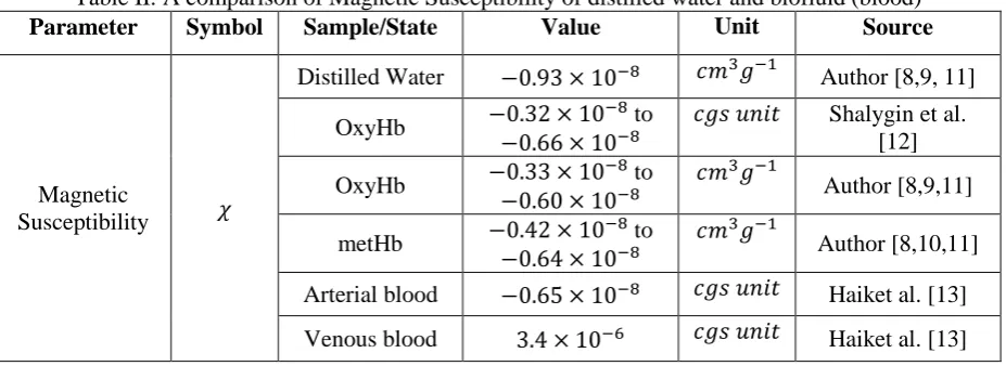
Table I shows the data on the physical properties of biofluid (blood). The table presents values of viscosity, velocity, flow rate, density, and electrical conductivity. These results are found in agreement with the literature. Table II shows the data on magnetic susceptibility measured experimentally in this study by the author. The magnetic susceptibility of different states of blood oxygenated hemoglobin (oxyHb), oxygenated methemoglobin (metHb) and distilled water are also presented. Also, magnetic susceptibility of venous blood and arterial blood reported by Haik et. al., [13], and oxyHb reported by Shalygin et. al., [12] were also reported. The magnetic susceptibility values found in the present study are found to agree with the literature, which is evident for the diamagnetic susceptibility of oxygenated state of blood.
Table I: Data on average values of the physical properties of biofluid (blood)
Property Range Unit
Viscosity 0.043-0.054 P
Velocity 12.3 -19.1 𝑐𝑚 𝑠−1
Flow Rate 0.095-0.152 𝑐𝑚3𝑠−1
Density 1.034-1.051 𝑔 𝑐𝑚−3
Electrical Conductivity 0.69-0.88 𝑆 𝑚−1
Table II: A comparison of Magnetic Susceptibility of distilled water and biofluid (blood)
Parameter Symbol Sample/State Value Unit Source
Magnetic
Susceptibility 𝜒
Distilled Water −0.93 × 10⁻⁸ 𝑐𝑚3𝑔−1 Author [8,9, 11]
OxyHb −0.32 × 10
−8 to
−0.66 × 10⁻⁸
𝑐𝑔𝑠 𝑢𝑛𝑖𝑡 Shalygin et al.
[12]
OxyHb −0.33 × 10
−8 to
−0.60 × 10⁻⁸
𝑐𝑚3𝑔−1
Author [8,9,11]
metHb −0.42 × 10
−8 to
−0.64 × 10⁻⁸
𝑐𝑚3𝑔−1
Author [8,10,11]
Arterial blood −0.65 × 10⁻⁸ 𝑐𝑔𝑠 𝑢𝑛𝑖𝑡 Haiket al. [13]
Venous blood 3.4 × 10⁻⁶ 𝑐𝑔𝑠 𝑢𝑛𝑖𝑡 Haiket al. [13]
4. DISCUSSION & CONCLUSIONS:
One of the basic constituents of the human body is blood, which proves to be a basic nourishing factor in our body. When a biofluid such as blood, subjected to the magnetic field, the blood particles flowing perpendicular to the applied field are repelled in the opposite direction, which is due to the presence of induced Lorentz force. As a result, the electromagnetic induction produced in the fluid slow down the flow rate, and flattens the velocity profile, while stretching it in the direction of the applied field. These effects heightened, when magnetic field strength increases.
Besides, magnetic effects on blood may be used in providing valuable information to the effects of magnetic resonance imaging (MRI) scanners. It is reported that when patients exposed to the magnetic field (1 to 4 T) in MRI scanning, resulted in a significant increase in blood viscosity, along with some side effects such as nausea, sleepiness, and vertigo.
It is observed that the applied magnetic field influences the electrocardiogram (ECG) of the human cardiac rhythm, which indicates the significance of the induced voltages. The electrical conductivity of blood is dependent on its velocity, increases marginally by the external magnetic field.
Moreover, the viscosity of the biofluid under the presence of magnetic field is found to increase significantly. The magnetic susceptibility of oxygenated state of blood is found to be diamagnetic. Thus, blood as a biofluid helps in understanding the possible health effects of magnetic fields.
REFERENCES:
1. TZirtzilakis, E. E (2005): A mathematical model for blood flow in magnetic field. Physics of Fluids, 17(1), 1-15. 2. Pauling, L; and Coryell, C. D (1936): The magnetic properties and structure of the hemochromogens and related
substances. Proc. Natl. Acad. Sci. USA., 22, 159-163.
3. Higashi, T;Yamagishi, A;Takeuchi, T;,Kawaguich, N;Sagawa, S;Onishi, S; and Date, M (1993): Orientation of erythrocytes in a strong magnetic field. Blood, 82 (4), 1328-1334.
4. A. Yamagishi, A (1990): Biological systems in high magnetic field. J. Magn.Magn. Mater., 90, 43-46.
5. Haik, Y; Pai, V; and Chen, C. J (2001): Apparent viscosity of human blood in high static magnetic field. J. Magn. Magn.Mater., 225, 180-186.
6. N. Nakona, N; Ostuka, J; Tasaki, A (1972): Biochem. Biophys.Acta., 278, 355.
7. Basharath Ali, M. Ibraheem Altaf & Adeel Ahmad (2007): An introduction to Human blood. J. Pure & Appl. Phys., 19 (2), 149.
8. Abdul Moiz Mohammed (2008), Studies on Magnetic properties of Human Blood and its constituents, M. Phil. diss. in Physics, Anna Malai University, India
9. Mohammed Abdul Moiz & Adeel Ahmad (2011): Magnetic susceptibility of normal human blood. J. Pure & Appl. Phys., 21(3), 395-399.
10. Abdul Moiz Mohammed & Adeel Ahmad (2015): Magnetic properties of Methemoglobin. Intl. J. Sci. Environ. Tech., 4(6), 1661 – 1665.
11. Abdul Moiz Mohammed (2016): A Study on Influence of Magnetic field on Blood. Int. Arch. App. Sci. Technol; 7 (1), 61-65.
12. Shalygin, A. N; Norina, S.B; Kondorsky, E.I (1983): Behavior of erythrocytes in high gradient magnetic field. J. Magn. Magn.Mater., 31, 555-556.
13. Haik, Y; Pai, V; and Chen, C.J (199): Biomagnetic fluid dynamics at interfaces. Cambridge University Press, Cambridge, 439-452.