Research Article
HE
4
has a High Diagnostic Value
to Detect Epithelial Ovarian Cancer
HE
4Memiliki Nilai Diagnostik yang Tinggi
untuk Mendeteksi Keganasan Ovarium Tipe Epitel
Bismarck J Laihad1, Hariyono Winarto1, Bambang Sutrisna2 1Division of Gynecology Oncology, Department of Obstetrics and Gynecology
2Department of Epidemiology
Faculty of Medicine University of Indonesia Jakarta
INTRODUCTION
Ovarian cancer is the second most common gyne-cologic malignancy. In Indonesia, based on the re-ports from 13 pathology laboratories in 2002, ovarian cancer ranked as the third (829 cases) most common from all malignancy in women, after cervical and breast cancer. In 2012, based on Jakarta cancer registry, ovarian cancer is the third leading female cancer with the incidence 4.27 in
100,000 women.1-4
Poor life expectancy in ovarian cancer is due to the lack of early-stage findings, causing most ovar-ian cancer cases to be found in advanced stages. Until now, there is no single biomarker which
could be used to predict ovarian cancer.5 CA-125,
as one of the most commonly used biomarker in epithelial ovarian cancer (EOC), is detected only in 50-60% of early-stage epithelial ovarian carcinoma
(EOC) patients.6
Abstract
Objective: To find out the diagnostic value of CA125 and HE4 as a tumor marker, and also RMI and ROMA as a malignancy predictor in patients with ovarian tumors.
Methods: This study was a diagnostic study using cross-sectional design.This study was performed in Jakarta from November 2010 to May 2011. One hundred and twenty eight serum samples of patients diagnosed with ovarian tumors were collected before undergoing surgery in Dr. Cipto Mangunkusumo General Hospital. The CA125 and HE4 levels were then examined. The histopathological examina-tion of tissue specimens were performed in Department of Pathol-ogy Anatomy in RSCM. For statistical analysis, we used a 2x2 table to produce ROC-AUC curve.
Results: The median value of HE4 and CA125 serum concentrations was higher and more significant on patients with ovarian malig-nancy than patients with benign ovarian tumor (p<0.05). Using the cut-off standard, HE4 had the highest accuracy value (76.5%). On the premenopausal group, HE4 and ROMA had the same AUC value, that is 85.0 % (95% CI: 0.73-0.96), whereas on the postmenopausal group, ROMA had the highest AUC value of 96.9 % (95% CI: 0.92-1.00).
Conclusion: HE4 has a high diagnostic value as a single tumor marker to detect epithelial ovarian cancer and its combination with CA125 (ROMA) gives an even better result.
[[Indonse J Obstet Gynecol 2013; 1-4: 209-14]
Keywords: epithelial ovarian cancer, human epididymis protein 4, risk of ovarian malignancy algorithm, tumor marker
Abstrak
Tujuan: Untuk menentukan nilai diagnosis penanda tumor CA 125 dan HE4, dan juga RMI dan ROMA sebagai prediktor keganasan pada tumor ovarium.
Metode: Penelitian ini adalah suatu studi diagnostik yang menggu-nakan metode potong lintang. Sejak November 2010 hingga Mei 2011, terdapat 128 pasien yang didiagnosis tumor ovari di RSCM. Dilakukan pengumpulan serum untuk memeriksa kadar CA125 dan HE4 dari 128 pasien tersebut. Pemeriksaan histopatologi dilakukan oleh Departe-men Patologi Anatomi RSCM. Kemudian, data yang didapat diolah dengan analisis tabel 2x2 dan kurva ROC-AUC.
Hasil: Nilai median dari konsentrasi serum HE4 dan CA125 ditemukan lebih tinggi pada pasien dengan keganasan ovari dibandingkan de-ngan pasien dede-ngan tumor jinak ovari (p<0,05). Penanda tumor HE4 memiliki nilai akurasi yang tertinggi berdasarkan nilai batas standar. Dalam grup perempuan pre-menopause, HE4 dan ROMA memiliki nilai AUC yang serupa pada 85% (95% CI: 0,73-0,96), sedangkan pada grup perempuan post-menopause, ROMA menunjukkan nilai AUC yang ter-tinggi pada skor 96,9% (95% CI: 0,92-1,00).
Kesimpulan: HE4 sebagai sebuah penanda tumor memiliki nilai diag-nostik yang tinggi untuk mendeteksi keganasan ovarium tipe epitel, dan kombinasi antara HE4 dan CA125 (ROMA) memberikan hasil yang lebih baik dibandingkan penggunaan satu penanda tumor HE4. [Maj Obstet Ginekol Indones 2013; 1-4: 209-14]
Kata kunci: human epididymis protein 4, kanker ovarium tipe epitel, penanda tumor, risk of ovarian malignancy alogarithm
Recently, several studies indicated that the
com-bined use of biomarkers such as CA125 and HE4
could improve the sensitivity and specificity of EOC
detection. HE4 serum marker has a high sensitivity
to detect an early stage ovarian cancer. Combina-tion of both markers is even more accurate than
the use of these markers individually.5,7
Although there are several scoring systems or methods to predict ovarian malignancy, the defi-nite method has not been established yet. Moore et al introduced a malignancy prediction method known as ROMA (Risk of Ovarian Malignancy Al-gorithm), which was worked out by combining the
results of CA 125 and HE4 examinations. Predictive
Probability Index (PPI) of ROMA had an accuracy
value up to 93.8%.5,8 However, Van Gorp et al
(2011) found that HE4 and ROMA were not
supe-rior to a single CA125 examination in predicting
ovarian malignancy.9
Based on the above background, this study aims
to compare the diagnostic value of CA125 and HE4
markers, and their combination in Risk Malignancy Index (RMI) and ROMA in predictingthe risk of ovarian malignancy in patients with ovarian tu-mors before undergoing surgery at Dr. Cipto Ma-ngunkusumo General Hospital (RSCM) in Jakarta, Indonesia.
MATERIALS AND METHODS
This was a cross sectional study, conducted at RSCM and Prodia Clinic Laboratory Jakarta from November 2010 to May 2011. The research popu-lation was all patients who came to RSCM and di-agnosed with ovarian tumors and met the inclusion criteria. The inclusion criteria were premenopausal or postmenopausal women, diagnosed as having ovarian tumors through physical examination/gy-necology and transvaginal ultrasound, and the tu-mor was considered respectable. Patients with his-topathological result of non-epithelial ovarian tu-mor, history of oophorectomy, history of previous ovarian cancer treatment, and pregnancy were ex-cluded from the study. Afterwards, blood samples were collected and stored in a -20° C temperature, and were analyzed using ARCHITECT plus i 2000 SR tool which measure the quantity of CA125 and
HE4. The pathologist from RSCM then conducted
the histopathological analysis of the tissue speci-mens.
Diagnostic method of pre-surgery patients with pelvic masses for the prediction of ovarian cancer is based on the value of CA-125 serum, ultrasound morphology (U) and menopause status (M). RMI = U x M x the value of CA-125, where ultrasound score = 1 if there is no morphological abnormalities
or found one, U= 3 if found ≥ 2 morphological
pic-ture. Menopause status score is M=1 on
pre-menopause and M=3 on post pre-menopause. Score ≥
200 was classified as malignant risk.
ROMA is an algorithm used to predict the risk of ovarian malignancy in patients with pelvic masses, so that patients can be stratified as low risk and
high risk based on the value of CA-125 and HE4.
Premenopausal women is classified as high risk when the Probability Prediction (PP) is more than 7.4 %, while postmenopausal women is classified as high risk when the PP is more than 25.3 %.
Data was analyzed using 9.2 Stata program. The statistical analysis aimed to obtain the value of sen-sitivity, specificity, PPV, NPV, and accuracy. An-other analysis on menopausal status and stage of epithelial ovarian cancer using ROC curve was also performed to obtain the value of AUC with 95% confidence interval calculations,. This study
com-pared the ROC and AUC value of CA125, HE4, RMI
and ROMA to the staging method in FIGO, with p value <0.05.
RESULTS
From November 2010 to May 2011, there were 128 patients at RSCM that met the inclusion and exclusion criteria. From those 128 patients, 61 pa-tients (47.66%) had benign ovarian tumor, 50 (39.06%) had malignant tumor, and the other 17 were borderline (13.28%). From 61 cases of be-nign ovarian tumors, the most common type was endometriosis (26 cases (42.62%)), followed by mucinous cystadenoma with 18 cases (29.51%), then serous cystadenoma and seromucinous (29.51 % and 4.92 %). For the malignant cases (epithelial ovarian cancer), the most common his-tological types were serous cystadenocarcinoma 19 cases (38%), followed by endometrioid with 14 cases (28%), mucinous with 8 cases (16%), clear cell with 7 cases (14%), and carcinosarcoma with 2 cases (4%).
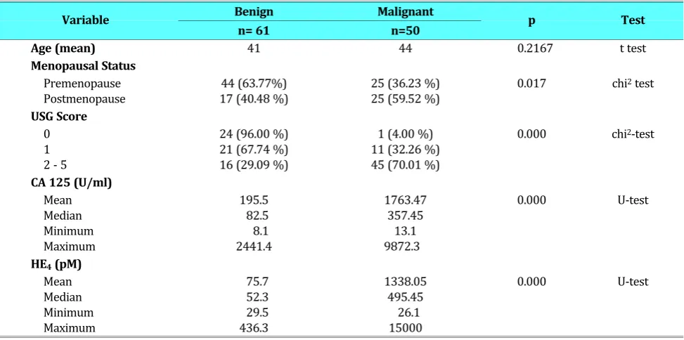
There were significant differences between be-nign and malignant groups on the menopausal
value. Median value of HE4 and CA125 serum
con-centration was significantly higher in patients with EOC compared to those with benign ovarian tumor, with p value < 0.05 (Table 1).
The diagnostic value of sensitivity, specificity, PPV, NPV, positive and negative like hood ratio (LR+ and LR-), as well as accuracy of tumor mark-ers in predicting the ovarian malignancy are pre-sented in Table 2. By using standard cut-off values,
HE4 had the highest accuracy value (76.5%),
fol-lowed by RMI, ROMA, and the last CA125 at 75.6%, 65.7%, and 56.7%, respectively.
As can be seen in Figure 1, HE4 and ROMA in
the premenopausal group had the same AUC value at 85.0% (95% CI: 0.73-0.96), whereas in the post-menopausal group ROMA had a higher AUC value
at 96.9% (95% CI: 0.92-1.00), followed by HE4
(93.9%). CA125 and RMI had a same AUC value at 93.6%. Furthermore, ROMA had the highest AUC
value of 90.5% followed by HE4, RMI, and CA125
respectively 89.9%, 87.3%, dan 82.0%.
Table1. Distribution of Age, Menopausal Status, USG Score, CA125, HE4on Benign and Malignant Ovarian Tumors
Variable Benign Malignant p Test
n= 61 n=50
Age (mean) 41 44 0.2167 t test
Menopausal Status
Premenopause Postmenopause
44 (63.77%) 17 (40.48 %)
25 (36.23 %) 25 (59.52 %)
0.017 chi2 test
USG Score
0 1 2 - 5
24 (96.00 %) 21 (67.74 %) 16 (29.09 %)
1 (4.00 %) 11 (32.26 %) 45 (70.01 %)
0.000 chi2-test
CA 125 (U/ml)
Mean Median Minimum Maximum
195.5 82.5 8.1 2441.4
1763.47 357.45 13.1 9872.3
0.000 U-test
HE4 (pM)
Mean Median Minimum Maximum
75.7 52.3 29.5 436.3
1338.05 495.45
26.1 15000
0.000 U-test
Table 2. Diagnostic Value of CA125, HE4, RMI and ROMA based on cut-off Standard
Marker StandardCut-off
Diagnostic Value
Sensitivity Specificity PPV NPV LR + LR – Accuracy
HE4 70 90.0 % 65.6 % 68.2 % 88.9 % 2.61 0.15 76.5 %
CA125 35 96.0 % 24.6 % 51.1 % 88.2 % 1.27 0.16 56.7 %
RMI 200 88.0 % 65.6 % 67.7 % 87.0 % 2.56 0.18 75.6 %
ROMA 7.4 / 25.3 94.0 % 42.6 % 57.3 % 89.7 % 1.64 0.14 65.7 %
0.
00
0.
25
0.
50
0.
7
5
1.
00
S
e
n
sit
iv
ity
0.00 0.25 0.50 0.75 1.00 1-Specificity
he4 ROC area: 0.899 ca125 ROC area: 0.8198 rmi ROC area: 0.8731 roma ROC area: 0.9046 Reference
Pre and Post Menopausal
A
DISCUSSION
The sensitivity of CA125 to detect EOC based on the determined cut-off standard (35 U/ml) was very high, reaching 96%. On the other hand, the specificity value of CA125 was very low (24.6%);
compared to HE4 with sensitivity value of 90% and
65.6 % specificity value. Hellstrom et al showed that there was no significant difference in
sensitiv-ity value of HE4 and CA125 in differentiating
ma-lignant and benign tumor. However, the specificity
of HE4 was significantly higher than that of
CA125.10 The very low value of CA125 specificity
on this research was because the mean and median values of CA125 from all benign tumors samples in this research were above the value of cut-off
0. 0 0 0. 2 5 0. 5 0 0. 75 1. 00 Se n s it iv it y
0.00 0.25 0.50 0.75 1.00
1-Specificity
he4 ROC area: 0.9249 ca125 ROC area: 0.8744 rmi ROC area: 0.909 roma ROC area: 0.9101 Reference
Stage III - IV
F
Figure 1. ROC Curve of CA125, HE4, RMI and ROMA
based on menopausal status and FIGO stage. (F) Malignant vs Benign on stage III-IV patients.
0. 0 0 0. 25 0. 5 0 0. 75 1. 00 S e n s it iv ity
0.00 0.25 0.50 0.75 1.00 1-Specificity
he4 ROC area: 0.8491 ca125 ROC area: 0.7414 rmi ROC area: 0.8082 roma_pre ROC area: 0.85 Reference
Premenopausal
Figure 1. ROC Curve of CA125, HE4, RMI and ROMA
based on menopausal status and FIGO stage. (B) Malignant vs Benign on premenopausal patients.
B
0. 00 0. 25 0. 50 0. 75 1. 00 S e n s it ivit y0.00 0.25 0.50 0.75 1.00 1-Specificity
he4 ROC area: 0.9388 ca125 ROC area: 0.9365 rmi ROC area: 0.9365 roma_post ROC area: 0.9694 Reference
Postmenopausal
C
Figure 1. ROC Curve of CA125, HE4, RMI and ROMA
based on menopausal status and FIGO stage. (C) Malignant vs Benign on postmenopausal patients.
0. 00 0. 2 5 0.5 0 0. 7 5 1. 0 0 S e n s itivi ty
0.00 0.25 0.50 0.75 1.00
1-Specificity
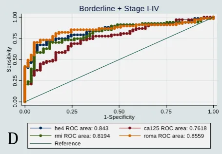
he4 ROC area: 0.843 ca125 ROC area: 0.7618 rmi ROC area: 0.8194 roma ROC area: 0.8559 Reference
Borderline + Stage I-IV
Figure 1. ROC Curve of CA125, HE4, RMI and ROMA
based on menopausal status and FIGO stage. (D) Malignant (including borderline) vs Benign on all patients.
D
0. 0 0 0. 2 5 0. 5 0 0.75 1.00 S e n s it iv it y0.00 0.25 0.50 0.75 1.00
1-Specificity
he4 ROC area: 0.8568 ca125 ROC area: 0.7308 rmi ROC area: 0.8145 roma ROC area: 0.8645 Reference
Stage I - II
Figure 1. ROC Curve of CA125, HE4, RMI and ROMA
based on menopausal status and FIGO stage. (E) Malignant vs Benign on stage I-II patients.
standard, as presented on Table 1. The standard
cut-off value for HE4 in this research was 70 mol/l,
based on a study by Moore et al (2008)7 and a
rec-ommendation of insert KIT ARCHITECT HE4
re-agent used in this research.
Holcomb et al, compared the ability of CA125 vs
HE4, and concluded that HE4 was more superior in
specificity compared to CA125. Similarly, according
to Van Gorp et al, HE4 had a higher specificity value
than CA125 using the cut-off standard.9,12
Several studies on CA125 and HE4 by Moore at
al (2008), Huhtinen at al (2009), Nolen et al (2010), Holcomb et al (2011), and Chang et al (2011), stated that a combination of CA125 and
HE4 could further improve the diagnostic ability to
differentiate malignant and benign tumors among patients with adnexal masses before surgery. Moore at al (2009) introduced ROMA (Risk of Ovarian Malignancy Algorithm), a stratification of risk in women with pelvic masses without involv-ing ultrasound. ROMA is considered more sensitive than RMI and calculated by combining the results
of CA125 and HE4.7,8,11-14 Using standard cut-off
value, HE4 and RMI are proven to have a higher
accuracy value than ROMA and CA125 (Table 2).
The AUC value of HE4 and ROMA is the highest
in all patients, both premenopausal group and postmenopausal group, compared to that of RMI and CA125. Montagnana et al also compared the
AUC values of HE4, CA125, and ROMA on pre and
postmenopausal groups, and concluded that HE4
and ROMA showed excellent ability only in the postmenopausal group, but not in the
pre-menopausal group.15
In this study, HE4 and ROMA in the
pre-menopausal group have the same AUC value at 85% (95% CI: 0.73-0.96). On the other hand, Van Gorp et al’s study compared the AUC values among
ROMA, HE4 and CA125, and stated that the ability
of HE4 and ROMA was not higher than a single
CA125 as tumor marker to predict ovarian malig-nancy. This was based on the comparison of ROC-AUC values in all patients (pre and
post-menopause) on ROMA (89.8%) vs HE4 (85.7%) vs
CA125 (87.7%), that after being tested statistically, there were not any significant differences among
the three (p>0,005).9
Advanced stage EOC (Figure 1.E), resulted in
higher AUC values for ROMA, HE4 and RMI than
those in early stage (Figure 1. F). This results was
supported by Gorp et al, and Moore et al, where
the AUC values for ROMA, HE4 and RMI were
higher in advance stage EOC patients than those
with early stage disease.11,17 Furthermore, CA125
had a low diagnostic value in early stages EOC, as stated by Sasarolidan Moore, where elevated levels of CA125 were only found in 50-60% of early stage
EOC cases.7,16,17
After comparing ROMA to RMI in 457 patients, Moore et al found that the AUC for ROMA was sig-nificantly higher than RMI in all stages of epithelial ovarian cancer. Moore et al, concluded that ROMA had a higher diagnostic value than RMI clinically
and statistically.18
This study found that HE4 as a new tumor
marker has a higher diagnostic value than CA125. Moreover, when the two are combined, such as in ROMA algorithm, it shows a better ability as a pre-dictor of epithelial ovarian cancer.
CONCLUSION
HE4 as a single tumor marker has a high diagnostic
value in detecting epithelial ovarian malignancy. It has a better specificity and accuracy compared to
CA125. However, combination of HE4 and CA125
(ROMA) shows a better ability as ovarian malignancy
predictor compared to a single HE4 marker.
CONFLICT OF INTEREST
The Author has no conflict of interest.REFERENCES
1. Aziz MF. Gynecological cancer in Indonesia. J Gynecol Oncol, 2009; 20(1): 8-10.
2. Boyle P, B Levin. World Cancer Report, WHO, Editor. Inter-national Agency for Research on Cancer: Switzerland. 2008 3. Hennessy BT, RL Coleman, M. Markman. Ovarian cancer.
Lancet, 2009; 374(9698): 1371-82.
4. Wahidin M, Noviani R, Hermawan S et al. Population-based cancer registration in Indonesia. Asian Pac J Cancer Prev. 13(4):1709-10.
5. Havrilesky LJ, Whitehead CM, Rubatt JM et al. Evaluation of biomarker panels for early stage ovarian cancer detection and monitoring for disease recurrence. Gynecol Oncol, 2008; 110(3): 374-82.
7. Moore RG, Brown AK, Miller CM et al. The use of multiple novel tumor biomarkers for the detection of ovarian carci-noma in patients with a pelvic mass. Gynecol Oncol, 2008; 108(2): 402-8.
8. Moore RG, McMeekin DS, Brown AK et al. A novel multiple marker bioassay utilizing HE4 and CA125 for the prediction of ovarian cancer in patients with a pelvic mass. Gynecol Oncol, 2009; 112(1): 40-6.
9. Van Gorp T, Cadron I, Despierre E et al. HE4 and CA125 as a diagnostic test in ovarian cancer: prospective validation of the Risk of Ovarian Malignancy Algorithm. Br J Cancer, 2011; 104(5): 863-70.
10. Hellstrom I, Raycraft J, Hayden-Ledbetter M et al. The HE4 (WFDC2) protein is a biomarker for ovarian carcinoma. Cancer Res, 2003; 63(13): 3695-700.
11. Chang X, Ye X, Dong L et al. Human epididymis protein 4 (HE4) as a serum tumor biomarker in patients with ovarian carcinoma. Int J Gynecol Cancer. 2011. 21(5): 852-8. 12. Holcomb K, Vucetic Z, Miller MC et al. Human epididymis
protein 4 offers superior specificity in the differentiation of benign and malignant adnexal masses in premenopausal women. Am J Obstet Gynecol. 2011. 205(4): 358 e1-6. 13. Huhtinen K, Suvitie P, Hiissa J et al. Serum HE4
concentra-tion differentiates malignant ovarian tumours from ovarian endometriotic cysts. Br J Cancer, 2009; 100(8): 1315-9.
14. Nolen B, Velikhokatnaya L, Marrangoni A et al. Serum biomarker panels for the discrimination of benign from ma-lignant cases in patients with an adnexal mass. Gynecol On-col. 2011. 117(3): 440-5.
15. Montagnana M, Danese E, Ruzzenente O et al. The ROMA (Risk of Ovarian Malignancy Algorithm) for estimating the risk of epithelial ovarian cancer in women presenting with pelvic mass: is it really useful? Clin Chem Lab Med. 2011. 49(3): 521-5.
16. Maharaj AGJI, Menon U. Principles and Practice of Gyne-cologic Oncology. 5th Ed. Development And Identification Of
Tumor Markers, ed. M.M. In Barakat R R, Randall M E. 2009; 145-50.
17. Sasaroli D, G Coukos and N Scholler. Beyond CA125: the coming of age of ovarian cancer biomarkers. Are we there yet? Biomark Med, 2009; 3(3): 275-88.