Evaluation of Peri-implant Soft and Hard Tissues in
Titanium Implants in Immediate and Delayed Cases:
A Comparative Study
1Joseph Edward, 2Ajith Samson, 3Joju George, 4Prakash G Dhanya ORIgInal ReseaRCh
in Titanium Implants in Immediate and Delayed Cases: A Com-parative Study. Int J Oral Care Res 2017;5(4):298-304.
Source of support: Nil
Conflict of interest: None
INTRODUCTION
Since the 1980s, bone-anchored dental implants have become a well-established and predictable treatment for restoring missing teeth. The implant placement protocol was to wait following tooth extraction to allow adequate bone maturation to support the implant. Rationale behind this was to reduce the portability of infection, provide a more stable base for implant, and increase the amount of keratinized gingiva. Today, many researchers and clini-cians are focusing on ways to achieve these successful results while simplifying and shortening the treatment process.1 The advantage is that it allows patients to regain function and natural-looking teeth more quickly, and also this helps to maintain the soft and hard tissue architecture. In the 1980s, the University of Tubingen advocated the procedure as the technique of choice for Tubingen and Munchen ceramic implants.2 In the clas-sification of implants according to timing of placement given by Wilson,3,4 the terms immediate, recent, delayed, and mature are used to describe the timing of implant placement in relation to soft tissue healing and predict-ability of guided bone regeneration procedures. Garber and Belser5 have described three scenarios for the timing of implant placement following extraction. Immediate placement occurs at the time of tooth extraction, staged placement occurs at least 8 weeks following extraction, and delayed placement is performed 3 months or more following extraction. Gomez-Roman et al6 defined imme-diate implants as occurring between 0 and 7 days after tooth extraction. Zitzman et al7 considered implant place-ment as delayed when it occurred between 6 weeks and 6 months after extraction. Mayfield,8 in his classification given in 1999, used the terms immediate, delayed, and late to describe time intervals of 0 week, 6 to 10 weeks, and 6 months or more after extraction respectively.
As the debate in timing of implant placement increased, the following new classification based on morphologic, dimensional, and histologic changes that 1Professor and Head, 2,3Assistant Professor, 4Consultant
1,4
Department of Oral and Maxillofacial Surgery, Azeezia College of Dental Sciences and Research, Kollam, Kerala, India
2Department of Oral and Maxillofacial Surgery, Government
Dental College, Alappuzha, Kerala, India
3
Department of Oral and Maxillofacial Surgery, Indira Gandhi Institute of Dental Sciences, Ernakulam, Kerala, India
Corresponding Author: Joseph Edward, Professor and Head Department of Oral and Maxillofacial Surgery, Azeezia College of Dental Sciences and Research, Kollam, Kerala, India, Phone: +919446572285, e-mail: [email protected]
ABSTRACT
Background and objective: Immediate implant placement may preserve the alveolar anatomy, and helps to maintain the bony crestal height. There are not enough studies addressing other outcome measures that determine the quality of survival, such as the peri-implant hard and soft tissue integrity. The objective of this study was to evaluate and compare the peri-implant soft tissues, crestal bone resorption, and peri-implant bone healing in immediate and delayed implants in the esthetic zone of maxilla.
Materials and methods: Selective sampling was done among the patients who reported to our center for implant placement. A total sample size of 100 implant sites in the esthetic zone of maxilla were selected from 77 patients (52 females and 25 males) with 50 sites in immediate implant group and 50 in delayed implant group. Samples from both groups were evaluated on follow-ups at 1st week, 1st month, 3rd month, and 6th month postopera-tively. Evaluation was based on (1) assessment of peri-implant soft tissue by means of implant esthetic score, (2) radiographic assessment of peri-implant crestal bone loss, (3) peri-implant bone densitometry by gray-scale assessment of radiograph.
Results: Unpaired t-test was performed for each parameter, which showed a very high level of significance for implant esthetic score, high significance for gray scale assessment, and no significance for peri-implant crestal bone loss.
Interpretation and conclusion: The present study showed that immediate implants had better esthetic and functional outcome in terms of peri-implant soft tissue and peri-implant bone healing. But there were no statistically significant data suggesting lesser alveolar crestal bone loss in immediate implants.
Keywords: Bone densitometry, Delayed implants, Immediate implants, Implant esthetic score.
Evaluation of Peri-implant Soft and Hard Tissues in Titanium Implants
follow tooth extraction was proposed by Hämmerle et al9 and accepted at the Third ITI Consensus Confer-ence 2004, Type I: Immediate placement: an implant is placed immediately in an extraction socket as part of the same procedure with no healing of bone or soft tissues. Type II: Early placement with some soft tissue healing (typically 4–8 weeks of healing) the postextrac-tion site has healed with soft tissue coverage of the alveolus but without significant bone healing. Type III: Early placement with partial bone healing: (typically 12–16 weeks of healing) the postextraction site has both healed with soft tissues and a significant degree of bone healing. Type IV: Late placement (more than 6 months after extraction) implant placement in a fully healed edentulous site.
Immediate implants have numerous advantages as they reduce the number of surgical procedures, morbid-ity, and overall treatment time. The criteria behind the long-term success of implants are stable bone support, minimal degree of inflammation, fully functional implant-supported crown. According to the studies, peri-implant bone loss usually occurs most during the first year of placement, thereafter the bone loss is less.10,11 Level of peri-implant mucosa and level of peri-implant marginal bone are strongly related. Therefore, the loss of peri-implant marginal bone could affect the peri-peri-implant mucosa and hence the esthetic outcome.
To date, several reviews have been published regard-ing the clinical outcome of immediate and conventional implant-supported single-tooth restorations in partially edentulous patients. Most of these reviews have mainly converged on implant survival and addressed to a lesser degree other outcome measures that determine the quality of survival. In a recent Cochrane system-atic review, the success, complications, esthetics, and patient satisfaction among different timing of implant placement after tooth extraction, such as immediate, immediate–delayed, and delayed were evaluated.12 Two studies of parallel group design, comparing immediate and delayed implant placement were included in this review. The meta-analysis of the two trials did not show any statistically significant difference between the two groups. Based on the few underpowered trials, it was concluded that there was insufficient evidence to deter-mine possible advantages or disadvantages of immediate or delayed implants.13
Therefore, the purpose of the present study is to compare between implants placed in fresh extraction sockets (immediate implants) and implants placed in healed extraction sites (delayed implants) in esthetic zone of maxilla, by peri-implant soft tissue and hard tissue analysis, thereby assessing the functional and esthetic outcome.
MATERIALS AND METHODS
Present study is a quantitative experimental study, to compare immediate and delayed implants placed in esthetic zone of maxilla. After getting approval from insti-tutional committee (ICE/IRB number ACDS/1624/12), selective sampling was done among the patients who came to the Department of Oral & Maxillofacial Surgery, Azeezia College of Dental Science and Research, for implant placement, from May 2012 to March 2015. A total sample size of 100 implant sites in the esthetic zone of the maxilla were selected from 77 patients (52 females and 25 males) who needed implant placement in the age range of 18 to 35 years. Forty-three patients (30 females and 13 males) were selected in the delayed implant group, in whom 50 healed partial edentulous spaces were identi-fied. Thirty-four (34) patients (12 males and 22 females) who needed extraction of tooth in the esthetic zone of maxilla were included in the immediate implant group. Indications for tooth extraction were untreatable carious lesions, endodontic treatment failure, tooth fracture, and other factors that could result in a hopeless prognosis.
Prior to implant placement and subsequent restora-tion, thorough medical history was taken from all the patients. Each case was precisely evaluated by thorough examination of intraoral tissues and periapical radio-graphs. The inclusion criteria were cases with fresh extraction sockets of maxillary teeth in the esthetic zone as experimental group and healed extraction sites in esthetic zone of maxilla, with a minimum postextraction period of 3 months as control group. Apparently healthy individuals within the age range of 18 to 35 years with sufficient alveolar bone height and width, and those who can maintain satisfactory oral hygiene were included in the study. Patients with any medical compromise or syndromes associated with bones, with history of any oral mucosal diseases, osseous defects, extraction sockets with deficient labial plates, severe periodontal problems, and cases with history of chronic alcoholism, smoking, or chewing habits were excluded. Consent was obtained from all the patients. Patients from both groups were placed under antibiotics for a minimum of 3 days prior to the procedure.
ligament space and gradually advanced toward the apex of the tooth, with care taken to preserve the thin labial wall (Fig. 1). After severing the periodontal ligament, and when the tooth is luxated enough, it is removed using narrow-beaked forceps. If the tooth or root stump was not luxated enough, one or two K-files or reamers of appropriate size were threaded into the root canal to engage the tooth, and the tooth was removed by manipulating the files using forceps. Reflection of flap was avoided as far as possible to preserve the vascular supply of periosteum covering the bone, so as to minimize bone resorption. After completion of extraction, the socket was debrided and then evaluated. A caliper was used to measure the maximum width of the extracted tooth root. An implant of diameter slightly larger than the maximum width of tooth root was selected. A rough estimation of the socket length was done by mea-suring the root length. A guide drill was placed into the socket, and by means of radio visio graph (RVG) the socket length was measured, and an implant of length 2 to 3 mm greater than the socket length was selected.
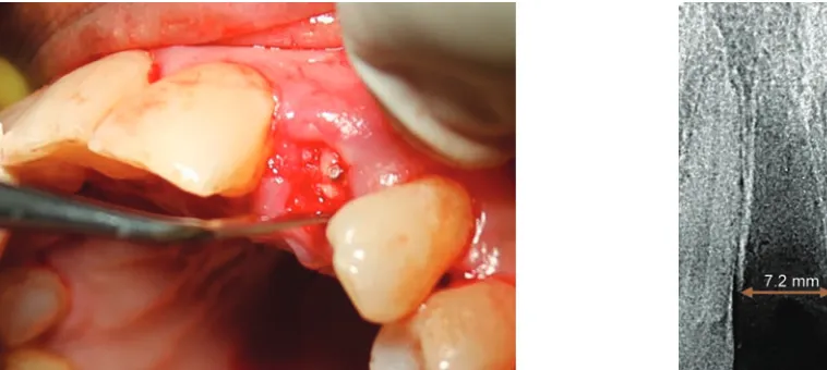
In delayed implant group, appropriate implant size was selected after RVG-based measurement of the alveo-lar bone (Fig. 2). Diameter of the implant was selected so as to retain a minimum of 1 mm of peri-implant alveo-lar bone in all dimensions. Length of the implant was selected, comparable to adjacent tooth root length. An envelope mucoperiosteal flap was given in all delayed implant cases to reflect the soft tissue. Appropriate drill-ing was performed for the placement of implants in both groups. The selected implants were screwed into the prepared site using a manual torque wrench, so as to maintain an insertion torque of 20 to 40 N. All implants were placed equicrestally. The mucoperiosteal flaps were repositioned to attain passive soft tissue primary closure
(diclofenac 50 mg three times a day), and chlorhexidine (0.2%) mouthwash for 5 days. The sutures were removed 1 week postoperatively.
METHOD OF EVALUATION
Samples were evaluated for soft tissue and hard tissue parameters on follow-ups at 1st week, 1st month, 3rd month, and 6th month postoperatively. Evaluation was based on assessment of peri-implant soft tissue by means of implant esthetic score, radiographic assessment of peri-implant crestal bone loss, and peri-implant bone densitometry by gray scale assessment of radiograph.
Peri-implant soft tissue was assessed by implant esthetic scoring,13 where the following parameters were looked in to: presence and stability of the mesial and distal papilla, ridge stability buccopalatally, texture of the peri-implant soft tissue, color of the peri-implant soft tissue, and gingival contour. The scoring was done according to the criteria proposed by Testori et al13: (A) Presence and stability of the mesiodistal papilla—0 = no papilla, 1 = papilla does not fill the entire space but is esthetically acceptable in harmony with adjacent teeth, 2 = total fill of papilla. The dimensional stability of the papilla was assessed by measuring the vertical distance from the apex of the mesial and distal papilla to the imagi-nary line connecting the cementoenamel junction of the two adjacent teeth. The height of the mesial and distal papilla was periodically measured with reference to this line (Fig. 3). (B) Ridge stability buccopalatally—0 = width with ridge loss, 1 = width-maintained ridge stability was measured in millimeters of buccal resorption with respect to adjacent natural teeth from the first follow-up to the 6 months follow-up. (C) Texture of the peri-implant soft tissue—0 = complete loss of texture, 1 = does not look
Evaluation of Peri-implant Soft and Hard Tissues in Titanium Implants
like healthy tissue, but some texture still maintained, 2 = looks like healthy gingival tissue around the natural teeth. (D) Color of the peri-implant soft tissue—0 = com-pletely different color from healthy tissue, 1 = does not look like healthy tissue but still esthetically acceptable, 2 = looks like healthy gingival tissue around the natural teeth. (E) Gingival contour—0 = evident asymmetry from the accepted parameters of scalloping, 1 = signs of asymmetry but esthetically acceptable, 2 = harmonious gingival contour.
Peri-implant crestal bone loss assessment: all implants were placed equicrestal (in level with the lower one among mesial and distal alveolar crests), so it was helpful in measuring the crestal bone loss with respect to platform of the implant. Crestal bone loss was measures based on analysis of RVGs (Fig. 4) taken on 1st week, 1st month, 3rd month, and 6th month follow-ups. Peri-implant bone gray scale assessment: the radiographic images taken on 1st week, 1st month, 3rd month, and 6th month follow-ups were enhanced on a gray scale with Photoshop 8. The optical density curves were adjusted to a percentage of
gray scale, where the least digital number (zero) corre-sponds to the most radiopaque point (the dense implant core) and the highest number (100) corresponds to the most radiolucent point (air). Thus, the images which were taken in different times were simulated in optical density curves and can be compared. Numerical average of four points in the middle third of the implant was taken for comparison. For standardization, the four points consid-ered were between the fourth and sixth threads of the implant body (cortical threads not counted) on mesial and distal aspects (Fig. 5).
DATA ANALYSIS
The values obtained for each implant sites were tabu-lated into a chart. For ease of statistical analysis, the immediate implant group was designated as 1 and the delayed implant group as 2. The values obtained by assessing each parameter at every follow-ups were entered separately, and for statistical analysis IBM Sta-tistical Package for the Social Sciences statistics software, version 20, was used, an unpaired t-test was applied to compare the two groups. Unpaired t-test was performed for each parameter, which showed a very high level of significance for implant esthetic score, high significance for gray scale assessment, and no significance for peri-implant crestal bone loss.
RESULTS
Seventy-seven patients who met inclusion criteria agreed to participate in follow-up study and had been treated with 100 dental implants. The values obtained from both groups showed some difference between them with better results for immediate implant group. Out of 50 immediate implants placement, 48 implants were successful, 2 implants had to be removed because
Fig. 3: Implant esthetic scoring Fig. 4: Assessment of peri-implant crestal bone loss
Peri-implant Esthetic Score
The mean values of peri-implant esthetic score obtained for immediate implant group (Group I) at 1st week, 1st month, 3rd month, and 6th month follow-ups were 7.2, 6.6, 6.6, and 6.5 respectively. For the delayed implant group (group II), the values were 5.6, 4.5, 4.5, and 4.5 at the corresponding follow-ups. A line graph plotted with these values showed a definite difference between the two groups (Graph 1). After applying the unpaired
t-test for the 1st week, 1st month, 3rd month, and 6th month values, the t-values obtained were 5.367, 9.0, 9.0, and 8.485 respectively, with 18 degrees of freedom. The p value was 0.000 in each category, showing a very high level of significance.
Peri-implant Crestal Bone Loss
The mean crestal bone loss in millimeters for immediate implant group (group I) was 0.22 (±0.092) at 1st week, 0.62 (±0.132) at 1st month, 0.85 (±0.178) at 3rd month, and 1.05 (±0.242) at 6th month follow-ups. For the delayed implant group (group II), the crestal bone loss in millimeters was 0.26 (±0.084) at 1st week, 0.69 (±0.185) at 1st month, 0.93 (±0.221) at 3rd month, and 1.13 (±0.283) at 6th month. On statistical analysis, the t-values for 1st week, 1st month, 3rd month, and 6th month were –1.014, –0.974, –0.891, and –0.680, all with 18 degrees of freedom. At none of the values the test was found to be significant with p values 0.324, 0.343, 0.385, and 0.505 in the chronological order of values obtained at each review. Although the line diagram plotting (Graph 2) the peri-implant crestal bone loss of immediate and delayed implant groups showed some
dif-(Gray Scale Assessment)
The mean values of gray scale values obtained were 51.35 (±3.587), 44.80 (±5.062), 41.075 (±6.058), and 39.425 (±6.607) for the immediate implant group (group I), and 63.45 (±12.371), 59.60 (±11.335), 54.675 (±9.399), and 48.275 (±6.697) for the delayed implant group (group II) in the 1st week, 1st month, 3rd month, and 6th month follow-ups respectively. A line diagram plotted with values of both the groups showed considerable difference (Graph 3). On performing an unpaired t-test for the variable in both groups, the t-values obtained were –2.971, –3.77, –3.946, and –2.975 at the corresponding intervals. The level of significance was high for every interval (0.008, 0.001, 0.001, and 0.008).
Graph 1: Implant esthetic score. Line graph shows a definite
difference between the two groups
Graph 2: Peri-implant crestal bone loss. The line diagram plotting the peri-implant crestal bone loss of immediate and delayed implant groups showed some difference, but the statistical test showed it
to be not significant
Graph 3: Peri-implant gray scale assessment. Line diagram plotted with values of both the groups showed considerable difference. The
Evaluation of Peri-implant Soft and Hard Tissues in Titanium Implants
DISCUSSION
In the anterior zone, the success of implant therapy is not only determined by high survival rates, but even more by the quality of survival, dictated by a mixture of several factors. Preferably, the appearance of the peri-implant soft tissue should be in harmony with the mucosa around the adjacent teeth and the implant crown should be in balance with the neighboring dentition. When considering the heights of interimplant papillae for instance, studies with conventional implant placement indicated that these papillae might show inadequacy for complete enclosure of the interimplant area with soft tissue, thereby failing to duplicate the interproximal soft tissue appearance of the adjacent teeth. This deficiency may affect the esthetic outcome unfavorably.
In the present study, the comparison of the implant esthetic score showed that the immediate implants have a definitely better esthetic outcome when compared with the delayed implants. The mean values in both groups showed considerable difference and statistically was proven with very high significance. This was in well accordance with the studies of Block et al,14 with 26 immediate and 29 delayed implants, where they found an average of 1 mm less recession in immediate implants than in delayed implants. There were also studies con-tradicting the present study results, such as the study by Lindeboom et al15 in 2006 with 25 immediate and 25 delayed implants, from which they concluded that there was no difference between the peri-implant soft tissue recession in immediate and delayed implants. The pro-gressive involution of the alveolar bone begins following tooth loss, and it is accompanied by a reduction in both the quality and quantity of hard and soft tissues. Experimen-tal animal researches and clinical studies demonstrated that the immediate implant placing reduces alveolar resorption.16,17 Moreover, this surgical procedure also allows a better final rehabilitation because it facilitates both morphological ridge contour preservation and accurate prosthetic implant installation, maintaining the natural tooth angle. There are also important benefits because the treatment time is reduced. Indeed, alveolar wound healing coincides with implant osseointegration and the patient can achieve the reinstatement of his/her edentulousness swiftly and by means of a single surgi-cal exposure. Thus, immediate placement of implants into fresh extraction sockets should have the potential to increase the patients’ acceptance of the procedure. The immediate implant procedure preserves bone and soft tissue structures necessary for implant placement. In contrast, as to delayed implant placement, pressure from prosthetic restorations during the healing time may decrease alveolar bone width and height, thereby
decreasing the bone volume required for proper implant placement.
The present study evaluated the peri-implant crestal bone loss and also the peri-implant bone healing by assessing bone density by means of gray scale assessment of postoperative radiographs. The results showed sig-nificant differences between the peri-implant gray scale assessment values of the immediate and delayed implant groups at all follow-ups. There was no significant differ-ence in the crestal bone loss between the two groups. The data obtained from evaluation of peri-implant alveolar crestal loss in immediate and delayed implant groups were plotted on a line diagram, which showed some dif-ference (Graph 2). But statistically there was no difdif-ference between the two groups. This result was similar to that obtained for Crespi et al18 and Block et al.14 Lindeboom et al15 did a comparative study on mesial and distal bone loss on immediate and delayed implants and found that the bone loss in the mesial aspect was less for immediate implants, but bone loss on the distal aspect was compa-rable in both groups. On the contrary, Palatella et al19 found that bone loss is more in immediate implant place-ment (0.54 ± 0.5 mm) than in delayed implant placeplace-ment (0.46 ± 0.54 mm), whereas Schropp et al,20 after a study in 23 immediate and 23 delayed implants, had found that mesial bone loss was more in delayed implant group, but distal bone loss was more for immediate implant groups. In the present study, no separate recording of the mesial and distal bone loss was done, instead the maximum bone loss was recorded.
Considering the alveolar bone loss which had occurred at the delayed implant sites during the period of healing from the time of tooth extraction to the time of implant placement, which is beyond the scope of the present study, the cumulative alveolar bone loss could be much more in the delayed implant group. The results of peri-implant bone healing assessed by peri-implant gray scale analysis showed that bone healing was better in immediate implants when compared with delayed implants. These results were comparable with the studies of Vignoletti et al,21 where they found that early healing occurs in implants placed in fresh extraction sockets. Paolantonio et al22 demonstrated osseointegration in implants placed simultaneously in fresh extraction sockets and healed bone of 48 individuals and found that the clinical outcome and degree of osteointegration of immediate implants does not differ from implants placed in healed, mature bone.
CONCLUSION
Extraction sites in the anterior maxilla present the great-est rgreat-estorative challenges due to the need for high-quality esthetic results. Immediate implants could be an answer to all these requirements. According to the present study, we can conclude that immediate implants showed better esthetic and functional outcome in terms of peri-implant soft tissue and peri-implant bone healing, but there was no statistically significant evidence showing the lesser crestal bone loss in case of immediate implants.
REFERENCES
1. Fonseca R, Marciani R, Turvey T. Oral and maxillofacial surgery. 2nd ed. Elsevier; 2009. p. 511-524.
2. Schulte W, Kleineikenscheidt H, Lindner K, Schareyka R. The Tubingen immediate implant in clinical studies. Deutsche Zahnarztliche Zeitschrift 1978 May;33(5):348-359, 319-325. 3. Wilson TG Jr, Schenk R, Buser D, Cochran D. Implants placed
in immediate extraction sites: a report of histologic and his-tometric analyses of human biopsies. Int J Oral Maxillofac Implants 1998 May-Jun;13(3):333-341.
4. Wilson TG Jr, Carnio J, Schenk R, Cochran D. Immediate implants covered with connective tissue membranes: human biopsies. J Periodontol 2003 Mar;74(3):402-409.
5. Garber DA, Belser UC. Restoration-driven implant placement with restoration—generated site development. Compend Contin Educ Dent 1995 Aug;16(8):796, 798-802, 804.
6. Gomez-Roman G, Kruppenbacher M, Weber H, Schulte W. Immediate post extraction implant placement with root-analog stepped implants: surgical procedure and statisti-cal outcome after 6 years. Int J Oral Maxillofac Implants 2001;16(4):503-513.
7. Zitzman NU, Scharer P, Marinello CP. Factors influencing the success of GBR. Smoking, timing of implant placement, implant location, bone quality and provisional restoration. J Clin Periodontol 1999 Oct;26(10):673-682.
8. Mayfield LJA . Immediate, delayed and late submerged and transmucosal implants. In: Lindhe J (ed). Proceedings of the 3rd European Workshop on Periodontology: Implant Den-tistry. Berlin: Quintessenz, 1999. pp. 520-534.
9. Hämmerle CH, Chen ST, Wilson TG Jr. Consensus statements and recommended clinical procedures regarding the place-ment of implants in extraction sockets. Int J Oral Maxillofac Implants 2004;19(suppl.):26-28.
10. Smith DE, Zarb GA. Criteria for success of osseointegrayed endosseous implants. J P Prosthet Dent 1989 Nov;62;567-573.
1986 Summer;1(1):11-25.
12. Esposito M, Grusovin MG, Polyzos IP, Felice P, Worthington HV. Timing of implant placement after tooth extraction: imme-diate, immediate-delayed or delayed implants? A Cochrane systematic review. Eur J Oral Implantol 2010 Autumn; 3(3):189-205.
13. Testori T, Bianchi F, Del Fabbro M, Capelli M, Zuffetti F, Berluc-chi I, TasBerluc-chieri S, Francetti L, Weinstein RL. Implant aesthetic score for evaluating the outcome: immediate loading in the aesthetic zone. Pract Proced Aesthet Dent 2005 Mar;17(2): 123-130.
14. Block M, Finger I, Castellon P, Lirettle D. Single tooth imme-diate provisional restoration of dental implants: technique and early results. J Oral Maxillofac Surg 2004 Sep;62(9): 1131-1138.
15. Lindeboom JA, Tijiook Y, Kroon FH. Immediate placement of implants in periapical infected sites: a prospective randomized study in 50 patients. Oral Surg Oral Med Oral Pathol Oral Radiol Endod 2006 Jun;101(6):705-710.
16. Devlin H, Sloan P. Early bone healing events in the human extraction socket. Int J Oral Maxillofac Surg 2002 Dec;31(6): 641-645.
17. Karagianes MT, Westerman RE, Hamilton AI, Adams HF, Wills RC. Investigation of long-term performance of porous-metal dental implants in nonhuman primates. J Oral Implantol 1982;10(2):189-207.
18. Crespi R, Capparé P, Gherlone E, Romanos GE. Immediate versus delayed loading of dental implants placed in fresh extraction sockets in the maxillary esthetic zone: a clinical comparative study. Int J Oral Maxillofac Implants 2008 Jul-Aug;23(4):753-758.
19. Palattella P, Torsello F, Cordaro L. Two-year prospective clinical comparison of immediate replacement vs. immedi-ate restoration of single tooth in the esthetic zone. Clin Oral Implants Res 2008 Nov;19(11):1148-1153.
20. Schropp L, Kostopoulos L, Wenzel A. Bone healing following immediate versus delayed placement of titanium implants into extraction sockets: a prospective clinical study. Int J Oral Maxillofac Implants 2003 Mar-Apr;18(2):189-199.
21. Vignoletti F, de Sanctis M, Berglundh T, Abrahamsson I, Sanz M. Early healing of implants placed into fresh extraction sockets: an experimental study in the beagle dog. II: ridge alterations. Department of Periodontology, Universidad Complutense de Madrid, Madrid, Spain; 2002.