IJPAR |Vol.8 | Issue 1 | Jan – Mar - 2019
Journal Home page: www.ijpar.com
Research article Open Access
Formulation and characterization of
RH
-Vascular Endothelial Growth
Factor loaded Solid lipid nanoparticles for chronic wounds healing
Mr.A.Sathyaraj*
1, Dr.D.Ramashekar Reddy
2 *1Research Scholar, Dept of Pharmacy, Krishna University, Machilipatnam
2Assistant Professor, Dept of Chemistry, Krishna University, Machilipatnam
*Corresponding Author: Mr.A.SathyarajEmail: [email protected]
ABSTRACT
Vascular endothelial growth factor (VEGF) is one of the most potent proangiogenic growth factors in the skin, and the amount of VEGF present in a wound can significantly impact healing. In this research study, rhVEGF (recombinant Vascular Endothelial Growth Factor) was epitomized solid lipid nanoparticles. In vitro tests were attempted in fibroblasts and keratinocytes to decide the bioactivity of the typified rh-VEGF and to gauge the cell take-up ability of the lipid nanoparticles, and their effectiveness was compared with that of a few intra-lesional organizations of a higher measurement of free rhVEGF and that of a solitary intra-lesional organization of rh-VEGF-loaded solid lipid nanoparticles created in our research facility. Mending was assessed as far as wound conclusion, recuperation of the incendiary stage, and re-epithelisation review.
INTRODUCTION
Since their first portrayal in the1990s, lipid nanoparticles, basically solid- lipid nanoparticles (SLN) have turned out to be intense medication conveyance frameworks that have pulled in much consideration and enthusiasm as effective and non-harmful transporters for different dynamic mixes [1, 2]. Both lipid nanoparticles are reasonable for the assurance of medications against debasement, upgrade tranquilize soundness against light, oxidation, or hydrolysis, and give the supported arrival of dynamic mixes [3]. The fundamental contrast amongst SLN is builds the stacking limit and diminishes spillage of the epitomized tranquilize amid capacity [1, 2, 4]. Lipid
nanoparticles are generally utilized through various courses of organization for example, parenteral, oral, visual, and aspiratory organization) due to their superb averageness, biodegradability, and low harmfulness. These nanoparticles are additionally reasonable conveyance frameworks for the topical treatment of skin maladies since they give high medication fixations in the treated skin zone. Moreover, foundational site effects may be lessened compared with the oral or parenteral organization courses because of the lower fundamental medication bioavailability gave by topical organization [5, 6]. In addition, their little molecule estimate and lipidic creation guarantee close contact between the nanoparticles and the
skin, the discharge of the typified tranquilizes in a controlled way, and an expansion in their living arrangement time in the skin [7]. These particles additionally indicate occlusive properties that expansion skin hydration and upgrade sedate entrance [2, 8, 9]. Surprisingly, these particles permit the organization of hydrophilic sedates on the skin, which may enhance the topical treatment of skin infections [2, 10]. These qualities make the utilization of lipid nanoparticles as medication conveyance frameworks a fascinating and reasonable system for the treatment of chronic wounds. Truth be told, the occurrence of incessant wounds is ascending as an outcome of the maturing populace, making the change of ceaseless injury treatment a noteworthy medicinal services issue [11– 13]. Ceaseless injuries, for example, diabetic, venous, and weight ulcers, are those that are not ready to accomplish anatomic and useful trustworthiness of the harmed territory following a month and a half of standard medicinal treatment. Sadly, the topical organization of polymeric miniaturized scale and nanospheres was not found to demonstrate the normal accomplishment because of the quick spillage of the proteins from the injury site, bringing about the need to manage them once every day [24, 25]. To overcome these impediments and considering the reasonableness of lipid mixes for topical organization, the embodiment of GF into lipid nanoparticles may advance their organization as far as measurement, conveyance example, and wellbeing. Hence, these particles are introduced as a promising option for perpetual injury treatment.
MATERIALS AND METHODS
Lipid nanoparticle preparation
SLN-rhVEGF is prepared through the emulsification and ultrasonication-based method [27, 28]. For SLN-rhVEGF preparation, 10 ml of a 1% (w/v) Tween® 80 aqueous solution was mixed with 2 ml of a dichloromethane solution containing 0.1% (w/v) rhVEGF and 5% (w/v) Precirol® ATO 5. Immediately after mixing, the mixture was emulsified for 30 s at 50 W. This step produced an o/w emulsion that was then stirred for 2 h to extract the organic solvent and allow particle formation. The SLN were then collected by centrifugation at 2,500 rpm for 10 min using a 100-kDa molecular
weight cut-off centrifugal filter unit and washed three times with MilliQ water. After the addition of trehalose as a cryoprotectant at a concentration of 15% (w/w) of the weighed lipid, the SLNs were freeze-dried. The target loading of rhVEGF in SLN-rhVEGF was 1% (w/w). For the cell experiments, fluorescent lipid nanoparticles were prepared by ncorporating 0.5% (w/w) Nile Red into the lipidic phase and following the method previously described.
Sterilization by Gamma ( γ)-Irradiation of
lipid nanoparticles
SLN-rhVEGF is sterilised by γ-irradiation, the nanoparticles were placed in 5 ml glass vials and covered with dry ice to ensure a low temperature. A dose of 25 kGy from 60Co was used to ensure effective sterilisation in accordance with European Pharmacopeia recommendations [30]. The effect of the sterilisation process on the nanoparticle properties and the encapsulated rhVEGF was then studied.
Nanoparticle characterization
filled in as a kind of perspective. The samples were put away at 32 ºC for 48 h and weighted toward the start and toward the finish of the analysis to get the
water misfortune due to vanishing. The occlusion factor (F) was figured utilizing the accompanying condition using the following equation:
Estimation
of
rhVEGF
encapsulation
Efficiency
The encapsulation Efficiency (EE) was figured by implication by estimating the free rhVEGF (non-epitomized) evacuated through the filtration/centrifugation procedure depicted in Section 2.1. Each sample was then weakened 1:10,000 with DPBS arrangement containing 0.05% (v/v) Tween® 20 and 0.1% (w/v) BSA. The
measure of free rhVEGF was evaluated utilizing a financially accessible Sandwich Enzyme-Linked Immunosorbent measure unit for human VEGF) following the producer's guidelines. The embodiment proficiency was surveyed utilizing the accompanying condition. All of the tests were performed in triplicate, and the results are reported as the means ± S.D.
In vitro release studies
The release study was conducted by incubating 32 mg of SLN-rhVEGF (corresponding to ~200 μg of rhVEGF), in 2 ml of 0.02 M phosphate-buffered saline (PBS) for three days. At selected intervals, the release medium was removed by filtration/ centrifugation and replaced by the same quantity of PBS [31]. The system was maintained under orbital rotation at 25 rpm, pH 7.4, and 37 ± 0.5 °C. The amount of rhVEGF was assayed by ELISA using the protocol. The results are given in terms of the cumulative percentage of rhVEGF released over time.
In vitro cell culture studies
Cell culture
Balb/C 3T3 A31 fibroblasts were cultured on Dulbecco's modified Eagle's medium (DMEM) supplemented with 10% (v/v) foetal calf serum (FCS) and 1% (v/v) penicillin–streptomycin at 37 °C in a humidified incubator with a 5% CO2
atmosphere. Human foreskin fibroblasts were cultured on DMEM supplemented with 15% (v/v) foetal bovine serum (FBS), 1% (v/v) L-glutamine, and 1% (v/v) penicillin–streptomycin at 37 ºC in a 5% CO2 atmosphere. The cells were grown on DMEM supplemented with 10% (v/v) FBS, 1% (v/v) L-glutamine, and 1% antibiotic–antimycotic at 37 ºC in a 5% CO2 atmosphere.
Effect of rhVEGF-loaded nanoparticles on
cell proliferation
The absorbance was then read at 450 nm and at 650 nm as the reference wavelength. The absorbance was directly proportional to the number of living cells in culture.
Cellular uptake of SLN-rhVEGF
A total of 50,000 Balb/C 3T3, HFF and HaCaT were cultured separately on cover slips in their respective complete culture medium for 24 h. The medium was then replaced with the following assay mediums: 25 μg of SLN-Nile Red-rhVEGF in complete DMEM. After 1 h of incubation, cells were washed twice with cold uptake buffer (0.14 M NaCl, 2 mM K2HPO4 and 0.4 mM KH2PO4) and
once with cold acid buffer (0.26 M citric acid monohydrate, 80 mM sodium citrate and 85 mM KCl) and fixed with 3.7% para-formaldehyde. Nuclei were then stained with DAPI (500 ng/ml) and cover slips were mounted into the slides for examination in a fluorescence microscope (nuclear stain: DAPI excitation/emission 358/461 nm and particle stain: Nile Red excitation/ emission 530/605 nm).
RESULTS AND DISCUSSION
Nanoparticle characterization
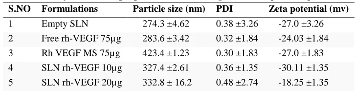
As depicted in Fig. 1, the SLN with incorporated rhVEGF showed similar mean sizes: 332.45 ± 16.62. Remarkably, gamma sterilisation did not cause any significant change in the mean size or in the PDI of the lipid nanoparticles. The reconstitution of dry particles for the size measurements did not present any inconvenience, suggesting the absence of particle fusion or aggregation. The analysis of the zeta potential revealed that both formulations presented a similar surface charge of approximately 34 mV. In addition, as described by Pardeike et al., the data shown in Table 1,2 demonstrated that the SLN-rhVEGF preparation method resulted in improved
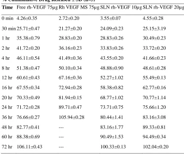
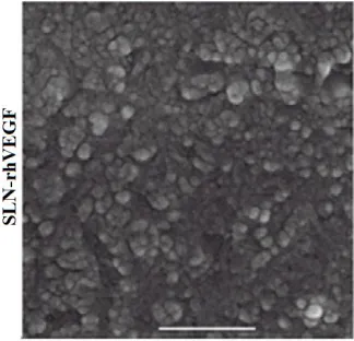
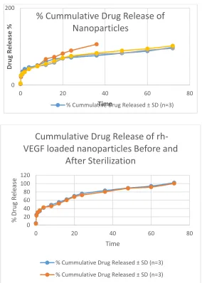
drug entrapment because the EE values 92.3 ± 2.2%, respectively (p b 0.001) [2]. These characteristics, together with the avoidance of the use of dichloromethane in the preparation of the SLN, may make SLN-rhVEGF a better alternative if it’s in vivo effectiveness is proved. Fig. 1 shows SEM and TEM photographs of the nanoparticles. In both formulations, the particles showed a smooth surface and similar size. The in vitro rhVEGF release profiles displayed in Fig. 2 show that both formulations presented similar release behaviours: an initial release (burst release) related to the percentage of surface-associated protein (SAP; 25.15 ± 3.19% for SLN-rhVEGF), a fast release phase from 8 h to 24 h (~75% of the total rhVEGF), and a slower release phase from 24 to 72 h ending with the release of the total amount of rhVEGF. Moreover, it is worth mentioning that the in vitro release profile of the γ-sterilised nanoparticles did not differ from that of the non-sterilized ones. These data suggest the suitability of the final sterilization of SLN-rhVEGF using gamma radiation. As depicted in Table 1, the nanoparticle characterization also revealed that the pH values of both formulations were within the range suitable for topical formulations (between 4 and 7), i.e., do not compromise the antimicrobial skin-barrier function observed with increased pH [42]. Moreover, the results from the occlusivity test described in Table 1 showed that the SLN were more occlusive. It should be noted that the occlusive properties of these formulations may allow their accumulation in the stratum corneum, resulting in the release of rhVEGF in a sustained manner and likely enhancing their therapeutic effect [4]. In addition, the rheological studies also revealed for the SLN dispersions showed viscosity values of approximately 7 × 10³ Pa · s at 5 rpm and a pseudo-plastic behavior (decreasing viscosity with an increase in the shear rate).
Table 1: Physiochemical properties of the investigated solid lipid nanoperticles S.NO Formulations Particle size (nm) PDI Zeta potential (mv)
1 Empty SLN 274.3 ±4.62 0.38 ±3.26 -27.0 ±3.26
2 Free rh-VEGF 75µg 283.6 ±3.42 0.32 ±1.84 -24.03 ±1.84
3 Rh VEGF MS 75µg 423.4 ±1.23 0.30 ±1.83 -27.0 ±1.83
4 SLN rh-VEGF 10µg 327.4 ±2.61 0.36 ±1.35 -30.11 ±1.35
Table 2: Evaluation of solid lipid nanoparticles S.NO Formulations Encapsulation efficiency %
1 Empty SLN 67.23 ± 8.50
2 Free rh-VEGF 75µg 71.64 ± 4.80
3 rh VEGF MS 75µg 56.43±0.24
4 SLN rh-VEGF 10µg 77.98 ± 9.40
5 SLN rh-VEGF 20µg 83.2 ± 4.50
Table 3: In-Vitro drug release of solid lipid nanoparticles
% Cummulative Drug Released ± SD (n=3)
Time Free rh-VEGF 75µgRh VEGF MS 75µgSLN rh-VEGF 10µgSLN rh-VEGF 20µg
0 min 4.26±0.35 2.72±0.20 3.55±0.07 4.55±0.28
30 min 25.71±0.47 21.27±0.20 24.09±0.23 25.15±3.19
1 hr 35.38±0.79 28.83±0.20 28.83±0.26 30.49±0.23
2 hr 41.72±0.20 36.16±0.23 33.83±0.26 33.72±0.20
4 hr 46.11±0.54 41.49±0.36 43.55±0.20 41.66±0.23
8 hr 51.38±0.47 50.10±0.34 48.88±0.90 48.61±0.28
12 hr 60.61±0.43 67.16±0.36 52.27±1.02 55.49±0.13
16 hr 67.55±0.34 72.94±0.28 58.38±0.82 62.77±0.16
20 hr 70.33±0.49 81.94±0.15 68.77±1.02 70.77±1.14
24 hr 71.72±0.28 89.71±0.47 73.71±0.75 75.66±1.20
36 hr 76.66±0.27 105.94±0.28 80.44±1.41 83.16±3.08
48 hr 82.77±0.41 --- 83.16±1.77 89.33±0.81
60 hr 88.38±0.69 --- 90.49±1.53 94.49±0.34
72 hr 106.11±0.43 --- 100.33±0.13 102.04±0.20
Table-4: results of the characterization of the developed formulations
PARAMETER SLN-rhEGF 20 µg
Encapsulation efficiency (%) 83.2 ± 4.50
PH 5.78 ± 0.12
Occlusion factor (%) 18.08 ± 1.31
Viscosity at 5 rpm (Pa · s) 6.67 × 10⁻³ ± 1.15 × 10⁻⁴ The data are shown as the means ± S.D.
Table-5: in-vitro drug release of solid lipid nanoparticles before and after sterilization S.NO Time
(min)
Before sterilization SLN rh-VEGF 20µg
After sterilization SLN rh-VEGF 20µg
1 0 min 4.55±0.28 3.46±0.28
2 30 min 25.15±3.19 23.02±3.19
4 2 hr 33.72±0.20 35.28±0.20
5 4 hr 41.66±0.23 42.56±0.23
6 8 hr 48.61±0.28 46.84±0.28
7 12 hr 55.49±0.13 52.64±0.13
8 16 hr 62.77±0.16 60.36±0.16
9 20 hr 70.77±1.14 68.58±1.14
10 24 hr 75.66±1.20 74.44±1.20
11 36 hr 83.16±3.08 82.62±3.08
12 48 hr 89.33±0.81 88.46±0.81
13 60 hr 94.49±0.34 92.65±0.34
14 72 hr 102.04±0.20 100.26±0.20
Table-6: Re-epithelisation grade of all of the studied groups. The data are shown as the means± S.D. Evaluation parameter Inflammation Score
Day-8 Day-15
Untreated control 0.129 ± 0.36 2.53±1.34
Empty SLN 1.478 ± 0.898 2.99±8.98
Free rhVEGF 75 µg 1.78±0.81 2.87±0.97
MS-rhVEGF 75 µg 2.21±0.75 2.93±0.95
SLN- rhVEGF 10 µg 2.95±1.18 3.62±0.97
SLN- rhVEGF 20 µg 2.94±0.58 3.41±0.44
SEM of SLN rh-VEGF Nanoparticles
TEM of SLN rh-VEGF Nanoparticles
Figure -2: Invitro Drud Release of Nanoparticles
Figure -3: Invitro Drug release Before & After Sterilization
0 200
0 20 40 60 80
D
ru
g R
e
le
ase
%
Time
% Cummulative Drug Release of
Nanoparticles
% Cummulative Drug Released ± SD (n=3)
0 20 40 60 80 100 120
0 20 40 60 80
%
Dru
g
Re
le
ase
Time
Cummulative Drug Release of
rh-VEGF loaded nanoparticles Before and
After Sterilization
% Cummulative Drug Released ± SD (n=3)
Figure-4: Invitro proliferation Assay – Measured using the CCK-8 colourimetric assay in Balb/C 3T3 Fibroblasts.
Figure-5: Invitro proliferation Assay – Measured using the CCK-8 colourimetric assay in HFF Fibroblasts.
Figure-7:Cellular uptake of SLN rh-VEGF
IN VITRO CELL CULTURE STUDIES
Effect of rhVEGF-loaded nanoparticles on
cell proliferation
The proliferation assays performed using Balb/c 3T3 fibroblasts, HFF fibroblasts, and HaCaT keratinocytes demonstrated the mitogenic effect of rhVEGF. The results, which are reported in Fig. 4,5,6, indicated that the groups treated with rhVEGF (SLN-rhVEGF, SLN-rhVEGF γ, and free rhVEGF) exhibited significantly higher cell proliferation than the control groups (0.2% FCS, empty SLN) in all of the studied cell lines, illustrating that the nano-encapsulation process did not affect the biological activity of the growth factor (p b 0.05). These differences, as shown in Fig. 6, were detectable starting from 24 h after treatment and became more evident at 48 and 72 h after treatment in the Balb/c fibroblasts and HaCaT keratinocytes. However, because the growth rate may depend on the cell line, the differences in HFF fibroblasts were not perceptible for the first 48 h (Fig. 6). In addition, no differences were found between the lipid nano-particle-treated groups, regardless of sterilization. These data suggest that gamma sterilization is a potential alternative to aseptic processing, which will facilitate future scale-up processes by reducing manufacturing costs. Nevertheless, the most striking result that emerged from these data is that the encapsulation of rhVEGF into nanoparticles, regardless of irradiation increased the mitogenic capability of all cell lines compared with that obtained with free rhVEGF: these differences were significant throughout the study for HaCaT keratinocytes and
fluorescence observed after the addition of the nanoparticles was a result of their cellular uptake and not due to the release of dye.
Cellular uptake of SLN formulations
Because the nanoparticles are intended to be administered topically on damaged skin and will thus be in contact with skin keratinocytes and fibroblasts, the nanoparticle cellular uptake was evaluated in the studied cells lines. The fluorescence microscope images shown in Fig. 4 revealed a high uptake of SLN-rhVEGF in all of the cell lines. Both formulations were distributed throughout the cytoplasm and around the cellular membrane; in contrast, none of the formulations were detectable in the nucleus. From a qualitative perspective found the uptake capacity of SLN-rhVEGF in the studied cell lines. As described in the literature, the rapid fading of Nile Red in hydrophilic environments is due to its lipophilic nature, which avoids the release of dye into cell culture medium [48]. Therefore, the fluorescence observed after the addition of the nanoparticles was a result of their cellular uptake and not due to the release of dye. In fact, the nanoparticle cellular uptake of SLN has been described by several researchers [49–51]. It is noteworthy to mention that the cellular uptake shown in Fig. 4 may be
related to the increased cell proliferation observed. In fact, as has been previously reported, the internalisation of the active compound into the cell cytoplasm is one of the possible strategies associated with bioactivity enhancement [46].
CONCLUSIONS
In the current work, empty SLN, 75 μg of free rhVEGF, 10 μg rhVEGF and 20 μg SLN-rhVEGF were developed, and their effectiveness was tested. The in vitro experiments revealed no loss of activity after the encapsulation or sterilization processes, which indicate that gamma sterilization is a suitable process for the final sterilisation of lipid nanoparticles. In the results demonstrate that the bioactivity of the nano formulations was higher in all of the studied cell lines than that of 75 μg free rhVEGF. In addition, based on its EE and the lack of solvent utilization, 20 μg NLC-rhVEGF is a better alternative than 75 rh free VEGF further animal studies are required to demonstrate the dose–response differences between 10 μg and 20 μg of nanoparticle-rhVEGF. Overall, these results suggest the promising future clinical application of SLN rhVEGF-loaded lipid nanoparticles, for the treatment of chronic wounds.
REFERENCES
[1]. Emerich, D. F.; Thanos, C. G. The pinpoint promise of nanoparticle-based drug delivery and molecular diagnosis. Biomol. Eng., 23, 2006, 171-184.
[2]. Ramanavicius, A.; Kausaite, A.; Ramanaviciene, A. Polypyrrole-coated glucose oxidase nanoparticles for biosensor design. Sens. Actuat. B: Chem., 111-112, 2005, 532-539.
[3]. Jamieson, T.; Bakhshi, R.; Petrova, D.; Pocock, R.; Imani, M.; Seifalian, A. M. Biological applications of quantum dots. Biomaterials, 28, 2007, 4717-4732.
[4]. Goldberg, M.; Langer, R.; Jia, X. Nanostructured materials for applications in drug delivery and tissue engineering. J. Biomater. Sci. Polym. Ed., 18, 2007, 241-268.
[5]. Farokhzad, O. C.; Langer, R. Impact of nanotechnology on drug delivery. ACS Nano, 3, 2009, 16-20.
[6]. Blasi, P.; Giovagnoli, S.; Schoubben, A.; Ricci, M.; Rossi, C. Solid lipid nanoparticles for targeted brain drug delivery. Adv. Drug Deliv. Rev., 59, 2007, 454-477.
[7]. Liu, W.; Hu, M.; Liu, W.; Xue, C.; Xu, H.; Yang, X. Investigation of the carbopol gel of solid lipid nanoparticles for the transdermal iontophoretic delivery of triamcinolone acetonide acetate. Int. J. Pharm., 364, 2008, 135-141.
[8]. Sung, J. C.; Pulliam, B. L.; Edwards, D. A. Nanoparticles for drug delivery to the lungs. Trends Biotechnol., 25, 2007, 563-570.
[9]. Tan, A.; De La Peña, H.; Seifalian, A. M. The application of exosomes as a nanoscale cancer vaccine. Int. J. Nanomed., 5, 2010, 889-900.
[11]. Ye, J.; Wang, Q.; Zhou, X.; Zhang, N. Injectable actarit-loaded solid lipid nanoparticles as passive targeting therapeutic agents for rheumatoid arthritis. Int. J. Pharm., 352, 2008, 273-279.
[12]. Chung, Y. I.; Tae, G.; Hong Yuk, S. A facile method to prepare heparin functionalized nanoparticles for controlled release of growth factors. Biomaterials, 27, 2006, 2621-2626.
[13]. Musumeci, T.; Ventura, C. A.; Giannone, I.; Ruozi, B.; Montenegro, L.; Pignatello, R.; Puglisi, G. PLA/PLGA nanoparticles for sustained release of docetaxel. Int. J. Pharm., 325, 2006, 172-179.
[14]. Haley, B.; Frenkel, E. Nanoparticles for drug delivery in cancer treatment. Urol. Oncol., 26, 2008, 57-64. [15]. Maeda, H.; Bharate, G. Y.; Daruwalla, J. Polymeric drugs for efficient tumor-targeted drug delivery based on
EPR-effect. Eur. J. Pharm. Biopharm., 71, 2009, 409-419.
[16]. Iyer, A. K.; Khaled, G.; Fang, J.; Maeda, H. Exploiting the enhanced permeability and retention effect for tumor targeting. Drug Discov. Today, 11, 2006, 812-818.
[17]. Brasnjevic, I.; Steinbusch, H. W. M.; Schmitz, C.; Martinez-Martinez, P. Delivery of peptide and protein drugs over the blood-brain barrier. Prog.Neurobiol., 87, 2009, 212-251.
[18]. Silva, G. A. Nanotechnology approaches for drug and small molecule delivery across the blood brain barrier. Surg. Neurol., 67, 2007, 113-116.
[19]. Allard, E.; Passirani, C.; Benoit, J. P. Convection-enhanced delivery of nanocarriers for the treatment of brain tumors. Biomaterials, 30, 2009, 2302-2318.
[20]. Pasha, S.; Gupta, K. Various drug delivery approaches to the central nervous system. Expert Opin. Drug Deliv., 7, 2010, 113-135.
[21]. Kusuhara, H.; Sugiyama, Y. Efflux transport systems for drugs at the bloodbrain barrier and blood-cerebrospinal fluid barrier (Part 2). Drug Discov. Today, 6, 2001, 206-212.
[22]. Ulbrich, K.; Hekmatara, T.; Herbert, E.; Kreuter, J. R. Transferrin- and transferrin-receptor-antibody-modified nanoparticles enable drug delivery across the blood-brain barrier (BBB). Eur. J. Pharm. Biopharm., 71, 2009, 251-256.
[23]. Liu, Z.; Jiao, Y.; Wang, Y.; Zhou, C.; Zhang, Z. Polysaccharides-based nanoparticles as drug delivery systems. Adv. Drug Deliv. Rev., 60, 2008, 1650-1662.
[24]. Jain, A. K.; Khar, R. K.; Ahmed, F. J.; Diwan, P. V. Effective insulin delivery using starch nanoparticles as a potential trans-nasal mucoadhesive carrier. Eur. J. Pharm. iopharm., 69, 2008, 426-435.
[25]. Kim, S.; Kim, J. H.; Jeon, O.; Kwon, I. C.; Park, K. Engineered polymers for advanced drug delivery. Eur. J. Pharm. Biopharm., 71, 2009, 420-430.
[26]. Chen, C.; Yu, C. H.; Cheng, Y. C.; Yu, P. H. F.; Cheung, M. K. Biodegradable nanoparticles of amphiphilic triblock copolymers based on poly(3-hydroxybutyrate) and poly(ethylene glycol) as drug carriers. Biomaterials, 27, 2006, 4804-4814.
[27]. Liang, H. F.; Yang, T. F.; Huang, C. T.; Chen, M. C.; Sung, H. W. Preparation of nanoparticles composed of poly( [gamma]-glutamic acid)- poly(lactide) block copolymers and evaluation of their uptake by HepG2 cells. J. Control. Release, 105, 2005, 213-225.
[28]. Byrne, J. D.; Betancourt, T.; Brannon-Peppas, L. ActiveJournal targeting schemes for nanoparticle systems in cancer therapeutics. Adv. Drug Deliv. Rev., 60, 2008, 1615-1626.
[29]. Bajpai, A. K.; Shukla, S. K.; Bhanu, S.; Kankane, S. Responsive polymers in controlled drug delivery. Prog. Polym. Sci, 33, 2008, 1088-1118.
[30]. Ganta, S.; Devalapally, H.; Shahiwala, A.; Amiji, M. A review of stimuliresponsive nanocarriers for drug and gene delivery. J. Control. Release, 126, 2008, 187-204.
[31]. Rapoport, N. Physical stimuli-responsive polymeric micelles for anti-cancer drug delivery. Prog. Polym. Sci., 32, 2008, 962-990.
[32]. Pinto Reis, C.; Neufeld, R. J.; Ribeiro, A. J.; Veiga, F. Nanoencapsulation I. Methods for preparation of drug-loaded polymeric nanoparticles. Nanomedicine, 2, 2006, 8-21.
[33]. Bilati, U.; Allqmann, E.; Doelker, E. Development of a nanoprecipitation method intended for the entrapment of hydrophilic drugs into nanoparticles. Eur. J. Pharm. Biopharm., 24, 2005, 67-75.
[35]. Kim, D. H.; Martin, D. C. Sustained release of dexamethasone from hydrophilic matrices using PLGA nanoparticles for neural drug delivery. Biomaterials, 27, 2006, 3031-3037.
[36]. Jain, R. A. The manufacturing techniques of various drug loaded biodegradable poly(lactide-co-glycolide) (PLGA) devices. Biomaterials, 21, 2000, 2475-2490.
[37]. Feng, S. S.; Mei, L.; Anitha, P.; Gan, C. W.; Zhou, W. Poly(lactide)-vitamin E derivative/montmorillonite nanoparticle formulations for the oral delivery of Docetaxel. Biomaterials, 30, 2009, 3297-3306.
[38]. Chaloupka, K, Malam, Y, Seifalian, A. M. Nanosilver as a new generation of nanoproduct in biomedical applications. Trends Biotechnol., 28, 2010, 580-8.
[39]. Pinto Reis, C.; Neufeld, R. J.; Ribeiro, A. n. J.; Veiga, F. Nanoencapsulation II. Biomedical applications and current status of peptide and protein nanoparticulate delivery systems. Nanomedicine, 2, 2006, 53-65.
[40]. Cohen-Sela, E.; Chorny, M.; Koroukhov, N.; Danenberg, H. D.; Golomb, G. A new double emulsion solvent diffusion technique for encapsulating hydrophilic molecules in PLGA nanoparticles. J. Control. Release, 133, 2009, 90-95.
[41]. Fonseca, C.; Simões, S.; Gaspar, R. Paclitaxel-loaded PLGA nanoparticles: preparation, physicochemical characterization and in vitro anti-tumoral activity. J. Control. Release, 83, 2002, 273-286.
[42]. Ceruti, M.; Crosasso, P.; Brusa, P.; Arpicco, S.; Dosio, F.; Cattel, L. Preparation, characterization, cytotoxicity and pharmacokinetics of liposomes containing water-soluble prodrugs of paclitaxel. J. Control.Release, 63, 2000, 141-153.
[43]. McCarron, P. A.; Marouf, W. M.; Quinn, D. J.; Fay, F.; Burden, R. E.; Olwill, S. A.; Scott, C. J. Antibody targeting of camptothecin-loaded PLGA nanoparticles to tumor cells. Bioconjug. Chem., 19, 2008, 1561-1569. [44]. Panyam, J.; Zhou, W. Z.; Prabha, S.; Sahoo, S. K.; Labhasetwar, V. Rapid endo-lysosomal escape of
poly(DL-lactide-co-glycolide) nanoparticles: implications for drug and gene delivery. FASEB J., 16, 2002, 1217-1226. [45]. Chang, J.; Jallouli, Y.; Kroubi, M.; Yuan, X. B.; Feng, W.; Kang, C. S.; Pu, P. Y.; Betbeder, D. Characterization
of endocytosis of transferrin-coated PLGA nanoparticles by the blood-brain barrier. Int. J. Pharm., 379, 2009, 285-292.
[46]. Gelperina, S.; Maksimenko, O.; Khalansky, A.; Vanchugova, L.; Shipulo, E.; Abbasova, K.; Berdiev, R.; Wohlfart, S.; Chepurnova, N.; Kreuter, J. Drug delivery to the brain using surfactant-coated poly(lactide-co-glycolide) nanoparticles: Influence of the formulation parameters. Eur. J. Pharm. Biopharm., 74, 2010, 157-63. [47]. Chavanpatil, M. D.; Patil, Y.; Panyam, J. Susceptibility of nanoparticleencapsulated paclitaxel to
P-glycoprotein-mediated drug efflux. Int. J. Pharm., 320, 2006, 150-156.
[48]. Xu, P.; Gullotti, E.; Tong, L.; Highley, C. B.; Errabelli, D. R.; Hasan, T.; Cheng, J. X.; Kohane, D. S.; Yeo, Y. Intracellular drug delivery by poly(lactic-co-glycolic acid) nanoparticles, revisited. Mol. Pharm., 6, 2008, 190-201.
[49]. Fujita, H.; Banno, H.; Yamanouchi, D.; Kobayashi, M.; Yamamoto, K.; Komori, K. Pitavastatin Inhibits Intimal Hyperplasia in Rabbit Vein Graft. J. Surg. Res., 148, 2008, 238-243.
[50]. Simosa, H. F.; Pomposelli, F. B.; Dahlberg, S.; Scali, S. T.; Hamdan, A. D.; Schermerhorn, M. L. Predictors of failure after angioplasty of infrainguinal vein bypass grafts. J. Vasc. Surg., 49, 2009, 117-121.
[51]. Kimura, S.; Egashira, K.; Nakano, K.; Iwata, E.; Miyagawa, M.; Tsujimoto, H.; Hara, K.; Kawashima, Y.; Tominaga, R.; Sunagawa, K. Local delivery of imatinib mesylate (STI571)-incorporated nanoparticle ex vivo suppresses vein graft neointima formation. Circulation, 118, 2008, S65-S70.
[52]. Li, J.; Loh, X. J. Cyclodextrin-based supramolecular architectures: Syntheses, structures, and applications for drug and gene delivery. Adv. Drug Deliv. Rev, 60, 2008, 1000-1017.
[53]. Du, Y. Z.; Xu, J. G.; Wang, L.; Yuan, H.; Hu, F. Q. Preparation and characteristics of hydroxypropyl- [beta]-cyclodextrin polymeric nanocapsules loading nimodipine. Eur. Polymer J., 45, 2009, 1397-1402.
[54]. Agueros, M.; Areses, P.; Campanero, M. A.; Salman, H.; Quincoces, G.; Penuelas, I.; Irache, J. M. Bioadhesive properties and biodistribution of cyclodextrin-poly(anhydride) nanoparticles. Eur. J. Pharm. Sci., 37, 2009, 231-240.