Effect of Continues Training and High Intensity Interval Training on
miR-29a and CTGF Gene Expression in Male Wistar Diabetic Rats’
Heart Tissue
Mania Roozbayani1, Maghsoud Peeri2*, Hamid Agha-Alinejad3, Mohammad Ali Azarbayjani4
Introduction
n recent decades, the number of people with diabetes is rising. International Diabetes Federation stated that365 million people suffered from diabetes in 2011 and is rising up to 552 million until 2030 (1).
The diabetic incidence rise is attributed to environmental and lifestyle changes (2). The
cardiac fibrosis is a common pathological process presented in a variety of diseases, including hypertension and diabetes. Diabetes and hypertension is the underlying cause of cardiovascular complications, including cardiac fibrosis (2-3). Cardiac fibrosis is one of the pathophysiological process that
I
1. Ph.D. Department of Exercise Physiology, Islamic Azad University, Central Tehran Branch, Iran.
2. Professor of Exercise Physiology, Department of Exercise Physiology, Central Tehran Branch, Islamic Azad University, Tehran, Iran.
3. Associate Professor of Exercise Physiology, Department of Exercise Physiology, Central Tehran Branch, Islamic Azad University, Tehran, Iran.
4. Professor of Exercise Physiology, Department of Exercise Physiology, Central Tehran Branch, Islamic Azad University, Tehran, Iran.
*Correspondence:
Maghsoud Peeri Faculty of Physical Education and Exercise Sciences, Central Tehran Branch of Islamic Azad University, Iran Zamin Blv., Shahrak. E. Gharb, Tehran, Iran.
Tel: (98) 912 112 44 34 Email: [email protected]
Received: 22 April 2017
Accepted: 24 June 2017
Published in July 2017
Abstract
Objective:High-intensity interval training (HIIT) and continues
aerobic training (CT) have cardio-protective effects in diabetic
rats. The functional role of microRNA in heart was studied.Only
miR-29a levels were found to correlate with cardiac fibrosis, This study tests the hypothesis that applying HIIT and CT cases
miR-29a increasing is associatedwith a reduction of connective
tissue growth factor (CTGF) induced cardiacfibrosis.
Materials and Methods: In this randomized controlled trial, 18 male diabetic rats were included. They were divided into 3 groups called as HIIT, CT and control. Exercise protocol was performed 5 days/week for 5 weeks. The miR-29a and CTGF synthesis were compared between the groups by real time- PCR. Results: Our results demonstrated that elevation of miR-29a
using HIIT (2.671.02, P=0.010,) or CT (1.790.49, P=0.002)
are effective in inhibiting CTGF (HIIT:0.17. 7, P
=0.000-CT:0.390.27, 0.000 -induced cardiac fibrosis, suggesting that these types training would be selected as a new adjunctive therapy in the heart fibrosis- derived diabetic.
Discussion:The HIIT and CT showed increased levels of miR-29a compared CT group which is shown to decrease CTGF level resulting into lowered fibrosis of heart tissue in diabetic patients. Keywords: miR-29a, CTGF Gene, High intensity interval training, Heart, Diabetes mellitus
IRANIAN JOURNAL OF DIABETES AND OBESITY, VOLUME 8, NUMBER 3, AUTUMN 2016 143 accompanied by myocardial stiffness, low
pumping capacity of the heart and overall lead to heart failure (4,5).
Connective tissue growth factor (CTGF) is a matricellular and multifunctional glycoprotein binding to heparin in all fibrosis conditions almost enriched. CTGF regulates cell proliferation, extracellular matrix remodeling and angiogenesis. CTGF can act on the cells that has been secreted it when synthesis of extracellular matrix (ECM) increases in normal fibroblasts to a level seen in cells of fibrotic lesions in an autocrine manner (6,7). However no curative treatment for cardiac fibrosis was found. The miR-29a implication in biological processes like fibrosis is important (8). The miR-29 family members were detected to target various professional genes and their pathways, regulating fibrosis and ECM remodeling in organs and tissues as kidney, liver, lung and heart (9-12). A functional link between miRNAs and cardiomyocyte fibrosis induced diabetic has remained unclear. A safe strategy that can be used to reduce cardiac fibrosis in diabetic patients is important.
It is less known about the events by which exercise could affect disturbances in cardiac fibrosis signaling pathways and activity of miR-29a in the diabetic male rats. Thus, it is important to know whether HIIT and CT might be beneficial in attenuating CTGF-induced disturbances in expression of proteins involved in cardiac fibrosis signaling pathways and miR-29a in the heart of diabetic male rats. The objective of this study was to determine the effects of continues training and high intensity interval training on miR-29a and CTGF gene expression in male wistar diabetic rats’ heart tissue.
Materials and Methods
Experimental
Twenty four diabetic mature male Albino Wistar rats were studied. They were supplied by the Pasteur Institute of Iran (Tehran, Iran), and housed under standard laboratory conditions in pairs in a 12 h–12 h light–dark
cycle, at 22 ± 2 °C room temperature, with humidity of 50±10% and the same nutrition. Their maintenance weight was 260±10 gr. Rats had unrestricted access to standard rat chow (pellet) and tap water. Total time spent at the laboratory was 5 weeks for all rats. They became diabetic by 50 mg/kg streptozotocin solution in citrate buffer injected intraperitoneal. During practice the animals did not receive any medical treatment. One week after injection, exercises were started by 5 sessions from low to normal speed. Then randomly rats were divided into 3 groups. Control group: The control group (8 rats) did not participate in any exercise program, but to create the same environmental conditions five times a week they were immobilized on a treadmill for 10 to 15 minutes per session. High intensity interval training (HIIT) group (8 rats): Each session consisted of 25 minutes of running on a treadmill by HIIT protocol that implemented by Roglic et al (16).The protocol consisted of: Warm up for five minutes with an intensity of 30-40% of VO2max then four times intervals (three minutes 85 to 90 percent VO2max intensity and one minute recovery with 30 to 35 percent of VO2max intensity between intervals), then five-minute cool-down with an intensity of 30-40% of VO2max (13).
Continuous aerobic training (CT) group (8 rats): Each session included 40 minutes of continuous aerobic training program running on a treadmill that consist of: five-minute warm-up with 30 to 40 percent of VO2max then 30 minutes running with 60- 65% VO2max and five-minute cool-down with 30 to 40 percent of VO2max.
Twenty four hours after the last training session, after an overnight fasting rats were anesthetized by intraperitoneal injection of ketamine (90 mg/ kg) and Xylazine (10 mg/kg).Then they were sacrificed, blood samples were collected directly from the hearts of rats and serum was isolated by centrifugation at 3000g, 10 ℃, for 4 min. The left ventricular heart tissue was removed, frozen in liquid nitrogen and stored for later
analysis immediately after washing in saline. cDNA synthesis was performed using 1µg of RNA using random hexamer primer and full reverse transcriptase enzyme.
Statistical analysis
To ensure the normal distribution of data Kolmogorov- Smirnov test was used. Then, to determine the significance of differences in the expression of each training group and the control group as well as to study the differences between the three groups T-independent test was used, in another way ANOVA test for homogeneity of variances and determining the position of significance in post hoc Tukey test was used as well. All statistical operations of research using SPSS version 22 with the significance level of
P<0.05 were considered.
This study confirmed by animal ethics committee of Tehran University (EC-00312).
Results
In this study, 24 diabetic male rats in three groups were studied for the gene expression of MIR29A and CTGF. Table one showed the average mean weight and serum glucose of
rats in 3 different groups. No changes in weight and glucose concentration were found applying HIIT and CT type exercise as compared to control group.
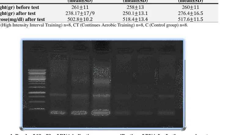
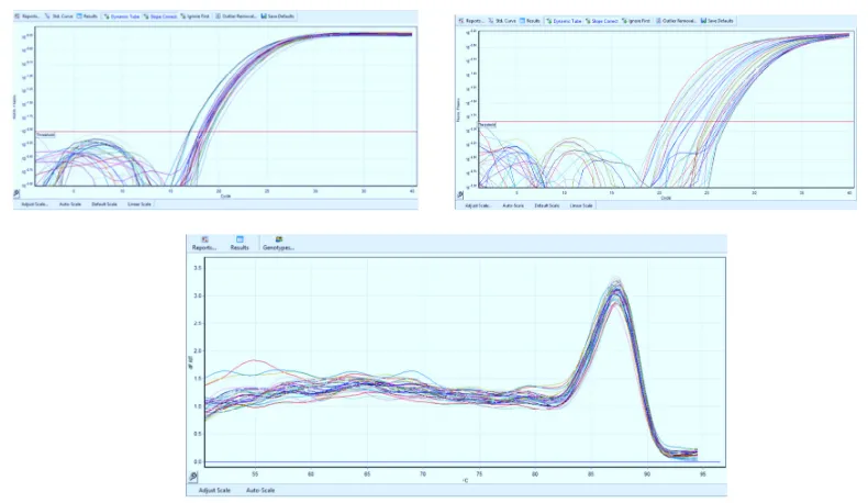
As can be seen average mean weight did not change significantly in three groups before and after exercise program. The glucose concentration also did not differ in groups under investigation. Electrophoresis was performed at a voltage of 120 for 60-90 minutes in order to detect 18s to 28s bands. Here the results indicated the good quality of RNA purified. In this study, qRT-PCR technique was used for gene expression assessment of miR-29a and CTFG. The gene expression curves were given for miR-29a and CTFG and the melting curve for GAPDH as follow.
The level of expression and melting were calculated using the standard formula for quantitative real time PCR. The sample curve for MIR29A and CTGF and GAPDH are given as above.
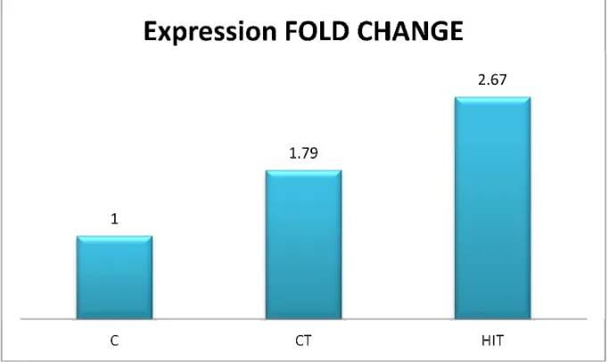
HIIT affected on the miR-29a expression was compared to controlled group. Mean fold changes of miR-29a expression in HIIT group versus control group was 2.67 (SD=1.02),
P=0.010 which was significant. CT affected
Table 1. The mean weightand standard deviation (SD) in three groups under investigation
Varible HIIT
(mean±SD)
CT (mean±SD)
C (mean±SD)
Weight(gr) before test 26111 25813 26011
Weight(gr) after test 238.1717/9 250.113.1 276.416.5
Glucose(mg/dl) after test 502.810.2 518.413.4 517.611.5
HIIT (High Intensity Interval Training) n=8, CT (Continues Aerobic Training) n=8, C(Control group) n=8.
Figure 1. Bands of 18s, 28s of RNA indicating proper purification of RNA for further experiments.
IRANIAN JOURNAL OF DIABETES AND OBESITY, VOLUME 8, NUMBER 3, AUTUMN 2016 145 on the miR-29a expression was compared to
controlled group, P=0.002. Mean fold changes
of miR-29a expression in HIIT group versus CT group was 1.79 (SD=0.49), P=0.002 which
was also shown to be significant (figure 3). One way ANOVA analysis showed that CTGF expression was significantly lower in HIIT and CT group compared to control group (0.17 . 7, P=0.000) and (0.39 0.27, P=0.000). One way ANOVA analysis showed
that CTGF expression was lower in HIIT group compared to CT group (P=0.037)
(fig-4).
The above results indicated that miR-29a concentration increased as compared to control group while CTGF concentration decreases considerably.
In summery it could be seen that miR-29a is increased in both HIIT and CT groups and CTGF decreased in both experimental group compared to control group, which indicated the positive effect of training applied in this experiment on lowering the fibrosis of heart tissues. We concluded considering the results that HITT and CT exercises will lower down the possibility of fibrosis by increasing mir29a as a controlling agent.
Discussion
The cardiac fibrosis induced by diabetes is an initial and important step in the development of cardiac dysfunction. In the present study we demonstrated that CTGF expresses significantly increased when the level of miR-29a is low.
The fifth leading cause of death globally is diabetes (14). Hyperglycemia induced cardiomyocytes intracellular changes with diabetes complications (15,16). In hyperglycemia, cellular metabolism and stable macromolecules acute changes during the time can result in chronic inflammation and tissue damage initiate, which induces organs dysfunction (17,18). This general process is modified by genetic determinants of individual susceptibility and independent accelerating factors (19-21).
Framingham in his concentration study on Heart stated that diabetes and HF initially are linked (Kannel et al., 1974). Diabetes increases N2.5-fold HF incidence (Nichols et al., 2004; Thrainsdottir et al., 2005; Aksnes et al., 2007). Diabetes-induced cardiac fibrosis is accompanied (and likely triggered) by the
Figure 2. A and B the expression curve for MIR29A and CTGF (different concentrations of miR-29a and CTGF respectively) and GAPDH melting curve are indicated here (C).
upregulation of transforming growth factor (TGF-β1), its receptor TGF-β receptor II, and its downstream mediator, CTGF (Mizushige et al., 2000; Way et al., 2002; Westermann et al., 2007; D'Souza et al., 2011).
By regulating the expression of target genes, miRNAs are involved in a variety of biological processes including cell cycle regulation, differentiation, development, metabolism, neuronal patterning and aging (22). Many studies have also demonstrated the cooperative nature ofthe CTGF and TGF-b in the animal models fibrosis promotion (23). The CTGF
plays an important role in the pathogenesis of chronic fibrotic diseases and causes EMT-like cell fate changes in vivo and in vitro (24,25). CTGF blockade inhibits extracellular matrix accumulation and EMT caused byangiotensin II and TGF-b (26,27).
In factCTGFisa CCN2 protein (28), the CCN proteins include of cyr61, nov, and ctgf are original details of participating regulator matri-cellular in cell signaling of internal and external (29). CCN proteins are secreted, extracellular matrix (ECM)-associated proteins that regulate such cellular processes as
Figure 3. miR-29a expression changes in HIIT and CT groups (The change was compared with the control group).
Figure 4. CTGF expression changes in HIIT and CT groups (The change was compared with the control group).
IRANIAN JOURNAL OF DIABETES AND OBESITY, VOLUME 8, NUMBER 3, AUTUMN 2016 147 adhesion, migration, mitogenesis,
differentiation, and survival. They also have been implicated in wound healing, tumorigenesis, and fibrotic and vascular diseases. Other perspectives on this family of proteins have emphasized their possible involvement in wound healing, fibrosis, and atherosclerosis (30). CTGF is both necessary and sufficient to initiate fibrosis in the presence of TGF-b and vice versa (31). Several studies show that how miRNAs and their relevant expressed target genes in experimental diabetic animals hearts are differentially and the miRNA levels deregulation in progressive heart failure are effective (32-35). The miRNAs are a plentiful class of about 17–25 nucleotides small noncoding RNAs, as many diverse biological processes original regulators. (36-38). They can express the protein-coding genes regulate at the post-transcriptional level through binding to the 3’untranslational region (3’UTR) of target mRNAs (38). The miRNAs by target genes expression are involved in a variety of biological processes including cell cycle regulation, differentiation, development, metabolism, neuronal patterning and aging (22). The miR-29 family contains of miR-29a, miR-29b and miR-29c that are encoded by two separate different loci, giving rise to bicistronic precursor pioneer miRs namely miR-29a/b1 and miR- 29b2/c (8). The miR-29 a and b (b1 and b2) in rat genome located on chromosome 4 (39)
L Lin et al stated that miR-29 be dedicated as prognostic marker and therapeutic target for hepatocellular carcinoma (HCC) (40). Sonnylal et al, stated that in vivo and in vitro CTGF expression in mesenchymal cells in the skin and lungs can cause changes in the differentiation program of adjacent epithelial cells. These changes might contribute to fibrogenesis (24). LB N et al. stated that Rat ̓s miR-29a and miR-29b in cardiovascular diseases causing is dramatically effective, miR-29a was one of the miRs of efficacious on children with Type 1 DM (41). Knabel M k et al, stated that a single systemic injection of
a miR-29a expressing adeno-associated virus prevent fibrosis in advanced fibrotic livers of carbon tetrachloride (CCL4). The miR-29 can be effective on diseases associated with fibrosis too (42). In Chuang & et al research, miR-29c function revealed with COL3A1 and DNMT3A expression inhibition. In this way miR-29c decreases a number of fibrosis cases pathways (43).
While exercise training prevents disease and recovers individuals health, there is debate in the literature regarding the optimal intensity of exercise. CT and HIIT have largely been employed to elicit functional improvements in aerobic capacity (44).
HIIT would be represented a time efficient clinically relevant tool to improve aerobic fitness in healthy individuals (45) and involves alternating between short bouts of high-intensity exercise and periods of lower intensities or inactivity (46). It has been recently shown that HIIT is safe, well tolerated, and leads to superior improvements in exercise capacity and fitness when compared to moderate intensity training (MIT) in various populations including those with obesity (47).
When compared to CT, HIIT offers similar health benefits, including improved insulin sensitivity, and endothelial function (48). Participants preferred HIIT over CT, which could potentially lead to higher compliance to the exercise regimen (49). Holloway Tanya M.,et al.in their research stated that both ET and HIIT would decrease left ventricular fibrosis, cross sectional area, and molecular markers of heart failure, and increase left ventricular capillarization similarly in Dahl/SS rats, providing clinical support for the use of HIIT (50). Many studies have successfully reduced diastolic stiffness by targeting the signaling factors involved in the development of fibrosis, such as TGF-β (51-53). In Tjønna ̓ et al. study, HIT was also found to be better than MIT in reducing cardiovascular risk factors in patients with metabolic syndrome (54).
In this study we described and evaluated the effects of 5 weeks HIIT and CT on miR-29a and CTGF gene expression in Male Wistar diabetic Rats’ Heart Tissue. In this way we reveal that by HIIT and CT, increasing miR-29a (fig-3) and decreases CTGF expression (fig-4). CTGF reduction is related with decreasing cardiac fibrosis. It would be one safe way for declining heart fibrosis- derive diabetic. Of course HIIT that involves alternating between short bouts of high-intensity exercise and periods of lower intensities or inactivity, can be perform in little time and with more comfortable and leads to superior improvements in exercise capacity and fitness. Hence HIIT implementation decreases heart fibrosis without drug tolerance
and acceptance. HIIT is better compatible for diabetic rats other than CT and it have better effects on cardiac function. Further studies can compare the HIIT and CT in human for use of this method in diabetic patients for better cardiac function and inhibit the diabetic derived from fibrosis. HIIT is well tolerated exercise and is effective in miR29a expression.
Conclusions
By the results of this study miR29a expression influence the CTGF expression which is a key factor in cardiac fibrosis. So HIIT particularly in diabetics can be a modality for lowering the risk of cardiac fibrosis.
References
1. Shaw JE, Sicree RA, Zimmet PZ. Global estimates of the prevalence of diabetes for 2010 and 2030. Diabetes research and clinical practice. 2010 Jan 31;87(1):4-14.
2. Shamhart PE, Luther DJ, Hodson BR, Koshy JC, Ohanyan V, Meszaros JG. Impact of type 1 diabetes on cardiac fibroblast activation: enhanced cell cycle progression and reduced myofibroblast content in diabetic myocardium. American Journal of Physiology-Endocrinology and Metabolism. 2009 Nov 1;297(5):E1147-53.
3. Eschalier R, Rossignol P, Kearney-Schwartz A, Adamopoulos C, Karatzidou K, Fay R, Mandry D, Marie PY, Zannad F. Features of Cardiac Remodeling, Associated With Blood Pressure and Fibrosis Biomarkers, Are Frequent in Subjects With Abdominal ObesityNovelty and Significance. Hypertension. 2014 Apr 1;63(4):740-6.
4. Kong P, Christia P, Frangogiannis NG. The pathogenesis of cardiac fibrosis. Cellular and Molecular Life Sciences. 2014 Feb 1;71(4):549-74. 5. Van Heerebeek L, Hamdani N, Handoko ML,
Falcao-Pires I, Musters RJ, Kupreishvili K, Ijsselmuiden AJ, Schalkwijk CG, Bronzwaer JG, Diamant M, Borbély A. Diastolic stiffness of the failing diabetic heart. Circulation. 2008 Jan 1;117(1):43-51.
6. Shi-Wen X, Leask A, Abraham D. Regulation and function of connective tissue growth factor/CCN2 in tissue repair, scarring and fibrosis. Cytokine & growth factor reviews. 2008 Apr 30;19(2):133-44. 7. Sonnylal S, Xu S, Jones H, Tam A, Sreeram VR,
Ponticos M, Norman J, Agrawal P, Abraham D, de Crombrugghe B. Connective tissue growth factor
causes EMT-like cell fate changes in vivo and in vitro. J Cell Sci. 2013 May 15;126(10):2164-75. 8. Papadopoulou AS, Serneels L, Achsel T,
Mandemakers W, Callaerts-Vegh Z, Dooley J, Lau P, Ayoubi T, Radaelli E, Spinazzi M, Neumann M. Deficiency of the miR-29a/b-1 cluster leads to ataxic features and cerebellar alterations in mice. Neurobiology of disease. 2015 Jan 31;73:275-88. 9. Li Z, Hassan MQ, Jafferji M, Aqeilan RI, Garzon
R, Croce CM, Van Wijnen AJ, Stein JL, Stein GS, Lian JB. Biological functions of miR-29b contribute to positive regulation of osteoblast differentiation. Journal of Biological Chemistry. 2009 Jun 5;284(23):15676-84.
10. Steele R, Mott JL, Ray RB. MBP-1 upregulates miR-29b, which represses Mcl-1, collagens, and matrix metalloproteinase-2 in prostate cancer cells. Genes & cancer. 2010 Apr;1(4):381-7.
11. Hawkins SM, Creighton CJ, Han DY, Zariff A, Anderson ML, Gunaratne PH, Matzuk MM. Functional microRNA involved in endometriosis. Molecular endocrinology. 2011 Mar 24;25(5):821-32.
12. Luna C, Li G, Qiu J, Epstein DL, Gonzalez P. Cross-talk between miR-29 and transforming growth factor-betas in trabecular meshwork cells. Investigative ophthalmology & visual science. 2011 May 1;52(6):3567-72.
13. Leandro CG, Levada AC, Hirabara SM, MANHAS-DE-CASTRO RA, De-Castro CB, Curi R, Pithon-Curi TC. APROGRAM OF MODERATE PHYSICAL TRAINING FOR WISTAR RATS BASED ON MAXIMAL OXYGEN CONSUMPTION. The Journal of
149
IRANIAN JOURNAL OF DIABETES AND OBESITY, VOLUME 8, NUMBER 3, AUTUMN 2016 Strength & Conditioning Research. 2007 Aug
1;21(3):751-6.
14. 14 - Roglic G, Unwin N, Bennett PH, Mathers C, Tuomilehto J, Nag S, Connolly V, King H. The burden of mortality attributable to diabetes. Diabetes care. 2005 Sep 1;28(9):2130-5.
15. Rubler S, Dlugash J, Yuceoglu YZ, Kumral T, Branwood AW, Grishman A. New type of cardiomyopathy associated with diabetic glomerulosclerosis. The American journal of cardiology. 1972 Nov 8;30(6):595-602.
16. Shen E, Diao X, Wang X, Chen R, Hu B. MicroRNAs involved in the mitogen-activated protein kinase cascades pathway during glucose-induced cardiomyocyte hypertrophy. The American journal of pathology. 2011 Aug 31;179(2):639-50. 17. Vassort G, Turan B. Protective role of antioxidants
in diabetes-induced cardiac dysfunction. Cardiovascular toxicology. 2010 Jun 1;10(2):73-86. 18. Turan B. Role of antioxidants in redox regulation of diabetic cardiovascular complications. Current pharmaceutical biotechnology. 2010 Dec 1;11(8):819-36.
19. Rubler S, Dlugash J, Yuceoglu YZ, Kumral T, Branwood AW, Grishman A. New type of cardiomyopathy associated with diabetic glomerulosclerosis. The American journal of cardiology. 1972 Nov 8;30(6):595-602.
20. Vassort G, Turan B. Protective role of antioxidants in diabetes-induced cardiac dysfunction. Cardiovascular toxicology. 2010 Jun 1;10(2):73-86.
21. Shen E, Diao X, Wang X, Chen R, Hu B. MicroRNAs involved in the mitogen-activated protein kinase cascades pathway during glucose-induced cardiomyocyte hypertrophy. The American journal of pathology. 2011 Aug 31;179(2):639-50. 22. Nicoloso MS, Sun H, Spizzo R, Kim H,
Wickramasinghe P, Shimizu M, Wojcik SE, Ferdin J, Kunej T, Xiao L, Manoukian S. Single-nucleotide polymorphisms inside microRNA target sites influence tumor susceptibility. Cancer research. 2010 Apr 1;70(7):2789-98.
23. Wang Q, Usinger W, Nichols B, Gray J, Xu L, Seeley TW, Brenner M, Guo G, Zhang W, Oliver N, Lin A. Cooperative interaction of CTGF and TGF-β in animal models of fibrotic disease. Fibrogenesis & tissue repair. 2011 Feb 1;4(1):4. 24. Sonnylal S, Xu S, Jones H, Tam A, Sreeram VR,
Ponticos M, Norman J, Agrawal P, Abraham D, de Crombrugghe B. Connective tissue growth factor causes EMT-like cell fate changes in vivo and in vitro. J Cell Sci. 2013 May 15;126(10):2164-75. 25. Burns WC, Twigg SM, Forbes JM, Pete J, Tikellis
C, Thallas-Bonke V, Thomas MC, Cooper ME, Kantharidis P. Connective tissue growth factor plays an important role in advanced glycation end product–induced tubular epithelial-to-mesenchymal
transition: implications for diabetic renal disease. Journal of the American Society of Nephrology. 2006 Sep 1;17(9):2484-94.
26. Rupérez M, Ruiz-Ortega M, Esteban V, Lorenzo Ó, Mezzano S, Plaza JJ, Egido J. Angiotensin II increases connective tissue growth factor in the kidney. The American journal of pathology. 2003 Nov 30;163(5):1937-47.
27. Chen L, ZHANG XL, ZHANG JD, LIU H. Influence of connective tissue growth factor antisense oligonucleotide on angiotensin II‐induced epithelial mesenchymal transition in HK2 cells. Acta Pharmacologica Sinica. 2006 Aug 1;27(8):1029-36.
28. 28-. Lau LF, Lam SC. The CCN family of angiogenic regulators: the integrin connection. Experimental cell research. 1999 Apr 10;248(1):44-57.
29. 29- Perbal B. CCN proteins: multifunctional signalling regulators. The Lancet. 2004 Jan 3;363(9402):62-4.
30. 30- Lau LF, Lam SC. The CCN family of angiogenic regulators: the integrin connection. Experimental cell research. 1999 Apr 10;248(1):44-57.
31. 31- Wang Q, Usinger W, Nichols B, Gray J, Xu L, Seeley TW, Brenner M, Guo G, Zhang W, Oliver N, Lin A. Cooperative interaction of CTGF and TGF-β in animal models of fibrotic disease.
Fibrogenesis & tissue repair. 2011 Feb 1;4(1):4. 32. 32- Goren Y, Kushnir M, Zafrir B, Tabak S, Lewis
BS, Amir O. Serum levels of microRNAs in patients with heart failure. European journal of heart failure. 2012 Feb 1;14(2):147-54.
33. 33- Elton TS, Martin MM, Sansom SE, Belevych AE, Györke S, Terentyev D. miRNAs got rhythm. Life sciences. 2011 Feb 28;88(9):373-83.
34. 34- Sakakibara M, Hirashiki A, Cheng XW, Bando Y, Ohshima K, Okumura T, Funahashi H, Ohshima S, Murohara T. Association of diabetes mellitus with myocardial collagen accumulation and relaxation impairment in patients with dilated cardiomyopathy. Diabetes research and clinical practice. 2011 Jun 30;92(3):348-55.
35. 35- Martín-Gallán P, Carrascosa A, Gussinyé M, Domínguez C. Biomarkers of diabetes-associated oxidative stress and antioxidant status in young diabetic patients with or without subclinical complications. Free Radical Biology and Medicine. 2003 Jun 15;34(12):1563-74.
36. 36- Bartel DP. MicroRNAs: genomics, biogenesis, mechanism, and function. cell. 2004 Jan 23;116(2):281-97.
37. 37- Kim DH, Sætrom P, Snøve O, Rossi JJ. MicroRNA-directed transcriptional gene silencing in mammalian cells. Proceedings of the National Academy of Sciences. 2008 Oct 21;105(42):16230-5.
38. 38- Bushati N, Cohen SM. microRNA functions. Annu. Rev. Cell Dev. Biol.. 2007 Nov 10;23:175-205.
39. 39- Kriegel AJ, Liu Y, Fang Y, Ding X, Liang M. The miR-29 family: genomics, cell biology, and relevance to renal and cardiovascular injury. Physiological genomics. 2012 Feb 15;44(4):237-44. 40. 40- Lin LL, Wang W, Hu Z, Wang LW, Chang J, Qian H. Negative feedback of miR-29 family TET1 involves in hepatocellular cancer. Medical Oncology. 2014 Dec 1;31(12):291.
41. 41- Nielsen LB, Wang C, Sørensen K, Bang-Berthelsen CH, Hansen L, Andersen ML, Hougaard P, Juul A, Zhang CY, Pociot F, Mortensen HB. Circulating levels of microRNA from children with newly diagnosed type 1 diabetes and healthy controls: evidence that miR-25 associates to residual beta-cell function and glycaemic control during disease progression. Experimental diabetes research. 2012 Jul 5;2012.
42. 42- Knabel MK, Ramachandran K, Karhadkar S, Hwang HW, Creamer TJ, Chivukula RR, Sheikh F, Clark KR, Torbenson M, Montgomery RA, Cameron AM. Systemic delivery of scAAV8-encoded MiR-29a ameliorates hepatic fibrosis in carbon tetrachloride-treated mice. PloS one. 2015 Apr 29;10(4):e0124411.
43. 43- Chuang TD, Khorram O. Mechanisms underlying aberrant expression of miR-29c in uterine leiomyoma. Fertility and sterility. 2016 Jan 31;105(1):236-45.
44. 44- Gibala MJ, McGee SL, Garnham AP, Howlett KF, Snow RJ, Hargreaves M. Brief intense interval exercise activates AMPK and p38 MAPK signaling and increases the expression of PGC-1α in human
skeletal muscle. Journal of applied physiology. 2009 Mar 1;106(3):929-34.
45. 45- Gillen JB, Gibala MJ. Is high-intensity interval training a time-efficient exercise strategy to improve health and fitness?. Applied Physiology, Nutrition, and Metabolism. 2013 Sep 27;39(3):409-12.
46. 46- Laursen PB, Jenkins DG. The scientific basis for high-intensity interval training. Sports Medicine. 2002 Jan 1;32(1):53-73.
47. 47- Coquart JB, Lemaire C, Dubart AE, Luttembacher DP, Douillard C, Garcin M. Intermittent Versus Continuous Exercise: Effects of Lower Exercise in Obese Women.
48. 48- Deighton K, Barry R, Connon CE, Stensel DJ. Appetite, gut hormone and energy intake responses to low volume sprint interval and traditional endurance exercise. European journal of applied physiology. 2013 May 1;113(5):1147-56.
49. 49- Sim AY, Wallman KE, Fairchild TJ, Guelfi KJ. High-intensity intermittent exercise attenuates ad-libitum energy intake. International journal of obesity. 2014 Mar 1;38(3):417-22.
50. 50- Holloway TM, Bloemberg D, da Silva ML, Simpson JA, Quadrilatero J, Spriet LL. High intensity interval and endurance training have opposing effects on markers of heart failure and cardiac remodeling in hypertensive rats. PloS one. 2015 Mar 24;10(3):e0121138.
51. 51- Litwin SE, Grossman W. Diastolic dysfunction as a cause of heart failure. Journal of the American College of Cardiology. 1993 Oct 1;22(4):A49-55. 52. 52- Grossman WI, Barry WH. Diastolic
pressure-volume relations in the diseased heart. InFederation proceedings 1980 Feb (Vol. 39, No. 2, pp. 148-155).
53. 53- Kass DA, Bronzwaer JG, Paulus WJ. What mechanisms underlie diastolic dysfunction in heart failure?. Circulation research. 2004 Jun 25;94(12):1533-42.
54. 54- Tjønna AE, Lee SJ, Rognmo Ø, Stølen TO, Bye A, Haram PM, Loennechen JP, Al-Share QY, Skogvoll E, Slørdahl SA, Kemi OJ. Aerobic interval training versus continuous moderate exercise as a treatment for the metabolic syndrome. Circulation. 2008 Jul 22;118(4):346-54.