Michał Szpinda, Anna Szpinda
Normative Growth Data for the External Diameters
of the External and Internal Iliac Arteries in Human
Fetuses – An Anatomical, Digital and Statistical Study
Normatywne dane na temat wzrostu średnic zewnętrznych tętnic
biodrowych zewnętrznych i wewnętrznych u płodów ludzkich – badanie
anatomiczne, cyfrowe i statystyczne
Department of Anatomy, Ludwik Rydygier Collegium Medicum in Bydgoszcz, Nicolaus Copernicus University, Bydgoszcz, Poland
Abstract
Background. Normative data on the diameters of the aorto-iliac segment are extremely useful in the diagnosis and monitoring of prenatal arterial variants and pathologies.
Objectives. The present study describes age-specific reference intervals and normal growth curves for the external diameters of the external and internal iliac arteries.
Material and Methods. Using anatomical dissection and digital-image analysis, the normal growth of the external diameters of the external and internal iliac arteries was studied in 124 spontaneously aborted human fetuses, aged 15–34 weeks.
Results. Neither sex differences nor laterality differences were found. The external diameters of the external iliac arteries increased from 0.31 ± 0.06 to 1.41 ± 0.31 mm on the right, and from 0.29 ± 0.04 to 1.37 ± 0.24 mm on the left, and generated the following growth curves of best fit: y = 0.665 – 0.056 x Age + 0.002 x Age2 ± 0.143 (R2 = 0.82) and y = 0.612 – 0.052 x Age + 0.002 x Age2 ± 0.118 (R2 = 0.86), respectively. The external diameters of the internal iliac arteries were found to be statistically larger than those of the external iliac arteries (p = 0.0000). The external diameters of the internal iliac arteries ranged from 0.44 ± 0.07 to 2.04 ± 0.43 mm on the right, and from 0.44 ± 0.06 to 1.83 ± 0.43 mm on the left, and modeled the following quadratic functions: y = 1.524 – 0.127 x Age + 0.004 x Age2 ± 0.242 (R2 = 0.74), and y = 1.391 – 0.117 x Age + 0.004 x Age2 ± 0.220 (R2 = 0.76), respectively. The right external iliac arteries (in 71% of the cases) and the right internal iliac arteries (in 65.3% of cases) were larger in external diameter.
Conclusions. The values of the external diameters of the external and internal iliac arteries are independent of sex. A strong trend towards higher values for the right external and internal iliac arteries is noted. The external diam-eter of the internal iliac artery is nearly 1.5 times greater than that of the external iliac artery. Surprisingly, normal growth of the external diameters of the external and internal iliac arteries follows quadratic functions (Adv Clin Exp Med 2012, 21, 2, 143–150).
Key words: external diameter, external iliac artery, internal iliac artery, human fetuses, quadratic regression.
Streszczenie
Wprowadzenie. Dane normatywne dotyczące średnic segmentu aortalno-biodrowego są bardzo użyteczne w roz-poznaniu i monitorowaniu wariantów i patologii tętnic w okresie prenatalnym.
Cel pracy. Praca ta opisuje specyficzne dla wieku przedziały wartości i krzywe normalnego wzrostu średnic zewnętrznych tętnic biodrowych zewnętrznych i wewnętrznych.
Materiał i metody. Za pomocą dysekcji anatomicznej i cyfrowej analizy obrazu zbadano prawidłowy wzrost śred-nic zewnętrznych tętśred-nic biodrowych zewnętrznych i wewnętrznych u 124 płodów człowieka pochodzących z poro-nień samoistnych i porodów przedwczesnych.
Wyniki. Nie stwierdzono różnic związanych z płcią płodu i stroną ciała (P > 0,05). Średnice zewnętrzne tętnic biodrowych zewnętrznych wzrastały od 0,31 ± 0,06 do 1,41 ± 0,31 mm po stronie prawej i od 0,29 ± 0,04 do 1,37 ± 0,24 mm po stronie lewej, generując krzywe wzrostu o najlepszym dopasowaniu odpowiednio: y = 0,665 – 0,056 x
Adv Clin Exp Med 2012, 21, 2, 143–150 ISSN 1899–5276
oRIgINAL PAPERS
Wiek + 0,002 x Wiek2 ± 0,143 (R2 = 0,82) i y = 0,612 – 0,052 x Wiek + 0,002 x Wiek2 ± 0,118 (R2 = 0,86). Średnice zewnętrzne tętnic biodrowych wewnętrznych były istotnie większe (P = 0,0000) niż tętnic biodrowych zewnętrz-nych. Średnice zewnętrzne tętnic biodrowych wewnętrznych wahały się od 0,44 ± 0,07 do 2,04 ± 0,43 mm po stronie prawej i od 0,44 ± 0,06 do 1,83 ± 0,43 mm po stronie lewej, generując odpowiednio następujące funkcje kwadrato-we: y = 1,524 – 0,127 x Wiek + 0,004 x Wiek2 ± 0,242 (R2 = 0,74) i y = 1,391 – 0,117 x Wiek + 0,004 x Wiek2 ± 0,220 (R2 = 0,76). Tętnice biodrowe wewnętrzne (65,3%) i zewnętrzne (71%) były silniejsze po stronie prawej.
Wniosek. Wartości średnic zewnętrznych tętnic biodrowych zewnętrznych i wewnętrznych nie zależą od płci. obserwuje się silny trend w kierunku większych wartości prawych tętnic biodrowych zewnętrznych i wewnętrz-nych. Średnica zewnętrzna tętnicy biodrowej wewnętrznej jest ok. 1,5 raza większa niż tętnicy biodrowej zewnętrz-nej. Prawidłowy wzrost średnic zewnętrznych tętnic biodrowych zewnętrznych i wewnętrznych następuje niespo-dziewanie zgodnie z funkcją kwadratową (Adv Clin Exp Med 2012, 21, 2, 143–150).
Słowa kluczowe: średnica zewnętrzna, tętnica biodrowa zewnętrzna, tętnica biodrowa wewnętrzna, płody człowie-ka, regresja kwadratowa.
It has been reported that the diameters of the human aorto-iliac segment increase quasi-linearly during gestation [1–11]. However, the current au-thors’ studies on the common iliac arteries [12] in-dicate that the external diameters increase in a qua-dratic fashion rather than growing proportionally. To date, neither anatomical studies nor Doppler ultrasonographic studies of normal growth curves for the external and internal iliac arteries have been reported in the medical literature.
Thus, the objectives of the present study were to establish the following:
age-specific reference intervals for the −
external diameters of the external and internal iliac arteries;
normal growth curves for each external −
diameter studied; and
the influence of sex and laterality (left-right) −
on the value of the external diameters examined.
Material and Methods
The examinations were carried out on 124 human fetuses of Caucasian origin of both sexes (60 males, 64 females) derived from spontaneous abortions or stillbirths, as described in a previ-ously published paper [11]. The legal and ethical aspects of the study were approved by the Nicolaus Copernicus University Research Ethic Committee (KB/217/2006). The sample was comprised of fe-tuses that were the outcome of intra-uterine growth restriction; the fetuses had no visible cardiovascu-lar anomalies. The fetal ages, which ranged from 15 to 34 weeks of gestation (Table 1), were accurately established on the basis of the following criteria: 1) the gestational age, based on measurements of the crown-rump length [13]; 2) the date of the begin-ning of the last maternal menstrual period; and 3) a combination of abdominal circumference, femur length and biparietal diameter, ascertained in early second-trimester ultrasound scans.
As described in a previous article [11], the arte-rial bed was filled with white latex LBS 3060 through a catheter Stericath (diameter of 0.5–1 mm), intro-duced by lumbar access into the abdominal aorta. The arterial bed filling was performed under con-trolled pressure of 50–60 mm Hg, using a SEP 11S syringe infusion pump (Ascor, Warsaw, Poland). All specimens were immersed in 10% neutral for-malin solution for 4–24 months for fixation, and then dissected at a magnification of 10 using a ste-reoscope with a Huygens ocular. Each fetus was dissected to expose its aorto-iliac segment. In situ,
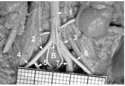
the common, external and internal iliac arteries were placed with a millimeter scale perpendicular to the optical lens axis, and then recorded using a Nikon Coolpix 8400 camera, digitalized to TIFF images (Fig. 1) and evaluated by a digital
image-Fig. 1. The abdominal aorta and iliac arteries in a female fetus aged 24 weeks: B – aortic bifurcation, 1 – abdominal aorta, 2 – right common iliac artery, 3 – left common iliac artery, 4 – right external iliac artery, 5 – right internal iliac artery, 6 – left external iliac artery, 7 – left internal iliac artery
analysis system (Leica QWin Pro 16, Cambridge, UK), which semi-automatically estimated the ex-ternal diameters of the exex-ternal and inex-ternal iliac arteries. Diameter measurements were derived by assuming that the filled iliac arteries were circular in cross section.
For each fetus the four following external di-ameters were evaluated:
1, 2: external diameters of the right and left ex-ternal iliac arteries, measured at their origins; and 3, 4: external diameters of the right and left in-ternal iliac arteries, measured at their origins.
In order to minimize measurement and ob-server bias, all the measurements were performed by one researcher. Each measurement was repeat-ed three times under the same conditions but at different times, and the mean of the three
mea-surements was then used. The differences between the repeated measurements, as well as the intra-observer variation, were evaluated by the Wilcox-on signed-rank test.
The values of the external diameters were correlated to fetal age in weeks in order to clarify their normative growth. The results obtained were assessed by a one-way ANoVA test for unpaired data and a post hoc RIR Tukey test. Regression analysis was used to derive the growth curves of the best fit for the plot for each external diameter against gestational age. Coefficients of determina-tion (R2) between particular external diameters
and fetal age were calculated. Results were consid-ered significant at p < 0.05.
Table 1. Distribution of the fetuses studied
Tabela 1. Liczebność badanych płodów
Fetal age (Wiek płodowy) Crown-rump length
(Długość ciemieniowo-siedzeniowa ) mm
Number
(Liczba) Sex (Płeć) Months
(Miesiące) weeks(Hbd-life) male female
mean SD min max
4 15 89.4 6.1 85.0 92.0 9 4 5
16 103.7 6.1 95.0 106.0 7 3 4
5
17 114.9 8.2 111.0 121.0 5 3 2
18 129.3 6.6 124.0 134.0 8 3 5
19 142.7 7.7 139.0 148.0 9 5 4
20 155.3 5.8 153.0 161.0 2 0 2
6
21 167.1 4.7 165.0 173.0 3 2 1
22 178.1 6.9 176.0 186.0 7 4 3
23 192.3 6.3 187.0 196.0 9 4 5
24 202.9 5.7 199.0 207.0 11 6 5
7
25 215.2 4.8 211.0 218.0 7 5 2
26 224.7 5.2 220.0 227.0 7 4 3
27 234.1 4.3 231.0 237.0 4 0 4
28 244.2 5.1 240.0 246.0 4 2 2
8
29 253.8 4.5 249.0 255.0 6 1 5
30 262.7 3.1 260.0 264.0 6 3 3
31 270.7 5.2 268.0 275.0 4 1 3
32 281.4 3.7 279.0 284.0 5 4 1
9 33 290.3 6.1 286.0 293.0 7 4 3
34 301.4 3.2 296.0 302.0 4 2 2
Results
No significant differences were found in the evaluation of intra-observer reproducibility of the measurements of the iliac arteries studied. Inter-observer variability was not assessed because all the measurements were carried out by the same observer.
The results obtained are presented in Table 2 and Figures 2–5. The statistical analysis of the di-ameters studied showed no sex differences, so the results are presented in Table 2 irrespective of sex.
Although the right-left differences for the whole group were not found to be statistically sig-nificant, the results for the right and left iliac arter-ies are presented separately, because of their great
inter-individual variability (Table 2) and a strong trend towards higher values on the right side.
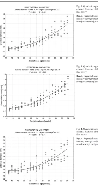
The values for external diameters of the ex-ternal iliac arteries increased from 0.31 ± 0.06 to 1.41 ± 0.31 mm on the right side, and from 0.29 ± 0.04 to 1.37 ± 0.24 mm on the left side for fetuses at the ages of 15 and 34 weeks, respective-ly. At first, linear patterns were generated as fol-lows: y = –0.572 + 0.052 x Age + 0.159 (R2 = 0.78;
p = 0.0000) for the right external iliac artery, and y = –0.553 + 0.050 x Age ± 0.135 (R2 = 0.72;
p = 0.0000) for the left external iliac artery. Howev-er, after several transformations it turned out that quadratic regressions were much better than other models, because the values of their coefficients of determination attained relatively greater values:
Table 2. External diameters of the external and internal iliac arteries related to fetal age
Tabela 2. Średnice zewnętrzne tętnic biodrowych zewnętrznych i wewnętrznych w zależności od wieku płodowego
Fetal age – weeks (Wiek płodowy – tygodnie)
n = 124 External diameters – mm (Średnice zewnętrzne – mm)
external iliac artery internal iliac artery
right left right left
mean
średnia SD meanśrednia SD meanśrednia SD meanśrednia SD
15 9 0.31 0.06 0.29 0.04 0.44 0.07 0.44 0.06
16 7 0.39 0.10 0.34 0.06 0.62 0.16 0.57 0.18
17 5 0.32 0.05 0.33 0.05 0.53 0.08 0.44 0.06
18 8 0.34 0.05 0.32 0.03 0.47 0.11 0.45 0.08
19 9 0.45 0.09 0.41 0.08 0.65 0.19 0.58 0.13
20 2 0.56 0.02 0.50 0.01 0.97 0.18 0.73 0.04
21 3 0.44 0.06 0.44 0.03 0.53 0.13 0.51 0.12
22 7 0.56 0.12 0.55 0.11 0.75 0.17 0.74 0.14
23 9 0.49 0.06 0.48 0.05 0.68 0.10 0.64 0.08
24 11 0.58 0.10 0.52 0.10 0.84 0.28 0.73 0.16
25 7 0.68 0.12 0.65 0.14 0.78 0.09 0.83 0.15
26 7 0.68 0.10 0.70 0.15 0.82 0.13 0.82 0.17
27 4 0.94 0.22 0.82 0.22 1.31 0.32 1.14 0.32
28 4 0.85 0.09 0.77 0.09 1.04 0.18 1.07 0.20
29 6 0.98 0.18 0.95 0.19 1.26 0.33 1.13 0.18
30 6 1.07 0.22 0.94 0.12 1.36 0.23 1.33 0.31
31 4 0.96 0.07 0.98 0.11 1.29 0.12 1.17 0.12
32 5 1.19 0.32 1.10 0.15 1.69 0.50 1.68 0.53
33 7 1.20 0.20 1.12 0.16 1.61 0.38 1.56 0.41
Fig. 2. Quadratic regression for the external diameter of the right external iliac artery
Ryc. 2. Regresja kwadratowa dla średnicy zewnętrznej tętnicy biod-rowej zewnętrznej prawej
Fig. 3. Quadratic regression for the external diameter of the left external iliac artery
Ryc. 3. Regresja kwadratowa dla średnicy zewnętrznej tętnicy biod-rowej zewnętrznej lewej
Fig. 4. Quadratic regression for the external diameter of the right internal iliac artery
0.82 and 0.86 respectively. Thus, the numerical data showed that external diameters of the right and left external iliac arteries modeled the growth curves of best fit as: y = 0.665 – 0.056 x Age + 0.002 x Age2 ± 0.143 (R2 = 0.82) – (Fig. 2) and y = 0.612
– 0.052 x Age + 0.002 x Age2 ± 0.118 (R2 = 0.86)
– (Fig. 3), respectively. In 71% of individuals the external diameter of the external iliac artery was found to be larger on the right side.
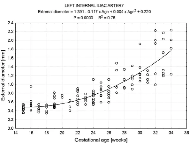
In the material under examination, the ex-ternal diameters of the inex-ternal iliac arteries were found to be statistically larger (p = 0.0000) than those of the external iliac arteries. The external di-ameters of the internal iliac arteries varied from 0.44 ± 0.07 to 2.04 ± 0.43 mm on the right, and from 0.44 ± 0.06 to 1.83 ± 0.43 mm on the left in fetuses at the ages of 15 and 34 weeks. The linear models for their growth were: y = –0.685 + 0.067 x Age ± 0.271 (R2 = 0.67; p = 0.0000) and y = – 0.676
+ 0.065 x Age ± 0.248 (R2 = 0.69; p = 0.0000) for
the right and left internal iliac arteries, respective-ly. However, the relationships between their exter-nal diameters and fetal age were much better ex-pressed by the quadratic models: y = 1.524 – 0.127 x Age + 0.004 x Age2 ± 0.242 (R2 = 0.74) for the
right internal iliac artery (Fig. 4), and y = 1.391 – 0.117 x Age + 0.004 x Age2 ± 0.220 (R2 = 0.76) for
the left internal iliac artery (Fig. 5). In 65.3% of the specimens the right internal iliac artery was found to be larger in diameter.
Discussion
The present study describes the normative growth of external diameters of the external and internal iliac arteries in human fetuses. Because the
specimens had been fixed in neutral buffered forma-lin for 12–24 months before quantitative analysis, all the measurements were taken in situ to minimize, as much as possible, tissue shrinkage related to for-malin fixation. Such shrinkage had little influence on the measurements of the filled iliac arteries in situ, the wall of which was mainly composed of elas-tic connective tissue. Similarly, in an earlier study one of the authors found only 0.5–1.0% shrinkage
in situ in major fetal elastic arteries that had been filled with latex and then immersed in 10% neutral buffered formalin solution for 1–2 years [14].
Reference data for external diameters of the internal and external iliac arteries in human fe-tuses is scarce [11, 15]. Moreover, in the available literature prior to this study there is no informa-tion expressed by precise mathematical models concerning the normal growth of the external di-ameters of the internal and external iliac arteries during gestation. Therefore, in this study, a digital image-analysis system was used to provide com-pletely novel data on an increase in the external diameters of developing external and internal iliac arteries at gestational ages ranging from 15 to 34 weeks.
The measurements were similar in both sexes, in keeping with other authors in relation to the iliac [11, 12, 15] and other fetal arteries [1–10, 16, 17]. However, significant sex differences in arte-rial diameter arise as late as during the postnatal period, when the arterial diameters are found to be larger in males [18–21].
Normal age-specific reference intervals for ex-ternal diameters of the exex-ternal iliac arteries were constructed as follows: from 0.31 ± 0.06 to 1.41 ± 0.31 mm on the right, and from 0.29 ± 0.04 to 1.37 ± 0.24 mm on the left for the fetuses aged 15 and
Fig. 5. Quadratic regression for the external diameter of the left internal iliac artery
References
[1] Angelini A, Allan LD, Anderson RH, Crawford DC, Chita SK, Ho SY: Measurements of the dimensions of the aortic and pulmonary pathways in the human fetus: a correlative echocardiographic and morphometric study. Br Heart J 1988, 60, 221–226.
[2] Ursell PC, Byrne JM, Fears TR, Strobino BA, Gersony MW: growth of the great vessels in the fetus with cardiac defects. Circulation 1991, 84, 2028–2033.
[3] Hornberger LK, Weintraub RG, Pesonen E, Murilo-Olivas A, Simpson IA, Sahn C, Hagen-Ansert S, Sahn DJ:
Echocardiographic study of the morphology and growth of the aortic arch in the human fetus. observations related to the prenatal diagnosis of coarctation. Circulation 1992, 86, 741–747.
[4] Hyett J, Moscoso G, Nicolaides K: Morphometric analysis of the great vessels in early fetal life. Hum Reprod 1995, 10, 3045–3048.
[5] Achiron R, Golan-Porat N, Gabbay U, Rotstein Z, Heggesh J, Mashiach S, Lipitz S: In utero ultrasonographic measurements of fetal aortic and pulmonary artery diameters during the first half of gestation. Ultrasound obstet gynecol 1998, 11, 180–184.
[6] Castillo EH, Arteaga-Martinez M, Garcia-Pelaez I, Villasis-Keever MA, Aguirre OM, Moran V, Vizcaino A:
Morphometric study of the human fetal heart. I. Arterial segment. Clin Anat 2005, 18, 260–268.
[7] Achiron R, Zimand S, Hegesh J, Lipitz S, Zalel Y, Rotstein Z: Fetal aortic arch measurements between 14 and 38 weeks’ gestation: in utero ultrasonographic study. Ultrasound obstet gynecol 2000, 15, 226–230.
[8] Szpinda M, Brazis P, Elminowska-Wenda G, Wiśniewski M: Morphometric study of the aortic and great pul-monary arterial pathways in human foetuses. Ann Anat 2006, 188, 25–31.
[9] Szpinda M: Morphometric study of the ascending aorta in human fetuses. Ann Anat 2007, 189, 465–472.
[10] Szpinda M: The normal growth of the thoracic aorta in human foetuses. Folia Morphol 2007, 66, 131–137.
[11] Szpinda M, Szpinda A, Dombek M, Wiśniewski M, Daroszewski M: External diameters of the abdominal aorta and iliac arteries in human fetuses. Adv Clin Exp Med 2011, 20, 6, 691–698.
[12] Szpinda M, Szpinda A, Woźniak A, Daroszewski M, Mila-Kierzenkowska C: The normal growth of the com-mon iliac arteries in human fetuses – an anatomical, digital and statistical study. Med Sci Monit 2012, 18, 3, 109–116.
34 weeks, respectively. According to Özgüner and Sulak, values for the diameter of the external iliac artery were consistently greater on the right side, but without significant differences: 0.44 ± 0.07 mm
versus 0.44 ± 0.07 mm in the first trimester, 0.81 ± 0.1 mm versus 0.79 ± 0.1 mm in the second tri-mester, 1.39 ± 0.2 mm versus 1.35 ± 0.2 mm in the third trimester, and 2.36 ± 0.2 mm versus 2.29 ± 0.2 mm at full term [15].
Surprisingly, the results of the present study show that the normal growth of the external iliac arteries follows quadratic functions. The authors proved that the best fit correlation between the diameter of the external iliac artery and the gesta-tional age was a parabola defined by the quadratic regressions y = 0.665 – 0.056 x Age + 0.002 x Age2
± 0.143 (R2 = 0.82) on the right, and y = 0.612 –
0.052 x Age + 0.002 x Age2 ± 0.118 (R2 = 0.86) on
the left.
In the material under examination, the values for the diameter of the internal iliac artery were found to range from 0.44 ± 0.07 to 2.04 ± 0.43 mm on the right, and from 0.44 ± 0.06 to 1.83 ± 0.43 mm on the left. Thus, the present results are in close accordance with the findings of Özgüner and Sulak [15]. In their material, the external diameters of the right and left internal iliac arteries were re-spectively: 0.68 ± 0.1 mm versus 0.61 ± 0.1 mm in the first trimester, 1.30 ± 0.3 mm versus 1.33 ± 0.3 mm in the second trimester, 2.37 ± 0.4 mm versus
2.29 ± 0.4 mm in the third trimester, and 3.63 ± 0.4
mm versus 3.57 ± 0.4 mm at the full term, but with no significant laterality differences.
The normal increase in external diameter of the internal iliac arteries was expressed by the quadrat-ic functions y = 1.524 – 0.127 x Age + 0.004 x Age2
± 0.242 (R2 = 0.74) on the right side, and y = 1.391
– 0.117 x Age + 0.004 x Age2 ± 0.220 (R2 = 0.76) on
the left side. These findings indicate that in human fetuses, in contrast to adults, the external diame-ter of the indiame-ternal iliac ardiame-tery was nearly 1.5 times larger than that of the external iliac artery, which concurs with Özgüner and Sulak [15].
The current authors believe that a particular strength of this study is the large number (n = 124) of normal fetuses used to generate the growth curves. It is noteworthy that the growth curves ob-tained in the study are completely new; they can serve as a database for in utero examination of the iliac arteries, and can be helpful in the prenatal di-agnosis and monitoring of abnormalities and pa-thologies of the fetal iliac arteries.
[13] Iffy L, Jakobovits A, Westlake W, Wingate MB, Caterini H, Kanofsky P, Menduke H: Early intrauterine devel-opment: I. The rate of growth of caucasian embryos and fetuses between the 6th and 20th weeks of gestation. Pediatrics 1975, 56, 173–186.
[14] Szpinda M: Morphometric study of the great arteries of the thorax in human fetuses (in Polish). Habilitation thesis. Bydgoszcz: CM UMK; 2006, pp. 1–143.
[15] Özgüner G, Sulak O: Development of the abdominal aorta and iliac arteries during the fetal period: a morpho-metric study. Surg Radiol Anat 2011, 33, 35–43.
[16] Panagouli E, Lolis E, Venieratos D: A morphometric study concerning the branching points of the main arteries in humans: relationships and correlations. Ann Anat 2011, 193, 86–99.
[17] Pennington N, Soames RW: The anterior visceral branches of the abdominal aorta and their relationship to the renal arteries. Surg Radiol Anat 2005, 27, 395–403.
[18] Dixon AK, Lawrence JP, Mitchell JRA: Age-related changes in the abdominal aorta shown by CT. Clin Radiol 1984, 35, 33–37.
[19] Pearce WH, Slaughter MS, LeMaire S, Salyapongse N, Feinglass J, McCarthy WJ, Yao JST: Aortic diameter as a function of age, gender and body surface area. Surgery 1993, 114, 691–697.
[20] Lederle FA, Johnson GR, Wilson SE, Gordon IL, Chute EP, Littooy FN, Krupski WC, Bandyk D, Barone GW, Graham LM, Hye RJ, Reinke DB: Relationship of age, gender, race and body size to infrarenal aortic diameter. J Vasc Surg 1997, 26, 595–601.
[21] Fleischmann D, Hastie TJ, Danneger FC, Paik TS, Tillich M, Zarins CK, Rubin GD: Quantitative determination of age-related geometric changes in the normal abdominal aorta. J Vasc Surg 2001, 33, 97–105.
Address for correspondence:
Michał Szpinda
Department of Anatomy
Ludwik Rydygier Collegium Medicum in Bydgoszcz Karłowicza 24
85-092 Bydgoszcz Poland
Tel.: +48 52 585 37 05 E-mail: [email protected] Conflict of interest: None declared Received: 19.08.2011
![Annual report [of the Data Protection Commissioner of Ireland] presented to each of the Houses of the Oireachtas, pursuant to Section 14 of the Data Protection Acts 1988 and 2003](data:image/gif;base64,R0lGODlhAQABAIAAAP///wAAACH5BAEAAAAALAAAAAABAAEAAAICRAEAOw==)