Jowita Woźniak
1, a–f, alicja Kędzia
2, a, B, D–f, Krzysztof Dudek
3, a–D, fVariability of the Trunks and Divisions
of the Brachial Plexus in Human Fetuses
Zmienność pni i gałęzi splotu ramiennego płodów ludzkich
1 Department of Neurosurgery, academic Clinical Hospital in Wrocław, Poland 2 Department of Normal anatomy, Wroclaw Medical University, Poland
3 Institute of Machine Design and Operation, Wroclaw Technical University, Poland
A – research concept and design; B – collection and/or assembly of data; C – data analysis and interpretation;
D – writing the article; E – critical revision of the article; F – final approval of article; G – other
Abstract
Background. The brachial plexus is an important part of peripheral nervous systems. The studies of variation of the brachial plexus and its parts in the prenatal period are extremely rare.
Objectives. The goal of the study was to assess brachial plexus trunk variability and their divisions in human fetuses.
Material and Methods. Examinations were carried out on a total of 220 brachial plexuses derived from 110 fetuses aged 4–7 months, including 50 females (45.45%) in a CRL range of 80–233 mm. The following methods were incor-porated into the study: anthropological, dissection, digital image acquisition and statistical methods. Symmetry and sexual dimorphism was observed.
Results. Trunk variations were observed in 12 (5.45%) plexuses as well as divisions variants in 74 (33.6%) cases. Variants were equally common in both genders and on both sides of the body. Variants of individual trunks were of a similar percentage, usually an observed middle trunk formed by nerve roots C7 and C8. anterior division
of the middle trunk (aDMT) was the most variable division, observed in 63 (28.63%) cases.
Conclusions. Trunk variants are rare and anomalies of divisions were observed in one-third of cases, regardless of the side of the body and sex. Brachial plexus variation recognition is important from a clinical point of view (Adv Clin Exp Med 2013, 22, 3, 309–318).
Key words: brachial plexus, anatomy, variation, trunk, division, human fetus.
Streszczenie
Wprowadzenie. Splot ramienny jest istotną częścią obwodowego układu nerwowego. Badania zmienności splotu
ramiennego i jego poszczególnych części w okresie płodowym są nieliczne.
Cel pracy. Ocena zmienności pni i gałęzi splotu ramiennego u płodów ludzkich.
Materiał i metody. Zbadano 220 splotów ramiennych wypreparowanych ze 110 płodów w wieku 4–7 miesięcy,
w tym 50 płci żeńskiej (45.45%), w przedziale v-tub: 80–233 mm. W pracy zastosowano następujące metody: sek-cyjna, antropologiczna, cyfrowa akwizycja obrazów, program Image J, metody statystyczne. Oceniono symetrię i dymorfizm płciowy.
Wyniki. Warianty pni obserwowano w 12 (5,45%) splotach, a anomalie gałęzi w 74 (33,6%) przypadkach. Warianty występowały jednakowo często u obu płci i po obu stronach ciała. anomalie poszczególnych pni stwierdzono w podobnym odsetku, najczęściej obserwowano pień środkowy utworzony przez gałęzie przednie korzeni nerwów C7 i C8. Najbardziej zmienną gałęzią była gałąź przednia pnia środkowego (aDMT) w 63 (28,63%) splotach.
Wnioski. Warianty pni występowały rzadko, a anomalie gałęzi w co trzecim przypadku, niezależnie od strony ciała i płci. Wiedza na temat zmienności splotu ramiennego jest ważna z klinicznego punktu widzenia (Adv Clin Exp Med 2013, 22, 3, 309–318).
Słowa kluczowe: splot ramienny, warianty, pień, gałąź, płody ludzkie.
adv Clin Exp Med 2013, 22, 3, 309–318 ISSN 1899–5276
ORIgINaL PaPERS
perinatal period, traction and compression [1]. The traditional division of brachial plexus damage refers to the anatomical location of the injury and divides them into upper (Erb’s palsy or Erb-Duch-enne palsy), middle and lower (Klumpke’s palsy or Dejerine-Klumpke palsy). Klumpke’s palsy is a less favorable prognosis. Right-sided brachial plexus palsy occurs in about 60% percent of peri-natal brachial plexus injury cases, which could be the result of the asymmetry of the body with a pre-dominance of the right hand, observed in human ontogeny [2]. Microsurgical methods of paralysis treatment include neurolysis or autografts [3]. Surgical procedures should be selected on an indi-vidual basis [4]. a study by Nath et al. [5] describes the effects of treatment with muscle transfers. Mod Quad surgery, is composed of 4 elements: latissi-mus dorsi transfer in order to obtain external ro-tation of the arm and the teres major muscle to stabilize the shoulder, subscapularis muscle release for the elimination of the internal rotation of the arm and the decompression and neurolysis of the axillary nerve. The authors described the case of a boy who was operated on using the Mod Quad method before the age of 4 years. The overall re-sults were satisfactory, there was a significant im-provement in function for the paretic right upper limb. Noticeable improvement was also observed in the Mallet scale, which helped in assessing the degree of damage to the plexus, as well as the ev-eryday functioning of the patient [5]. Knowledge about the variability of the brachial plexus is an important part of clinical practice, and is helpful during complex reconstruction procedures. The study of the prenatal development of the brachial plexus and adjacent structures in the open litera-ture are rare. Lewis [6] in 1902 described the de-velopment of the human arm. according to Lewis, the first differentiation of mesenchymatakes place in the 4th week. Continuity of the spinal nerves forming the brachial plexus, C4, C5, C6, C7, C8 and T1, was underlined. The primordia of the dorsal nerves passes the arm and ends up at the height of the distal end of the humerus in the sheath of the forming primordia arm muscles. In about the 5th week (embryo length: 10.5–11 mm), the huge
40th day, the median, radial and ulnar nerves came to the level of the hand. On the 49th and 50th (Car-negie stages 20 and 21), a similar orientation and arrangement of the brachial plexus as in adults is observed. Woźniak et al. [8] evaluated the growth rate of the brachial plexus in human fetuses aged between 14–32 weeks. In most of the analyzed geometric parameters (diameter and length of the nerve), each of the plexus showed no asymmetry and sexual dimorphism. Weekly growth was dif-ferent for difdif-ferent parts of the brachial plexus, as well as correlations between variables. growth rate was uneven. It was faster in the initial period, between 14–18 weeks of gestation, and slower be-tween 24–28 weeks.Plexus growth was described by four mathematical formulas: linear regression, logarithmic function, the von Bertalanffy growth model and the gompertz curve [8]. The applied mathematical formulas proved useful in describ-ing its growth rate. Rodriguez-Niedenführ et al. [9–11] studied the embryological development of blood vessels of the upper limb and described its variants. That study was conducted on 112 embry-os in developmental stages 12–23 (in CRL range 3.5–30 mm). In stage 15, subclavian and axillary arteries were already differentiated. In stage 17, the brachial artery, and in 18, arteries at the level of the forearm were differentiated to reach the final mor-phological form in stage 21. Due to the small num-ber of studies of fetal morphological variations of the brachial plexus, the aim of the work was also to complement the thematic niche.
The main goal of the study was assessment of brachial plexus trunk variability and their divi-sions during the fetal period.
Material and Methods
A B
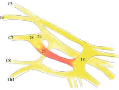
Fig. 1. anomalies of upper trunks, where: C4 – Th1 – nerve roots, 20 – lateral cord, 21 – anterior division of upper
trunk (aDMT), 22 – posterior division of upper trunk, 23 – anterior division of middle trunk, 28 – middle trunk (fig-ure courtesy of authors)
Ryc. 1. anomalie pnia górnego, gdzie: C5 – Th1 – korzenie splotu ramiennego, 20 – pęczek boczny, 21 – gałąź
przed-nia pprzed-nia górnego, 22 – gałąź tylna pprzed-nia górnego, 23 – gałąź przedprzed-nia pprzed-nia środkowego, 28 – pień środkowy (rycina ze zbiorów autorów)
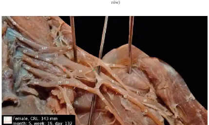
Fig. 2. Middle trunk was formed by C7 and C8 nerve roots while lower trunk created only Th1 root as well as
addition-al connection between anterior division of middle trunk (aDMT) and mediaddition-al cord (red star), where: C5 – Th1 –
bra-chial plexus roots, 8 – musculocutaneous nerve, 9 – axillary nerve, 10 – radial nerve, 11 – lateral root of median nerve, 12 – medial root of median nerve, 13 – median nerve, 14 – ulnar nerve, 16/17 – brachium and antebrachium medial cutaneous nerves, 18 – medial cord, 19 – posterior cord, 20 – lateral cord, 21 – anterior division of upper trunk, 22 – posterior division of upper trunk, 23 – anterior division of middle trunk (aDMT), 24 – posterior division of middle trunk, 25 – posterior division of lower trunk, 26 – anterior division of lower trunk, 27– upper trunk, 28 – middle trunk, 29 – lower trunk (figure courtesy of authors)
Ryc. 2. Pień środkowy uformowany przez korzenie C7 i C8 wraz z pniem dolnym utworzonym tylko przez korzeń
nerwu Th1 a także dodatkowe połączenie między gałęzią przednią pnia środkowego (aDMT) i pęczkiem
przyśrod-kowym (czerwona gwiazdka), gdzie: C5 – Th1 – korzenie splotu ramiennego, 8 – nerw mięśniowo-skórny, 9 – nerw
pachowy, 10 – nerw promieniowy, 11 – korzeń boczny nerwu pośrodkowego, 12 – korzeń przyśrodkowy nerwu pośrodkowego, 13 – nerw pośrodkowy, 14 – nerw łokciowy, 16/17 – nerwy skórne przyśrodkowe ramienia i przed-ramienia, 18 – pęczek przyśrodkowy, 19 – pęczek tylny, 20 – pęczek boczny, 21 – gałąź przednia pnia górnego, 22 – gałąź tylna pnia górnego, 23 – gałąź przednia pnia środkowego, 24 – gałąź tylna pnia środkowego, 25 – gałąź tylna pnia dolnego, 26 – gałąź przednia pnia dolnego, 27 – pień górny, 28 – pień środkowy oraz 29 – pień dolny (rycina ze zbiorów autorów)
fetuses did not reveal any visible developmental or genetic defects. The following methods were used in the study: anthropological and dissection meth-ods, digital image acquisition with use of a
anthropo-Fig. 3. additional connections of the middle trunk with anterior division of upper trunk (a) and medial cord (B), where: C5 – Th1 – nerve roots, 18 – medial cord, 20 – lateral cord, 21 – anterior division of upper trunk, 23 – anterior
division of middle trunk (aDMT), 24 – posterior division of middle trunk, 25 – posterior division of lower trunk, 26 – anterior division of lower trunk, 27 – upper trunk, 28 – middle trunk and 29 – lower trunk (figure courtesy of authors)
Ryc. 3. Dodatkowe połączenie pnia środkowego z gałęzią przednią pnia górnego (a) i pęczkiem przyśrodkowym (B), gdzie: C5 – Th1 – korzenie splotu ramiennego, 18 – pęczek przyśrodkowy, 20 – pęczek boczny, 21 – gałąź przednia
pnia górnego, 23 – gałąź przednia pnia środkowego, 24 – gałąź tylna pnia środkowego, 25 – gałąź tylna pnia dolnego, 26 – gałąź przednia pnia dolnego, 27 – pień górny, 28 – pień środkowy oraz 29 – pień dolny (rycina ze zbiorów auto-rów)
A B
Fig. 4. Variants in the form of an additional connection between the lateral cord (20) and medial root of the median nerve (12) – green stars as well as additional connection between lower trunk (29) and lateral cord (20) – red star (fig-ure courtesy of authors)
Ryc. 4. Wariant dodatkowego połączenia między pęczkiem bocznym (20) a korzeniem przyśrodkowym nerwu
pośrodkowego (12) – zielone gwiazdki oraz dodatkowe połączenie pomiędzy pniem dolnym (29) i pęczkiem bocznym (20) – czerwona gwiazdka (rycina ze zbiorów autorów)
logical method was based on an assessment of fetal age by measuring somatic CRL and vertex-plantare length. Image J was used for image processing and the creation of 3D models of the brachial plexus, which were helpful in the evaluation of topography and anatomical variability. Morphological varia-tions evaluation of the brachial plexus trunks and their divisions was done with a 0/1 system, where
Fig. 5. an additional connection between anterior division of upper trunk (21) and – anterior division of middle trunk (aDMT, 23) – red stars as well as connection between anterior division of middle trunk (23) and medial root of median nerve (12) (figure courtesy of authors)
Ryc. 5. Dodatkowe połączenie między gałęzią przednią pnia górnego (21) a gałęzią przednią pnia środkowego (aDMT, 23) – czerwone gwiazdki a także połączenie między gałęzią przednią pnia środkowego (23) a korzeniem przyśrodkowym nerwu pośrodkowego (12) (rycina ze zbiorów autorów)
Fig. 6. anterior division of middle trunk (aDMT, 23) was connected with medial cord (18), where: C5 – Th1
– brachial plexus roots, 18 – medial cord, 23 – anterior division of middle trunk (aDMT), 24 – posterior divi-sion of middle trunk and 28 – middle trunk (figure courtesy of authors)
Ryc. 6. gałąź przednia pnia środkowego (aDMT,
23) łączy się z pęczkiem przyśrodkowym (18), gdzie: C5 – Th1 – korzenie splotu ramiennego, 18 – pęczek
przyśrodkowy, 23 – gałąź przednia pnia środkowego, 24 – gałąź tylna pnia środkowego i 28 – pień środkowy (rycina ze zbiorów autorów)
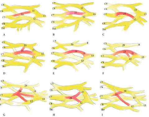
cases, a separate trunk was formed by the C4 and C5 nerve roots, giving two divisions (co-creating side and rear bunches). The C6 root created a separate connection to the anterior and posterior divisions (fig. 1a). In the next case (fig. 1B), the C4 and C5 roots formed a separate connection by dividing into two divisions, which then joined to the C6 nerve divisions to form anterior and posterior divisions which then joined to the anterior and posterior di-visions. The middle trunk in four cases was formed by the C7 and C8 nerve roots while the lower trunk created only the T1 root (fig. 2). additional connec-tions of the middle trunk with the anterior division of the upper trunk and medial cord (fig. 3) were ob-served only in individual cases. The lower trunk in 3 plexuses created an additional connection to the lateral cord. These anomalies were accompanied by variants in the form of an additional connection between the lateral cord and the medial root of the median nerve (fig. 4). anomalies within the divi-sions were observed in 74 plexuses (33.6%). Divi-sion of the upper trunk variant was found only in only one case: an additional connection between the anterior division of the upper trunk and the anterior division of the middle trunk (fig. 5). The anterior division of the middle trunk (aDMT) was the most variable. Its variants were observed in 63 cases. absence of the anterior division of the mid-dle trunk (aDMT) was observed in one case, and in 2 cases, this division was connected with the me-dial cord as shown in figure 6. The most common anatomical variations of the anterior division of the middle trunk (aDMT) are shown in figure 7. Nine morphological types were distinguished. figure 7a shows the connection of the anterior division of the middle trunk (aDMT) with the lateral root of the median nerve, which was observed in 3 cases. 7B shows a direct connection between the division and the median nerve, which was observed in 4 cases. 7C
Results
Fig. 7. Morphological types of anterior division of middle trunk (aDMT) variations, where: C5 – Th1 – brachial plexus
roots, 8 – musculocutaneous nerve, 11 – lateral root of median nerve, 12 – medial root of median nerve, 13 – median nerve, 20 – lateral cord as well as 23 – anterior division of middle trunk (aDMT) (figure courtesy of authors)
Ryc. 7. Morfologiczne warianty gałęzi przedniej pnia środkowego (aDMT), gdzie: C5 – Th1 – korzenie splotu
ramien-nego, 8 – nerw mięśniowo-skórny, 11 – korzeń boczny nerwu pośrodkowego, 12 – korzeń przyśrodkowy nerwu pośrodkowego, 13 – nerw pośrodkowy, 20 – pęczek boczny i 23 – gałąź przednia pnia środkowego (aDMT) (rycina ze zbiorów autorów)
D E F
G H I
Fig. 8. an additional connection between anterior division of middle trunk (aDMT, 23) and medial root of median nerve (12) – red stars (figure courtesy of authors)
Ryc. 8. Dodatkowe połączenie między gałęzią przednią pnia środkowego (aDMT, 23) i korzeniem przyśrodkowym
Fig. 9. Connection between anterior division of middle trunk (aDMT, 23) and lateral cord (20) – double lat-eral root of median nerve, where: C5 – Th1 – brachial
plexus roots, 11 – lateral root of median nerve, 12 – medial root of median nerve, 13 – median nerve, 20 – lateral cord and 23 – anterior division of middle trunk (aDMT) (figure courtesy of authors)
Ryc. 9. Połączenie między gałęzią przednią pnia środ-kowego (aDMT, 23) I pęczkiem przyśrodkowym (20) tworzące podwójny korzeń boczny nerwu pośrodko-wego, gdzie: C5 – Th1 – korzenie splotu ramiennego,
11 – korzeń boczny nerwu pośrodkowego, 12 – korzeń przyśrodkowy nerwu pośrodkowego, 13 – nerw pośrodkowy, 20 – pęczek boczny i 23 – gałąź przednia pnia środkowego (aDMT) (rycina ze zbiorów auto-rów)
Fig. 10. Variant of the posterior division of the middle trunk (24) – connection with medial cord (18) – red star as well as extra connection between lateral cord (20) and medial root of median nerve (12) – green star (figure courtesy of authors)
Ryc. 10. Wariant gałęzi tylnej pnia środkowego (24) – połączenie z pęczkiem przyśrodkowym (18) – czerwona gwiazdka oraz dodatkowe połączenie między pęczkiem bocznym (20) a korzeniem przyśrodkowym nerwu pośrodko-wego (12) – zielona gwiazdka (rycina ze zbiorów autorów)
shows a division connection with the median nerve, found in 1 case. In 7D, the anterior division of the middle trunk gives additional fibers to the median nerve, found in 1 case. In 7E, the aDMT creates a lateral root of the median nerve, and doesn’t con-nect to the lateral cord, which is formed only by the anterior division of the upper trunk. This variant was observed in 4 cases. 7f is similar to 7E, but the aDMT gives an additional fiber to the medial root of the median nerve. This variant was observed in 1 case. In 7g, in 3 cases, the aDMT joined to the lateral cord and gave two additional parts to the me-dial and lateral root of the median nerve. and in 7H, the aDMT divided into two parts and joined
Fig. 12. Connection between posterior division of lower trunk (25) and radial nerve (10) – red star and unusual con-struction of posterior cord (19) (figure courtesy of authors)
Ryc. 12. Połączenie między gałęzią tylną pnia dolnego (25) a nerwem promieniowym (10) – czerwona gwiazdka oraz nietypowa budowa pęczka tylnego (19) (rycina ze zbiorów autorów)
0 – normal 46 (92%) 55 (92%) 47 (94%) 60 (100%)
1 – morphological variant 4 (8%) 5 (8%) 3 (6%) 0 (0%)
Type of division (Typ budowy gałęzi)
0.795 0.770
0 – normal 33 (66%) 41 (68%) 32 (64%) 40 (67%)
1 – morphological variant 17 (34%) 19 (32%) 18 (36%) 20 (33%)
Fig. 11. Posterior division of middle trunk (24) and posterior division of lower trunk (25) anomaly, where: C5 – Th1 – brachial plexus roots, 11 – lateral root of
median nerve, 12 – medial root of median nerve, 13 – median nerve, 24 – posterior division of middle trunk, 25 – posterior division of lower trunk, 28 – mid-dle trunk, 29 – lower trunk (figure courtesy of authors)
Ryc. 11. anomalia gałęzi tylnej pnia środkowego (24) i gałęzi tylnej pnia dolnego (25), gdzie: C5 – Th1
trunk, in which both divisions gave additional fi-bers connecting with each other in a common trunk and then, after separation, joined the median nerve (fig. 11). In 8 cases, there were abnormalities of the posterior division of the lower trunk, in 4 cases, the division joined to the radial nerve and the posterior cord was formed by the posterior divisions of the upper and middle trunks (figure 12) and in other cases, the division originated from C8, not from the lower trunk (fig. 13).
Discussion
The variability of fetal brachial plexus was de-scribed by Uysal et al. [12]. They studied 200 bra-chial plexuses obtained from fetuses aged between 13 and 40 weeks of fetal life. It was found that in the 13th week the plexus is already completely formed. Morphological variants were observed in 53.5% of cases, more often on the right side and in male fetuses. Woźniak et al. [13] analyzed the variability of the nerve roots of the brachial plexus in 220 plexuses obtained from human fetuses aged between 4–7 months of gestation, including 50 fe-males. anomalies in this part of the plexus were observed in 15.9% of cases, mostly related to C4 spinal nerve roots. There was no asymmetry or sexual dimorphism. Trunk and division anomalies in our study were found in 5.45% and 33.6% of cases. anatomical variants of the individual trunks showed similar rates. It is interesting that in the case of divisions, the most variable (in 28.63%
of cases) was the anterior division of the middle trunk (aDMT). anomalies in other divisions were observed at a small percentage. In the available lit-erature, there were no reports of lower trunk pos-terior division variants described in the work: the connection of the posterior division of the lower trunk of the radial nerve accompanied by a dif-ferent construction of the posterior cord (created only through the anterior and posterior divisions of the middle trunk). figure 7 presents the anom-alies of the anterior division of the middle trunk (aDMT) in the form of nine morphological types. In type E and f, the side bunch is separated from the lateral root of the median nerve, so the nerve fibers originating from the nerve roots from C5 – C6 did not take part in the creation of the me-dian nerve. In this case it might be suspected that a person with type E and f construction of the bra-chial plexus may have saved a function of the me-dian nerve in Erb’s paralysis, the ability to turn the forearm or the behavior of the biceps reflex and its functions. In types C, f, g and H, additional nerve fibers from C7 nerve root supply the medial root of the median nerve, which can remain not insig-nificant in Klumpke-type damage. These observa-tions, however, require further investigation.
a small variation in tree trunks is supported by data from the literature. Uysal et al. [12] ob-served that the lower trunk was unformed in 9% of cases, while the upper only in 1%. In one case, creation of the lower trunk from rear divisions of the abdominal nerve Th1 and Th2 was found [12].
absence of the upper trunk was also observed by Villamere et al. [14], during a routine autopsy of a 55-year-old woman, on the left side. The nerve roots C5 and C6 were not united in the common trunk, but separately gave front and rear divisions which co-created side and rear cords. Nayak et al. [15] also described a variant of the upper trunk, which was formed by the nerve roots C5, C6 and C7. a comprehensive assessment of the variants of the brachial plexus in adults was conducted by Matejcik [16]. aDMT was also the most common variant in this author’s work.
anomalies of divisions occurred most often in the work of Uysal et al. [12], especially the ante-rior division of the middle trunk (aDMT) in 23% of cases. In the available literature, the importance of the knowledge of brachial plexus variations in clinical practice is emphasized [12, 16]. It seems to be invaluable in cases of treatment of the brachial plexus through reconstruction in both infants and adults. It was found that variants of trunks were rare while division anomalies were observed in one third of all cases, regardless of the side of the body and sex. Brachial plexus variations recogni-tion is important from a clinical point of view.
Fig. 13. Posterior division of lower trunk (25) origi-nated from C8, where: C5 – Th1 – brachial plexus roots,
25 – posterior division of lower trunk, 29 – lower trunk (figure courtesy of authors)
Ryc. 13. gałąź tylna pnia dolnego odchodzi od korze-nia nerwu C8, gdzie: C5 – Th1 – korzenie splotu
[7] Shinohara H, Naora H, Hashimoto R, Hatta T, Tanaka O: Development of the innervation pattern in the upper limb staged human embryos. acta anat 1990, 138, 265–269.
[8] Woźniak J, Kędzia A, Dudek K: Mathematical models of brachial plexus development during fetal period: clinical aspects. adv Clin Exp Med 2012, 21(2), 151–167.
[9] Rodriguez-Niedenführ M, Burton GJ, Deu J, Sañudo JR: Development of the arterial pattern in the upper limb of staged human embryos: normal development and anatomic variations. J anat 2001, 199, 407–417.
[10] Rodriguez-Niedenführ M, Vazquez T, Nearn L, Ferreira B, Parkin I, Sañudo JR: Variations of the arterial pat-tern in the upper limb revisited: a morphological and statistical study, with a review of the literature. J anat 2001, 199, 547–566.
[11] Rodriguez-Niedenführ M, Vazquez T, Parkin I, Sañudo JR: arterial patterns of the human upper limbs: update of anatomical variations and embryological development. Eur J anat 2003, suppl. 1, 21–28.
[12] Uysal II, Seker M, Karabulut AK, Büyükmumcu M, Ziylan T: Brachial plexus variations in human fetuses. Neurosurgery 2003, 53(3), 676–684.
[13] Woźniak J, Kędzia A, Dudek K: Variations of nerve roots of the brachial plexus in clinical aspects during foetal period. arch Perinat Med 2012, 18(2), 100–105.
[14] Villamere J, Goodwin S, Hincke M, Jalali A: a brachial plexus variation characterized by the absence of the superior trunk. Neuroanatomy 2009, 8, 4–6.
[15] Nayak S, Somayaji N, Vollala VR, Reghunathan D, Rodrigues V, Samuel VP, Malloor PA: a rare variation in
the formation of upper trunk of the brachial plexus – a case report. Neuroanatomy 2005, 4, 37–38.
[16] Matejcik V: aberrant formation and clinical picture of brachial plexus from the point of view of a neurosurgeon. Bratisl Lek Listy 2003, 104(10), 291–299.
Address for correspondence:
Jowita WoźniakDepartment of Neurosurgery
academic Clinical Hospital in Wrocław Borowska 213
50-556 Wrocław Poland
Tel.: +48 71 734 34 00
E-mail: [email protected]
Conflict of interest: None declared