Xiaoli Kang
A–D, Jun Yu
C, D, Yan Wei
B, F, Peiquan Zhao
E, FThe Effect of A
2AReceptor Antagonist (SCH 442416)
on the mRNA Expression of Kir 2.1 and Kir 4.1 Channels
in Rat Retinal Müller Cells Under Hypoxic Conditions
in Vitro
Wpływ antagonisty receptora A
2A(SCH 442416)
na ekspresję mRNA kanałów Kir 2.1 i Kir 4.1 w komórkach Müllera
u szczurów w warunkach hipoksji
in vitro
Department of Ophthalmology, Xinhua Hospital, Shanghai Jiao Tong University School of Medicine, Shanghai, China
A – research concept and design; B – collection and/or assembly of data; C – data analysis and interpretation;
D – writing the article; E – critical revision of the article; F – final approval of article; G – other
Abstract
Objectives. To investigate the situation of inwardly rectifying potassium channels Kir 2.1 and Kir 4.1 under hypo xic conditions, and whether the A2A receptor antagonist could modulate the mRNA expression of Kir 2.1 and Kir
4.1 channels in retinal Müller cells in vitro under hypoxic conditions.
Material and Methods. Müller cells were treated with 0.1 1, 10 and 100 µM of A2A receptor antagonist (SCH
442416) under hypoxic conditions for 24 h, and the expression of the Kir 2.1 and Kir 4.1 mRNA channels was examined using realtime polymerase chain reaction (qPCR).
Results. There were no significant changes in the mRNA expression of the Kir 2.1 or Kir 4.1 channels under hypoxic conditions compared with normal conditions after 24 h cultured in vitro. The mRNA expression of Kir 2.1 and Kir 4.1 channels treated with 0.1 µM SCH 442416 under hypoxic conditions were increased, but at higher concentrations of SCH 442416, the mRNA expression of the Kir 2.1 and Kir 4.1 channels decreased.
Conclusions. TheA2A receptor antagonist (SCH 442416) could increase mRNA expression of the Kir 2.1and Kir
4.1 channels in Müller cells to protect the retinal neurons in vitro under hypoxic conditions (Adv Clin Exp Med 2013, 22, 6, 825–829).
Key words: Müller cells, Kir 4.1 channels, Kir 2.1 channels, A2A receptor antagonist, hypoxia.
Adv Clin Exp Med 2013, 22, 6, 825–829 ISSN 1899–5276
ORIgINAl PAPERS
© Copyright by Wroclaw Medical University
Hypoxia certainly plays a critical role in reti nal disease and is a primary cause of central neu ronal damage. local hypoxia can cause hyperper meability and neovascularization of the retinal vasculature [1–3].
Müller cells are the major glial cells of the reti na. They have a wide array of responses to main tain homeostasis for neuronal and vascular ele ments [4, 5]. They can maintain the integrity of the bloodretinal barrier and clear metabolic waste via regulation of the homeostasis of extracellular pH and K+ ions. To avoid high K+ levels, which can
induce depolarization of retinal neurons, Müller cells can take up excess K+ from the extracellular
space, especially in the synaptic layers of the retina, and release a similar amount of K+ into spaces out
side of the neural retina, especially into the blood and the vitreous humor [6].
Müller cells mediate the mechanisms of K+
high levels of extracellular K+. The Kir 4.1 channels
are weaker Kir channels, allowing either “inward” or “outward” K+ currents depending on the con
centrations of extracellular K+ [7–11].
Adenosine is a reactive metabolite involved in cellular communication during periods of some pathological state. In the eyes, the levels of adeno sine increase due to retinal ischemia and/or hypo xia. Four adenosine receptor subtypes (A1, A2A, A2B,
and A3) have been identified. Recently, neuropro
tection of the A2A blockade has been found in some
animal models of some neurodegenerative disor ders, such as epilepsy and Huntington’s disease, and some excitotoxic conditions, such as ischemia and trauma [12–14]. The current authors’ previous studies also found that A2A receptor (A2AR) antago
nist could significantly upregulate the expression of glutamine synthetase and glutamate aspartate transporter, and maintain glutamate homeostasis under hypoxic conditions [15]. However, it was still not clear whether A2AR antagonist also could
regulate the expression of the Kir channels in Mül ler cells in hypoxia.
The current study focusses on investigating the situation of the Kir 2.1 and Kir 4.1 channels under hypoxic conditions, and whether A2AR antagonist
(SCH 442416) could regulate the mRNA expres sion of Kir 2.1 and Kir 4.1 channels in retinal Mül ler cells in vitro under hypoxic conditions.
Material and Methods
Drugs
A2AR antagonist (SCH 442416), 2(2Furanyl)
7[3(4methoxyphernyl)propyl]7Hpyrazolo[4,3e] [1,2,4]triazolo[1,5c]pyrimidin5amine was pur chased from Tocris Bioscience. The SCH 442416 was dissolved in a serumfree medium to make final concentrations of 0.1, 1, 10 and 100 µM.
Cell Culture
The eyeballs of postnatal day 0–3 Sprague Dawley rats were purchased from Shanghai labo ratory Animal Center CAS (SlACCAS), and each retina was dissected and stored in DHank’s solu tion (Anresco) on ice. All of the tissues were dis sociated and incubated for 15 min in PBS, which contained 0.125% trypsin (Anresco) at 37°C.
The tissues were then cultured in T75 flasks (in air containing 5% carbon dioxide, at 37°C). After the 1st outgrowth, the medium was refreshed every 48 h, and maintained in DMEM/F12 medium (gibco) containing glutamine (2 mM), streptomy cin (100 µg/ml), penicillin (100 U/ml) and 10% fetal bovine serum (FBS) (gibco).
After 8–11 days, the flasks were shaken at 37°C, 100 r/min for 1 h. By shaking, other kinds of cells (such as microgilal cells and retinal ganglion cells) were rinsed off, so that a purified Müller cell popu lation was obtained.
The cultures were incubated at 37°C for a 2nd passage. The experiments were performed after the 2nd passage when the confluence was 75–80 %.
Müller Cell Proliferation
in Normoxia or Hypoxia
Firstly, Müller cells were planted in sixwell plates at 5 × 10⁵/ml for 24 h. Secondly, the medium was
replaced with serum and various concentrations of SCH 442416 (0.1, 1, 10 and 100 µM) and the cultures were placed in hypoxic conditions (37°C, 94% nitro gen, 1% oxygen, 5% carbon dio xide) for 24 hours as different hypoxia groups. For the normoxia group, the medium was changed to serumfree DMEM and the cultures were placed in normal conditions (37°C, 20% oxygen, 5% carbon dioxide) for 24 h. After 24 h of incubation, the cells were analyzed.
Immunocytochemistry
Müller cells cultured under hypoxic condition for 24 h were fixed with 4% paraformaldehyde for 10 min. The cover slips were incubated in primary antibodies: antigFAP (Abcam, 1 : 200, polyclonal mouse antibody) and antigS (Abcam, 1 : 5000, polyclonal rabbit antibody) overnight at 4°C. Then the cover slips were immunolabeled with fluoros cein isothiocyanate FITC (Invitrogen, 1 : 200) or Cy3 (Biolegend, 1 : 200). The cells were then ob served by laser confocal microscopy (leica).
Analysis of mRNA Expression by
Real-Time PCR
RNA was isolated from Müller cells with Trizol reagent (Invitrogen). The cDNAs were reverse tran scribed according to the manufacturer’s instructions. Realtime PCR was used to analyze the mRNA ex pression of the Kir2.1 and Kir4.1 channels.
The primer sequences were as follows: Kir2.1 channels: sense 5’ gcctcctggttgctgttc3’, antisense 5’tggtggtctgcgtctcaat3’; Kir4.1channels: sense 5’ agttcgcacttcctatctaccg3’, antisense 5’ gggacgc cactttcacaa3’; βactin, sense 5’cccatctatgagggttac gc3’, antisense 5’tttaatgtcacgcacgatttc3’.
Statistical Analysis
Data are shown as the mean ± standard error (n = 4 for each group). The analyses were executed with SPSS 13.0 software. The data were analyzed by a oneway ANOVA test followed by an lSD test for multiple comparison. Differences were consid ered statistically significant when P < 0.05.
Results
Immunocytochemistry of Retinal
Müller Cells in Hypoxia
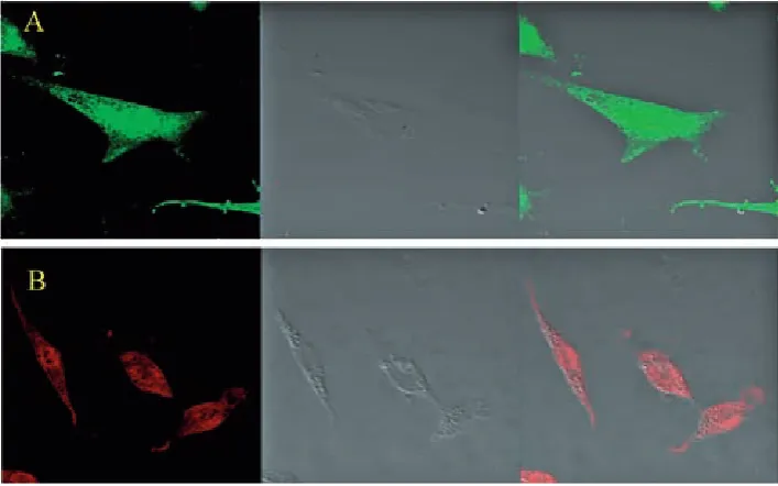
The figures showed positive labeling for gFAP and gS under hypoxic conditions; gFAP and gS were the molecular markers for Müller cells in the retina (Fig. 1), so those cells were identified as Müller cells.
gS, a molecular marker for Müller cells in the retina, has usually been used as a specific label for Müllercells. Fig. 1B shows the expressions of gS by immunocytochemistry staining. Müller cells scarcely express gFAP in a normal retina, but the expression gets stronger when the retina is dam aged [15–17]. In the current study, more than 90% of the cells showed positive markers for gFAP (Fig. 1A). The cells can be identified as Müller cells, and hypoxia induced the activation of Mül lercells.
gFAP (which is green) was labeled for Müller cells (A); gS (which is red) was labeled for Mül ler cells (B).
The effect of SCH 442416 on
mRNA
Expression of Kir 2.1 Channels in
Müller Cells
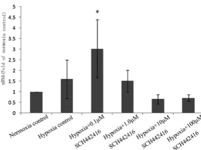
The PCR data showed that the mRNA expression of Kir 2.1 channels was significantly increased when Müller cells were treated with 0.1 µM SCH 442416 in hypoxia, compared with the normoxia control, the hypoxia control, and even with any another SCH 442416 concentration (1, 10 or 100 µM) (Fig. 2).
The mRNA levels of Kir 2.1 channels were sig nificantly increased when Müller cells were treat ed with 0.1 µM SCH 442416 in hypoxia, compared with the normoxia control, the hypoxia control, and even with any another SCH 442416 concen tration (1, 10 or 100 µM) (* p < 0.05).
The Effect of SCH 442416 on
the mRNA Expression of Kir 4.1
Channels in the Cultured Retinal
Müller Cells
The PCR data showed that the mRNA expres sion of Kir 4.1 channels was increased when Mül ler cells were treated with 0.1 µM SCH 442416 in hypoxia, compared with the normoxia control, the hypoxia control, and even any another SCH 442416 concentration (1, 10 and 100 µM) (Fig. 3).
The mRNA levels of Kir 4.1 channels were sig nificantly increased when Müller cells were treat ed with 0.1 µM SCH 442416 in hypoxia, compared with the normoxia control, the hypoxia control, and even any another SCH 442416 concentration (1, 10 and 100 µM) (* p < 0.05).
Discussion
The results of this study showed that: 1) hy poxia could induce the activation of Müller cells; 2) there were no significant changes in the mRNA expression of Kir 2.1 and Kir 4.1 channels in hy poxia, compared with normal conditions after 24 h of culturing in vitro; 3) Müller cells had increased mRNA expression of Kir 2.1, Kir 4.1 channels cul tured with 0.1 µM SCH 442416 under hypoxic conditions, but when the SCH 442416 concentra tion was further increased, the mRNA expression of the Kir 2.1 and Kir 4.1channels decreased (Fig. 2 and 3).
Hypoxia certainly plays a crucial role in reti nal diseases like retinal vascular occlusion, diabe tes and glaucoma [3, 18]. In a normal retina, there is little or no gFAP in the Müller cells, but it be comes strong when the retina is damaged. In the present study, it was found that hypoxia could re sult in a high expression of gFAP, from which it can be inferred that hypoxia could induce the acti vation of Müllercells.
In hypoxic conditions, activated neurons re lease K+, which can induce the depolarization of
neurons, thereby causing neuronal hyperexcitation resulting in excess release of neurotransmitters and glutamate toxicity. Müller cells can take up excess K+ from the extracellular space, and release an ap
propriate amount of K+ into spaces outside of the
retina. Kir channels localized in Müller cell mem branes are used to mediate extracelluar K+ [6].
Müller cells express different types of K+ chan
nels. Kir 2.1 channels are found in neuronabut ting membranes, and Kir 4.1 channels are found in membranes adjacent to the space outside the
neural retina [6]. The results of the present study indicate that the response of Kir 2.1and Kir 4.1 channels of Müller cells to hypoxia was not espe cially sensitive in vitro (Fig. 2 and 3). The data also showed that the mRNA expression of Kir 2.1 chan nels increased slightly under hypoxic conditions, compared with normal conditions (Fig. 2).
On the basis of the results, the authors pro pose 2 surmises. 1st: Kir 2.1 and Kir 4.1 channels were not the key regulatory proteins in the Mül ler cells under hypoxic conditions. 2nd: in the present study, the experimental hypoxic time was 24 h; maybe this was too short for the activation of Kir 2.1 and Kir 4.1 channels. The question needs to be investigated in further experiments.
The results of the study showed that 0.1 µM SCH 442416 could upregulate the mRNA expres sion of the Kir 2.1and Kir 4.1 channels of Müller cells in hypoxia. But when the SCH 442416 con centration was increased, the mRNA expression of the Kir 2.1 and Kir 4.1channels decreased. So the authors concluded that low concentrations of SCH 442416 could upregulate the mRNA expression of Kir 2.1 and Kir 4.1 channels, which may accelerate the clearance of K+ to protect the retinal neurons.
In recent years, A2AR antagonist has been
viewed as an attractive drug to treat neurolo gical disorders [19, 20]. On the basis of the cur rent results, the authors regard A2AR antagonist
as a novel choice for neuroprotection under hy poxic conditions. Of course, there are some prob lems that need to be resolved, such as the appropri ate drug concentration and the best hypoxic time. The authors plan to study these problems in future experiments.
Fig. 3. The mRNA expression of Kir 4.1 channels of Müller cells treated with SCH 442416 (0.1, 1, 10 and 100 µM)
Fig. 2. The mRNA expression of Kir 2.1 channels of Müller cells cultured with SCH 442416 0.1, 1, 10 and100 µM)
References
[1] Tretiach M, Madigan M, Wen L, Gillies M: Effect of Müller cell coculture on in vitro permeability of bovine retinal vascular endothelium in normoxic and hypoxic conditions. Neurosci lett 2005, 378, 160–165.
[2] Kitano S, Morgan J, Caprioli J: Hypoxic and excitotoxic damage to cultured rat retinal ganglion cells. Exp Eye Res 1996, 63, 105–112.
[3] Caprioli J, Kitano S and Morgan J: Hyperthermia and hypoxia increase tolerance of retinal ganglion cells to a noxia and excitotoxicity. Invest Ophthalmol Vis Sci 1996, 37, 2376–2381.
[4] Harada T, Harada C, Kohsaka S: MicrogliaMüller glia cell interactions control neurotrophic factor production during lightinduced retinal degeneration. J Neurosci 2002, 22, 9228–9236.
[5] Yu J, Zhong Y, Shen X, Cheng Y, Qi J ,Wang J:In vitro effect of adenosine A2A receptor antagonist SCH 442416 on the expression of glutamine synthetase and glutamate aspartate transporter in rat retinal Müller cells at elevated hydrostatic pressure. Oncol Rep 2012, 27, 748–752.
[6] Reichenbach A, Wurm A, Pannicke T, Landiev I, Wiedemann P, Bringmann A: Müller cells as players in retinal degeneration and edema. graefe’s Arch Clin Exp Ophthalmol 2007, 245, 627–636.
[7] Kofuji P, Biedermann B, Siddharthan Vl: Kir potassium channel subunit expression in retinal glial cells: implica tions for spatial potassium buffering. glia 2002, 39, 292–303.
[8] Bringmann A, Francke M, Pannicke T: Role of glial K+ channels in ontogeny and gliosis: a hypothesis based upon
studies on Müller cells. glia 2000, 29, 35–44.
[9] Chen K, Nicholson C: Spatial buffering of potassium ions in brain extracellular space. Biophys J 2000, 78, 2776–2797.
[10] Fakler B, Bond C, Adelman J: Heterooligomeric assembly of inwardrectifier K+ channels from subunits of differ
ent subfamilies: Kir2.1 (IRK1) and Kir4.1 (BIR10). Pflügers Arch 1996, 433, 77–83.
[11] Yu J, Chen C, Wang J, Cheng Y, Wu Q, Zhong Y, Shen X: In vitro effect of adenosine on the mRNA expression of Kir 2.1 and Kir 4.1 channels in rat retinal Müller cells at elevated hydrostatic pressure. Exp Ther Med 2012, 3, 617–620.
[12] Sebastião A, Ribeiro J: Triggering neurotrophic factor actions through adenosine A2A receptor activation: impli cations for neuroprotection. Br J Pharmacol 2009, 158, 15–22.
[13] Leite M, Wilhelm E, Jesse C: Protective effect of caffeine and a selective A2A receptor antagonist on impairment of memory and oxidative stress of aged rats. Exp gerontol 2010, 46, 309–315.
[14] Marcellino D, Lindqvist E, Schneider M: Chronic A2A antagonist treatment alleviates parkinsonian locomotor deficiency in MitoPark mice. Neurobiol Dis 2010, 40, 460–466.
[15] Yu J, Xin H, Wu Q, Wang J, Yu X, Zhao P: The effect of A2A receptor antagonist (SCH 442416) on the mRNA
expression of glutamine synthetase and glutamate aspartate transporter in rat retinal Müller cells under hypoxic conditions in vitro. Exp Ther Med 2012, 3, 803–806.
[16] Shen X, Zhong Y, Xie B: Pigment epithelium derived factor as an antiinflammatory factor against decrease of glutamine synthetase expression in retinal Müller cells under high glucose conditions. graefes Arch Clin Exp Ophthalmol 2010, 248, 1127–1136.
[17] Shen F, Chen B, Danias J: glutamateinduced glutamine synthetase expression in retinal Müller cells after short term ocular hypertension in the rat. Invest Ophthalmol Vis Sci 2004, 45, 3107–3112.
[18] Choi D: glutamate neurotoxicity and diseases of the nervous system. Neuron 1988, 1, 623–634.
[19] Pepponi R, Ferrante A, Ferretti R, Martire A, Popoli P: Regionspecific neuroprotective effect of ZM 241385 towards glutamate uptake inhibition in cultured neurons. Eur J Pharmacol 2009, 617, 28–32.
[20] Morelli M, Carta A, Jenner P: Adenosine A2A receptors and Parkinson’s disease. Handb Exp Pharmacol 2009, 193, 589–615.
Address for correspondence:
Peiquan Zhao
Department of Ophthalmology
Xinhua Hospital, Shanghai Jiao Tong University School of Medicine 1665 Kongjiang Road
Shanghai China 200092
Email: [email protected]
Conflict of interest: None declared