R E S E A R C H A R T I C L E
Open Access
Reduced PRC2 function alters male
germline epigenetic programming
and paternal inheritance
Jessica M. Stringer
1,2, Samuel C. Forster
3,4,5, Zhipeng Qu
6, Lexie Prokopuk
1,5, Moira K. O
’
Bryan
7, David K. Gardner
8,
Stefan J. White
9, David Adelson
6and Patrick S. Western
1,5*Abstract
Background:Defining the mechanisms that establish and regulate the transmission of epigenetic information from parent to offspring is critical for understanding disease heredity. Currently, the molecular pathways that regulate epigenetic information in the germline and its transmission to offspring are poorly understood.
Results:Here we provide evidence that Polycomb Repressive Complex 2 (PRC2) regulates paternal inheritance. Reduced PRC2 function in mice resulted in male sub-fertility and altered epigenetic and transcriptional control of retrotransposed elements in foetal male germ cells. Males with reduced PRC2 function produced offspring that over-expressed
retrotransposed pseudogenes and had altered preimplantation embryo cleavage rates and cell cycle control.
Conclusion:This study reveals a novel role for the histone-modifying complex, PRC2, in
paternal intergenerational transmission of epigenetic effects on offspring, with important implications for understanding disease inheritance.
Keywords: Germline, Epigenetic reprogramming, PRC2, H3K27me3, Paternal inheritance, Fertility
Background
Numerous studies have investigated the inheritance of physiological effects caused by environmental impacts on the parental genome, but the underlying epigenetic mechanisms regulating such inheritance are poorly understood [1,2]. It is well established that DNA methy-lation is passed through the germline (oocytes and sperm) to the following generation, where it influences gene activity, embryonic development and post-natal life [1, 3–5]. In addition, recent studies have demonstrated effects of histone demethylases on inheritance [6,7]. For example, zygotic over-expression of the Histone 3 lysine
27 (H3K27) demethylase, Kdm6b, demonstrated a role
for maternal H3K27 methylation in regulating DNA
methylation-independent imprinting [7]. Similarly,
increased levels of histone 3 lysine 4 dimethylation
(H3K4me2) in developing sperm resulted in paternally transmitted effects on health and development in mice [6]. In this study, we provide evidence that epigenetic inheritance in mice is also altered by a hypomorphic
mutation in embryonic ectoderm development (Eed), a
gene that is essential for H3K27 trimethylation
(H3K27me3).
H3K27me3 is mediated by Polycomb Repressive Com-plex 2 (PRC2), which is comprised of the essential protein
components EED, EZH2 and SUZ12 [8]. In mice,
complete loss of function of any of these components results in loss of PRC2 activity, global reduction in
H3K27me3 and embryonic lethality [9–12]. While
complete loss of Eed results in lethality at gastrulation [13], germ cell-specific deletion results in male sterility [14]. However, an N-ethyl-N-nitrosourea (ENU)-induced
hypomorphic allele, Eedl7Rn51989SB, compromises PRC2
function and is compatible with survival, although some foetuses are lost during gestation due to defective placen-tal development [13,15].Eedl7Rn5-1989SBmice carry a point mutation at nucleotide 1989 that disrupts function of one
* Correspondence:[email protected] 1
Centre for Reproductive Health, Hudson Institute of Medical Research, Clayton, Victoria 3168, Australia
5Molecular and Translational Science, Monash University, Clayton, Victoria
3168, Australia
Full list of author information is available at the end of the article
of the WD repeat domains in the EED protein. This hypo-morphic mutation does not abrogate the ability of EED to mediate H3K27 methylation as the Eedl7Rn5-1989SB allele can rescue H3K27 methylation in ES cells lacking theEed gene [16]. Moreover, despite low EED function, adult mice with the hypomorphic Eedl7Rn5-1989SBmutation are fertile [17], allowing the investigation of PRC2 in epigenetic inheritance.
During embryonic development, epigenetic information is reprogrammed in the germline to ensure transmission of the correct information to the next generation. This involves extensive reorganisation of histone modifications and the removal of almost all DNA methylation from
foetal germ cells [18–24]. In mice, removal of DNA
methylation is initiated in migrating germ cells at around embryonic day (E)9, but is not complete until E13.5, after the germ cells have entered the developing gonads. Entry of germ cells into the gonads coincides with the removal of DNA methylation from imprinting control regions (ICRs), non-imprinted intergenic and intronic sequences and from many transposable elements (TEs), including
LINE and SINE elements [18, 22–26]. During germline
reprogramming, LINE and SINE elements are likely repressed by mechanisms other than DNA methylation to prevent TE expression and consequent insertional muta-tions [18,26].
H3K27me3 broadly regulates developmental gene expression through its ability to repress target gene tran-scription. In foetal germ cells, H3K27me3 is enriched at
developmental genes and on the 5′ flanking regions of
some TEs, including LINE1 elements, intergenic regions, introns and imprint control regions [26–29]. Loss of function of the H3K9me3 methyltransferase SET domain Bifurcated 1 (SETDB1) in the developing male germline results in loss of DNA methylation, H3K9me3 and
H3K27me3 at a subset of TEs [26]. This suggests that
H3K27me3 functions with DNA methylation and
H3K9me3 to co-regulate specific TEs in the germline [26]. Similarly, in cultured embryonic stem cells, H3K27me3 represses TEs in the absence of DNA methylation, estab-lishing a functional requirement for H3K27me3 on these sequences [30].
H3K27me3 is enriched in foetal germ cells and in germ cells undergoing spermatogenesis [28, 29, 31, 32]. Moreover, H3K27me3 has been detected at developmen-tal gene promoters in mature sperm, indicating that H3K27me3 may be transmitted to offspring and that such genes are poised for activation in the preimplanta-tion embryo [33–36]. Another study showed retention of nucleosomes at repetitive sequences in sperm, including at LINE elements [37–39]. Together, these studies raise the possibility that PRC2 and H3K27me3 regulate TEs during germline reprogramming and may modulate epi-genetic inheritance in offspring. However, whether the
potential inherited effects are directly mediated by his-tone modifications in offspring, or involve other mecha-nisms such as DNA methylation or altered inheritance of RNAs is unknown.
The aim of this study was to determine whether PRC2 contributes to the regulation of paternal epigenetic inheritance in a mammalian model. Using the
hypo-morphic Eedl7Rn5-1989SB mice, we provide evidence that
PRC2 modulates H3K27me3 enrichment on TEs and re-presses retrotransposable LINE elements in the foetal male germline. Moreover, our data indicate that PRC2 is required in the paternal germline to regulate offspring development and repress a cohort of retrotransposed pseudogenes and related lincRNAs in offspring.
Results
Eedl7Rn5-1989SBmice are sub-fertile and provide a model for the study of epigenetic inheritance through the paternal germline
Since the primary aim of this study was to determine the role of EED in paternal epigenetic inheritance, we first assessed survival and male fertility in our colony of Eedl7Rn5-1989SB mice. While the expected proportions of Eedwt/wt and Eedwt/hypo offspring were produced, the
proportion of Eedhypo/hypo mice was significantly
re-duced (ratio 1:2:0.1), demonstrating that Eedl7Rn5-1989SB homozygosity reduces viability (Additional file1: Figure S1A). At E15.5 Eedwt/wt, Eedwt/hypoandEedhypo/hypo foe-tuses were recovered in a 1:2:0.6 ratio (Additional file1: Figure S1A). As few still-births or neonatal deaths were
observed, we concluded that most Eedhypo/hypoembryos
died during the second half of gestation, consistent with previous observations [13,17]. Despite the loss of some foetuses, these experiments confirmed the survival of Eedhypo/hypo males to adulthood, allowing the study of the male germline in a background of low EED function.
While previous studies found that homozygous
Eedl7Rn5-1989SBmice produced offspring [17], the level of fertility in these mice remained unknown. We therefore completed a fertility analysis to determine whether the
Eedhypomorphic mutation affected male germline
d
f
g
h
e
b
a
c
males produced no pups or small litters more frequently thanEedhypo/wtandEedwt/wtmales (Additional file1: Figure S1B; chi-square P= 1.8E−05), indicating sub-fertility in someEedhypo/hypomales. No difference was observed in the
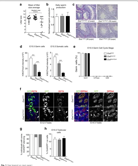
average daily sperm count between genotypes (Fig. 1b),
and there was no correlation between litter size or fre-quency and male age (Additional file1: Figure S1C).
Consistent with sporadic sub-fertility, testicular
morphology of Eedhypo/hypo males was also variable, but
consistent with fertility outcomes. In obviously
sub-fertile Eedhypo/hypo males, germ cells were reduced and vacuoles present in some testis cords, indicating that germ cells were lost through sloughing (Fig.1c). By con-trast, normal testicular morphology was observed in Eedhypo/hypo males that produced normal litter sizes (Fig. 1c). As a cohort (n= 20), abnormal testicular hist-ology was only observed in fourEedhypo/hypomales, with the remainder maintaining apparently normal testes with qualitatively normal spermatogenesis and weight. Com-bined, these data reflect the relatively mild sub-fertility and testicular phenotypes observed in these mice.
To determine whether the Eed hypomorphic
muta-tion affected testis formamuta-tion, we assessed the impact of reduced EED function on H3K27me3 levels and the development of germ and somatic cells in foetal testes. E12.5 and E15.5 were examined as they repre-sent the earliest stages of testis formation and male
germline development, and the completion of
PRC2-dependent reorganisation of H3K27me3 and initiation of DNA re-methylation in the paternal
germline, respectively [1, 3–5, 40]. While H3K27me3
was detected in germ cells of all genotypes by
im-munofluorescence (Additional file 1: Figure S2A), flow
cytometric assessment revealed significantly reduced global H3K27me3 levels in E15.5 germ and somatic cells in Eedhypo/wt and Eedhypo/hypocompared to Eedwt/ wt
testes (Fig. 1d, Additional file 1: Figure S2B). Re-duced levels of H3K27me3 were presumably due to
hypomorphic function of EED, as EED, EZH2 and SUZ12 were all detected in the germ and somatic cells of Eedwt/wt, Eedhypo/wt and Eedhypo/hypo testes (Additional file 1: Figure S2A).
Although H3K27me3 levels were reduced, the percent-age of Sertoli and germ cells in the gonad (Additional file1: Figure S2C-D), the proliferation of Sertoli cells (Add-itional file1: Figure S2E) and the entry of germ cells into mitotic arrest (Fig.1e) was unaffected. Similarly, qRTPCR analyses of a range of testis development genes involved in Sertoli, germ and steroidogenic cell development in E12.5 and E15.5 foetal testes (Additional file 1: Figure S2F), and flow cytometric analysis of SOX9 and AMH (Additional file 1: Figure S2G-H) revealed no differences
between Eedwt/wt Eedhypo/wt and Eedhypo/hypo testes.
Collectively, these data demonstrated that reduced EED function significantly affected male fertility and germline H3K27me3 levels. However, the majority of males were able to produce litters in which epigenetic inheritance could be effectively studied.
Reduced EED function resulted in stochastic silencing in male foetal germ cells
To facilitate isolation of germ cells, ourEedhypomorphic
mice carried a randomly integrated Oct4GFP transgene
that is robustly transcribed in all foetal germ cells until birth, but remains silent in somatic cells [41,42]. Initially, to confirm the veracity of the Oct4GFP transgene in this model, we used immunofluorescence to examine OCT4, DPPA4 and MVH in germ cells of E12.5Eedwt/wtEedhypo/ wt
and Eedhypo/hypo testes (Additional file 1: Figure S2I). Although OCT4, DPPA4 and MVH were detected in all
germ cells, we observed silencing of Oct4GFP in some
small patches of germ cells in some germ cells at E12.5 and E14.5 (Fig.1f ). However, this was not fully penetrant, as it affected ~ 60% ofEedhypo/hypoand ~ 10% ofEedwt/hypo individuals, and silencing was only evident in small num-bers of germ cells (Fig.1g). Indeed, analysis of FACS data
(See figure on previous page.)
Fig. 1Reduced EED function resulted in male subfertility and reduced litter size.aAverage litter size produced by wild-type female mice mated toEedwt/wt(n= 10),Eedhypo/wt(n= 13) andEedhypo/hypo(n =13) males mated to for two periods of 30 days. Each point represents the average of two, four or six litters per male. Average litter sizes produced byEedhypo/hypomales were more variable in size than fromEedwt/wtandEedhypo/wt
males (Bartlett’s testP= 0.0002).bNormalised daily sperm production ofEedwt/wt(n =3),Eedhypo/wt(n= 5), andEedhypo/hypo(n= 6) males (mean ± SEM, one-way ANOVA; no significant differences).cTestis histology from sub-fertile and fertileEedhypo/hypomales (top row) compared to their
revealed that there was no difference in the proportion of Oct4GFP-positive cells obtained from E15.5 foetal testes of Eedhypo/hypo, Eedwt/hypo and Eedwt/wt animals (Fig. 1h).
This was consistent with similar numbers of
MVH-positive germ cells in the testes of E15.5 Eedhypo/ hypo
, Eedwt/hypo and Eedwt/wt animals (Additional file 1: Figure S2C). Combined, these data demonstrated that al-though normal numbers of foetal germ cells were present in Eedhypo/hypo testes, occasional stochastic silencing of
Oct4GFP occurred in germ cells of males with reduced
EED function. As transgene silencing has been observed in other epigenetic models [43–45], we proposed that the stochastic Oct4GFP silencing in Eedhypo/hypo germ cells was indicative of an altered epigenetic state in the germ-line ofEedhypomorphic males.
H3K27me3 is required to repress LINE elements in male foetal germ cells
To determine whether epigenetic state was disrupted in E15.5 male foetal germ cells we used ChIP-seq analysis to assess H3K27me3 enrichment in germ cells isolated
from each of four single Eedhypo/hypo male embryos and
four Eedwt/wt male embryos. ChIP-seq yielded averages of 20,139,219 and 20,195,969 reads from theEedhypo/hypo and Eedwt/wt samples, respectively, of which 97.2–97.9% were alignable to the mm10 reference genome using
bowtie2 (Additional file 1: Figure S3). HOMER analysis
using a search region size of 1100 bp identified 60,933
and 55,453 H3K27me3 peaks in the Eedhypo/hypo and
Eedwt/wtsamples, respectively (Additional file2: Table S1 and Additional file3: Table S2).Importantly, comparison of our data to three similar datasets demonstrated sig-nificant overlap at known PRC2 targets (Additional file1: Figure S3G, Additional file 4: Table S3), demonstrating high specificity of the ChIP analysis. In addition, visual-isation of normalised read counts in the Eedhypo/hypoand Eedwt/wt germ cell samples demonstrated clear H3K27me3 enrichment in the 5-prime regions of PRC2 target genes and no enrichment on a constitutively
expressed gene Sdha, demonstrating sensitivity of the
assay (Additional file1: Figure S3H).
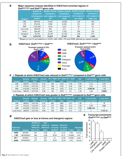
In addition, a high proportion of peaks were identified at repeat elements including LINE, LTR and SINE ele-ments in both Eedhypo/hypo and Eedwt/wt germ cell
sam-ples (Fig. 2a). We used hypergeometric testing to
determine whether the expected number of repeats was represented for each repeat category in the ChIP-seq data for Eedwt/wt and Eedhypo/hypo germ cells. In both Eedhypo/hypo and Eedwt/wt samples, SINE elements were substantially under-represented (fold enrichment = 1.31
and 1.22, respectively; p~ 0) in H3K27me3-enriched
peaks, but LINE elements were substantially
over-represented (fold enrichment = 0.51 and 0.61, re-spectively; P~ 0; Additional file 5: Table S4), suggesting
that LINE elements were preferentially captured in the ChIP seq assay, but SINE elements were not (Additional file 5: Table S4). LTRs were represented at expected ratios inEed -hypo/hypo
and Eedwt/wt samples (fold enrichment = 1.04 and 1.0, respectively; Additional file5: Table S4). Together, these data indicated strong enrichment of H3K27me3 not only at PRC2 target loci, but also at some repeat sequences, most notably LINE elements.
To investigate locus-specific variation in H3K27me3, we used HOMER to identify specific sequences with
dif-ferential H3K27me3 enrichment by comparing Eedhypo/
hypo
samples to Eedwt/wt samples (Additional file 1:
Figure S3I-J). As H3K27me3 has previously been impli-cated in repression of both coding and non-coding se-quences in foetal male germ cells [26, 28], we included repetitive sequences in our analyses. Samples were com-pared usingEedwt/wtas the baseline target and searching
for regions with a cumulative Poisson P value less than
0.0001 (sequencing-depth dependent) and ≥2-fold
reduction in H3K27me3 precipitated sequences. This
re-vealed 923 regions with≥2-fold reduction in H3K27me3
in Eedhypo/hypo comparedEedwt/wt germ cells (i.e. WT >
HOM). The reciprocal comparison using Eedhypo/hypo
samples as baseline andEedwt/wtsamples as target
iden-tified 1,158 regions with ≥2-fold increased H3K27me3
in Eedhypo/hypo germ cells (i.e. HOM > WT). Only 58 of these regions associated with coding genes (35 decreased and 23 increased, 2.84% of all differential peaks), while 1,951 LINE, LTR, SINE, intergenic and intronic se-quences were identified with significantly different (≥ 2-fold) levels of H3K27me3 (Fig. 2a; Additional file 6: Table S5 and Additional file7: Table S6). These included 120 LINE, 93 LTR, 56 SINE, 378 intergenic and 206
in-tronic with decreased (Fig. 2a) and 362 LINE, 187 LTR,
52 SINE, 377 intergenic and 120 intronic with increased (Fig.2a) H3K27me3 inEedhypo/hypocompared toEedwt/wt germ cells, representing > 97% of all differential ChIP peaks (Fig.2b). Using a more stringent analysis employ-ing edgeR, with a false discovery rate cut off ofP< 0.05, we identified 7 refGene annotated LINE1 loci with
sig-nificantly fewer reads mapped in Eedhypo/hypo compared
to theEedwt/wtsamples (Additional file8: Table S7), but no LINE1 elements with significantly more reads
mapped in Eedhypo/hypo compared to the Eedwt/wt
sam-ples. Together, these data indicated that subtle differ-ences in H3K27me3 regulation occurred predominantly at repetitive sequences, introns and intergenic regions in Eedhypo/hypogerm cells.
a
b
c
d
e
losing H3K27me3 inEedwt/wtandEedhypo/hypogerm cells relative to the total number of repeats in the genome. For example, LINE elements occupy 19% of all genome repeats (Fig. 2c (i)), and 18% of repeats were identified
with reduced H3K27me3 in Eedhypo/hypo compared to
Eedwt/wt germ cells which were LINE elements (Fig. 2c (i)). In contrast, 27% of repeat sequences were identified
with increased H3K27me3 in Eedhypo/hypo compared to
Eedwt/wt germ cells which were LINE elements, a
signifi-cantly higher proportion than the expected 19% (Fig. 2c
(ii)). Thus, LINE elements with increased H3K27me3 were significantly over-represented in Eedhypo/hypo germ cells (Fig.2c; fold enrichment 1.39,P~ 0), but LINE ele-ments with reduced H3K27me3 were detected at the
ex-pected frequency. LTRs were very moderately
over-represented in peaks with increased H3K27me3 in Eedhypo/hypo germ cells (Fig. 2c; observed 21%, expected 19%; enrichment ratio 1.09; P= 0.01). In contrast, SINE elements were under-represented in repeats with either reduced or increased H3K27me3, a result that was con-sistent with overall under-representation of SINE
ele-ments in both the Eedhypo/hypo and Eedwt/wt germ cell
H3K27me3 ChIPseq datasets (Fig. 2c). Although very
few low complexity repeats had altered H3K27me3 in the ChIPseq dataset, these sequences were over--represented in both H3K27me3 gain and loss categories (i.e. some low complexity sequences gained H3K27me3, while others lost H3K27me3).
For non-repetitive genomic sequences (e.g. intergenic, in-tronic and promoters), we determined whether the expected percentage of peaks was represented for each se-quence category in the ChIPseq data relative to the percent-age of the total genome occupied by each sequence category. For example, intergenic sequences occupy 30.93% of the genome, but were represented at normal frequency
in peaks with decreased (32.96%, P= 0.42, chi-square
analysis) but were over-represented in peaks with
in-creased H3K27me3 (41.17%, P< 0.0001; Fig. 2d). In
contrast, intronic sequences were under-represented
in peaks losing H3K27me3 in Eedhypo/hypo germ cells
but were normally represented in peaks gaining
H3K27me3 (Fig. 2d). Promoters, exons, 5′UTRs, 3′
UTRs, small RNAs, tRNAs, rRNAs and CpG islands were not significantly over- or under-represented in peaks with
either increased or decreased H3K27me3 in Eedhypo/hypo
compared toEedwt/wtgerm cells. Together, these data in-dicated that H3K27me3 was redistributed throughout the genome of Eedhypo/hypo compared to Eedwt/wt germ cells. Although this apparently resulted in increased H3K27me3 at some intergenic and repeat sequences, significant num-bers of LINE, LTR, SINE elements, intergenic and intronic sequences were detected with reduced H3K27me3.
Since H3K27me3 is a repressive modification, reduced H3K27me3 may result in increased transcription from the underlying sequence. To determine whether this was the case, RNA sequencing (RNA-seq) was performed to an aver-age depth of > 20 million reads per sample on FACS purified E15.5 male foetal germ cells using four independentEedhypo/ hypo
and four Eedwt/wt samples (Additional file 1: Figure S4A). Comparison of 1000 genes that were not differentially expressed indicated a high level of technical consistency be-tween the sample sets. (Additional file1: Figure S4B).
Simi-larly, principal component analysis revealed strong
correlation between the RNA-seq sample sets generated fromEedwt/wtgerm cells (Additional file1: Figure S4C), al-though, notably, there appeared to be greater variation
be-tween samples from Eedhypo/hypo germ cells. This was
reminiscent of the observed stochastic variation inOct4GFP expression in E15.5 germ cells inEedhypo/hypomice, but not inEedwt/wtmice. Consistent with the lack of differences in H3K27me3 enrichment at protein-coding genes, we observed no differences in expression of protein-coding genes inEedhypo/hypocompared toEedwt/wtgerm cells using a significance limit of P< 0.01 with Benjamini-Hochberg false detection correction. However, analysis of repetitive se-quences annotated using HOMER, including TEs,
re-vealed significant enrichment of RNA-seq reads
mapping to annotated LINE elements (P= 0.033,
Fisher Exact Test, Benjamini-Hochberg false detection correction), intergenic (P= 0.0364, Fisher Exact Test, Benjamini-Hochberg false detection correction) and
low complexity sequences (P= 0.025, Fisher Exact
Test, Benjamini-Hochberg false detection correction) in Eedhypo/hypo germ cells, although reads mapping to
(See figure on previous page.)
Fig. 2Reduced EED leads to epigenetic dysregulation of transposable elements in the paternal germline. aMajor sequence classes identified with H3K27me3 enrichment identified by ChIP-seq in FACS purified E15.5Eedwt/wt(n= 4) andEedhypo/hypo(n =4) germ cells.bGraphical summary of regions with loss or gain of H3K27me3 inEedhypo/hypogerm cells (n =4) compared toEedwt/wtgerm cells (n =4).cHypergeometric analysis of the expected and observed representation of sequences on which H3K27me3 was reduced (i) or increased (ii) inEedhypo/hypo(n =4) compared to
SINE sequences and protein coding sequences
remained unchanged (Fig. 2e). Although increased
transcription of LINE elements was observed as a
class in E15.5 Eedhypo/hypo germ cells compared to
controls, we could not identify specific LINE
sequences that were consistently dysregulated. Given
the stochastic variation observed in Oct4GFP
expres-sion (Fig. 1f, g), and the increased variation between Eedhypo/hypo germ cell samples in the RNA-seq data (Additional file 1: Figure S4C), a plausible explanation for this is that a specific LINE element may be affected at one loci in one cell, but not affected in
another cell, resulting in variation across the whole
cell population.
Despite this caveat, these combined RNA-seq and ChIP-seq data demonstrate that H3K27me3 was sub-stantially redistributed on LINE, SINE and LTR elements and on intergenic and intronic regions, but not on protein-coding genes in germ cells ofEedhypo/hypo mice. Moreover, although only a subset of retrotransposed LINE elements showed reduced H3K27me3, this class of repeats showed almost 10-fold increased global expres-sion inEedhypo/hypocompared toEedwt/wtgerm cells.
Paternal PRC2 regulates retrotransposed pseudogene silencing in offspring
The altered H3K27me3 enrichment, increased transcrip-tion of retrotransposed elements in foetal male germ
cells and the stochastic silencing of the Oct4GFP trans-gene was strongly suggestive of epitrans-genetic dysregulation in the developing male germ cells. We therefore estab-lished a model to investigate whether PRC2-mediated epigenetic dysregulation in the paternal germline might lead to inherited defects in offspring. We hypothesised that sperm developing from diploid germ cells with
reduced PRC2 function (Eedhypo/hypo) would have
dis-rupted epigenetic patterning and produce offspring with altered gene expression profiles. In this model, Eedhypo/ hypo
males produce Eedhypo sperm that develop in the
absence of normal EED, while Eedhypo/wtmales produce
Eedhypo sperm that develop in the presence of a normal
functioning Eed allele. Based on this differential EED
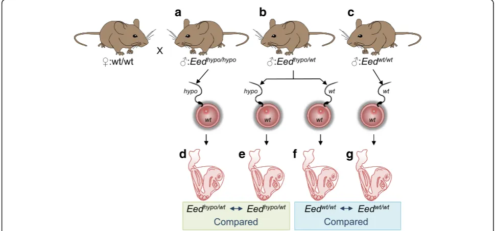
content, mating of these males with normal wild-type females would allow the detection of paternally transmit-ted epigenetic effects in the absence of any confounding maternal contributions. Critically, comparison of
off-spring with the sameEedhypo/wtgenotype produced from
Eedhypo/hypo and Eedhypo/wt fathers would reveal differ-ences in gene expression due to altered epigenetic
pat-terning in the sperm (Fig. 3). Similarly, epigenetic
differences could also exist in sperm produced byEedwt/ wt
andEedhypo/wtmales due to reduced EED function in the germline ofEedhypo/wtmales.
To test this model, independentEedhypo/hypo,Eedhypo/wt and Eedwt/wtmales were mated with Eedwt/wt females of the same background that had never been exposed to
a
d
e
f
g
b
c
Fig. 3Breeding and experimental plan to assess epigenetic inheritance. Wild-type (wt/wt) females that had never been exposed to theEedhypomorphic mutation were mated to eitherahomozygous (Eedhypo/hypo),bheterozygous (Eedhypo/wt) males (n =3 littermate pairs for each genotype) orcwild-type
(Eedwt/wt,n =2) males. Sperm in heterozygous and wild-type males develop with at least one fully functional copy ofEed(bandc), while sperm in
the Eed mutation (Fig. 3). Eedhypo/hypo and Eedhypo/wt brothers produced from three independent mating pairs
were used to sire Eedhypo/wt embryos. Precisely staged
and size matched E8.5 heterozygous and wild-type pro-geny were collected from each cross and photographed. Whole genome gene expression profiles of heterozygous progeny from Eedhypo/hypo andEedhypo/wtmales were de-termined using RNA-seq at a depth of ~ 40 million reads
per sample (n= 4 offspring from each group; 40.75±7
and 51.3±13 million reads per sample for Eedhypo/hypo or threeEedhypo/wtsires, respectively; Fig.4a).
Comparison of gene expression patterns in E8.5Eedhypo/ wt
embryos sired by three Eedhypo/hypo or three Eedhypo/wt
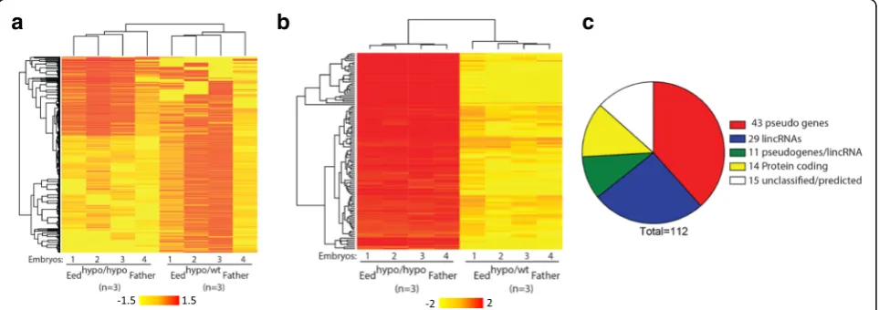
sibling males (Fig. 4a) identified 1986 differentially
expressed transcripts, representing at least 1851 genetically distinct genes separated by more than 5000 bp (P< 0.01, Benjamini-Hochberg false detection correction) (Fig.4b).
Of these genes, 779 exhibited greater than 1.5-fold
change in expression between Eedhypo/wt heterozygous
offspring produced by Eedhypo/hypo males and Eedhypo/wt males. These data demonstrated that there were tran-scriptional differences between Eedhypo/wt offspring that result from altered PRC2 function in the paternal germline.
To confirm these changes, Agilent 8x60K arrays were used to perform a technically independent assessment of gene expression in the same E8.5 embryos. A total of 234 differentially expressed transcripts, representing 128 distinct genes, were identified between the offspring from Eedhypo/hypo and Eedhypo/wt fathers (P< 0.01; ≥
2-fold change; Benjamini-Hochberg false detection
cor-rection) (Fig. 4c). Of these 128 genes, 112 were also
identified as differentially regulated in the RNA-seq ana-lysis (Additional file9: Table S8). Moreover, the direction of change (up- or downregulation) for the transcripts identified by array correlated with the RNA-seq analyses (R2= 0.87) (Additional file1: Figure S5A). Mapping ana-lyses revealed localization of these genes across all
auto-somes and the X chromosome (Additional file1: Figure
S5B). By contrast, comparison ofEedwt/wt embryos
pro-duced by Eedhypo/wt and Eedwt/wt fathers (n= 4 each) using Agilent 8x60K arrays did not identify any signifi-cant differentially expressed genes (Additional file 1: Figure S5C; cut-off: > 2-fold change and P< 0.01 with Benjamini-Hochberg false detection correction). This indicates that having at least one wild-type Eed allele is
sufficient to support normal paternal epigenetic
inheritance.
Gene ontology analysis of the 112 differentially expressed genes identified by the array and RNAseq ana-lyses revealed significant enrichment for processed retro-transposed pseudogenes (P< 4 × 10⁻⁷, Fisher Exact test) and lincRNAs (P< 0.05, Fisher Exact test) (Fig. 4d). Further examination using Retrofinder in UCSC (Retro-posed Genes V6, UCSC) identified 54 expressed retro-transposed sequences (pseudogenes and pseudogenes/
lincRNA; Fig. 4d). Typically, multiple independent
copies of the same parent gene were identified, indicat-ing that these pseudogenes are commonly regulated. In addition, GeneSpring analysis classified 40 probes as
c
a
b
Fig. 4Global transcription is altered in offspring ofEedhypo/hypomales.aHeatmap of 1986 differentially expressed transcripts (P< 0.01,≥1.5-fold change;
Benjamini-Hochberg false detection correction) detected using RNA-seq. Each column represents an RNA sample from an individual heterozygous embryo sired by either a homozygous (Eedhypo/hypo) or heterozygous (Eedhypo/wt) male. Three differentEedlittermate pairs (i.e.Eedhypo/hypoandEedhypo/wtbrothers)
were used to generate progeny for each genotype.bHeat map of 112 differently expressed genes detected using both Agilent 8x60K arrays and RNA-seq (P< 0.01;≥2-fold change; Benjamini-Hochberg false detection correction for arrays and RNAseq). Each column (1–4) represents an RNA sample from an individual heterozygous embryo sired by either anEedhypo/hypo or anEedhypo/wtmale.c Pie chart illustrating functional classification of
differentially regulated genes detected using micro-array and RNA-seq. Processed retrotransposed pseudogenes (P< 4 × 10⁻⁷, Fisher exact
test) and lincRNAs (P< 0.05, Fisher exact test) were significantly enriched in genes upregulated inEedhypo/wtprogeny of Eedhypo/hypo
lincRNAs, 11 of which were also classified as retrotran-sposed pseudogenes, consistent with the established abil-ity of pseudogenes to produce noncoding RNAs [46]. All of these retrotransposed genes and lincRNAs were
up-regulated in progeny of Eedhypo/hypo males compared to
progeny of Eedhypo/wt males (Fig. 4c), suggesting a pri-mary role for paternal EED in silencing these sequences in the offspring. With the exception of retrotransposed pseudogenes, no differences were detected in expression of other repetitive sequences, including LINE elements.
Paternal PRC2 alters preimplantation cleavage rates and cell cycle gene expression in offspring
To further investigate the role of paternal germline EED function in embryonic development, we analysed preim-plantation development. Zygotes produced byEedhypo/hypo andEedhypo/wtmales were cultured to blastocyst stage and
their development documented using automated
time-lapse photography of individual embryos. Embryos were imaged every 5 min facilitating measurement of cleavage rates and embryo development to blastocyst stage. Heterozygous embryos (n= 24) sired byEedhypo/hypo males underwent 2–4 cell cleavage ~ 3 h earlier than het-erozygous (n= 10,P= 0.0054) or wild-type embryos sired byEedhypo/wtmales (n= 12,P= 0.0240; Fig.5a). Consistent with this, time to develop from two-cell to eight-cell em-bryos was reduced compared to heterozygous and
wild-type embryos produced by Eedhypo/wt males. Time
from two-cell to blastocyst was also reduced in heterozy-gous embryos produced byEedhypo/hypomales but was not significantly different from wild-type embryos produced byEedhypo/wtmales (Fig.5a). Collectively, these data show
that preimplantation embryos from Eedhypo/hypo males
exhibit impaired development.
Consistent with this, RNA-seq analysis of eight-cell
embryos produced by Eedhypo/hypo and Eedwt/wt males
(n= 5 pools of ~ 10 embryos/sample; > 20 million
reads per sample; Fig. 5b, Additional file 1: Figure
S4D-F) revealed 157 transcripts with > 2-fold
increased expression and 109 transcripts with
de-creased expression in the offspring of Eedhypo/hypo
males (P< 0.01, Benjamini-Hochberg false detection
correction) (Fig. 5b; Additional file 10: Table S9 and
Additional file 11: Table S10). Examination of these
differentially expressed genes using GSEA identified KEGG_Cell_Cycle as the only significantly enriched pathway affected in the preimplantation progeny of Eedhypo/hypo and Eedwt/wt males (q= 0.00479). Included in this list were five genes, three of which regulate
DNA replication and cell cycle progression [Mad2l1
(11.8 fold down), Tdfp2 and Mcm3 (2.2-fold down)]
and two that regulate meiotic progression in oocytes [Pkmyt1 (4.7-fold up) and Sme1b (3.8-fold up)]
(Fig. 5b, Additional file 10: Table S9 and
Additional file 11: Table S10). In addition, six of the top 12 most highly upregulated genes in eight-cell embryos
produced by Eedhypo/hypo males were retrotransposed
pseudogenes. However, analysis using HOMER revealed no differences in the expression of LINE elements or other repetitive sequences.
To determine the impacts of depleting paternal EED on peri-natal development, post-natal day (PND) 5 offspring of Eedhypo/hypo and Eedhypo/wt males were weighed, mea-sured and fixed for histopathological analyses. PND5 pups
a
b
Fig. 5Preimplantation embryonic cleavage is advanced in offspring ofEedhypo/hypomales.aTiming of two- to four-cell, two- to eight-cell and
two-cell-blastocyst milestones in heterozygous (Eedhypo/wt, HET,n =23) offspring sired by homozygous (Eedhypo/hypo, HOM) males compared to
heterozygous (Eedhypo/wt, HET,n= 10) and wild-type (Eedwt/wt, WTn= 12) offspring sired by heterozygous (Eedhypo/wt) males mated to wild-type
produced by Eedhypo/hypo and Eedhypo/wt males (n= 2, for each genotype, each crossed with three wild-type females) were not significantly different in weight, crown-rump or nose-rump lengths (6–7 litters for each genotype, n= 56 and 48, respectively; litter sizes 7–9 pups, with one litter of 6 and 1 litter of 10) (Additional file 1: Figure S6). In addition, histopathological analyses examining 41 tissues were performed on PND5 male heterozygous offspring sired by three sets of sibling Eedhypo/hypo(n= 7 offspring) andEedhypo/wtmales (n= 6 offspring) and identified no ob-vious phenotypic differences.
Together, these data demonstrated that males lacking EED in the paternal germline produce offspring with altered transcriptional control of retrotransposed
pseu-dogenes and lincRNAs. Moreover, preimplantation
development was altered in these offspring, with more rapid cleavage and dysregulated control of cell cycle. Since retrotransposed sequences were also epigenetically dysregulated and over-expressed in the paternal germ-line, it is likely that reduced function of PRC2 in the pa-ternal germline explains the developmental differences inherited in the offspring ofEedhypo/hypomales.
Discussion
With the exception of DNA methylation, establishment of epigenetic information in the germline and its inherit-ance in the following generation is poorly understood. Recent studies in mice and humans have demonstrated differential enrichment of H3K27me3 at retained nucle-osomes in sperm, raising the possibility that PRC2 estab-lishes heritable epigenetic information that significantly affects paternal offspring [34–36]. Here we identify epi-genetic and transcriptional changes in the paternal germline ofEedhypo/hypo males during a key period of pa-ternal epigenetic programming. Moreover, offspring pro-duced by Eedhypo/hypo males were significantly different from offspring produced byEedhypo/wtcontrols at devel-opmental and molecular levels. Since these offspring were all heterozygous for theEedmutation, but were de-rived from sperm that developed with or without normal EED function, these observations provide prima facie evidence that PRC2 mediates epigenetic effects in the paternal germline that alter transcriptional and develop-mental outcomes in offspring. Consistent with roles for PRC2 in regulating intergenerational inheritance in Drosophila, C. elegans and Xenopus [47–51], our data support a role for PRC2 in regulating epigenetic inherit-ance in mammals. Moreover, a previous study
demon-strated that altered function of the H3K4me3
demethylase in sperm can mediate paternally transmit-ted transgenerational epigenetic inheritance in mice [6]. Together, these studies strongly indicate that epigenetic inheritance is influenced by histone-modifying enzymes in mammals.
Surprisingly, although survival and male fertility were
compromised in Eed hypomorphic animals, low EED
function did not significantly alter expression patterns of protein-coding genes in developing male foetal germ cells. Consistent with this, H3K27me3 was not depleted on protein-coding genes inEedhypo/hypo male foetal germ cells, perhaps explaining why developmental gene expression remained unaltered. Similarly, in a related study we reduced global H3K27me3 levels by 80% in male foetal germ cells using the anti-EZH2 drug, GSK126, to treat gonads cultured from E12.5 to E15.5, but no significant changes in transcription of coding genes were detected using expression arrays [40]. Com-bined, these studies indicate that despite enrichment of H3K27me3 on many developmental genes that are not expressed in germ cells [28], these genes appear to resist upregulation when EED or EZH2 function is compro-mised and/or H3K27me3 levels are reduced.
Although H3K27me3 enrichment and transcription of coding genes was unaffected in foetal germ cells ofEed -hypo/hypo
males, H3K27me3 was reduced on a substantial number of LINE, SINE and LTR elements. Moreover,
transcription of LINE elements was significantly
increased as a class inEedhypo/hypogerm cells, although it was not possible to identify individual TE sequences that were transcriptionally altered suggesting that variation in expression may occur at different LINE element loci on a cell to cell basis. Consistent with this, we observed
silencing of the Oct4GFP transgene in occasional
patches of germ cells in 60% of male Eedhypo/hypo
foe-tuses, indicating that activity of this transgene is subject to EED-sensitive stochastic cell to cell variation in male germ cells. Combining these observations, we propose that loss in H3K27me3 enrichment across LINE ele-ments in the male germ cells in Eedhypo/hypo mice leads to derepression of LINE elements, but this occurs in a stochastic pattern in individual cells. The impact of this across the cell population was manifest in significantly increased expression of LINE elements as a class across the cell population. Similar stochastic variation has pre-viously been demonstrated for epigenetic regulatory
mechanisms [52] and may be more pronounced in the
Eedhypo/hypo model than in global or tissue-specific complete loss of function (e.g.Eedknock out) models in which H3K27me3 is completely removed.
upregulated sequences in eight-cell progeny of Eedhypo/ hypo
males were also pseudogenes. Combined, these ob-servations provide evidence that PRC2 contributes to H3K27me3-mediated repression of LINE elements dur-ing epigenetic reprogrammdur-ing in the paternal germline.
Transposable elements constitute around 45% of the genome in mammals. Some of these elements retain potential transpositional activity and must be silenced to prevent their activation and random integration in the
genome [53, 54]. LINE elements encompass a group of
non-LTR retrotransposons which make up around 20% of the human genome and are common to many eukary-otes [55–57]. A subset of LINE elements still retains the ability for activity and random mutagenesis; hence, strict epigenetic silencing of these sequences is vital for
gen-ome integrity [58]. Retrotransposed pseudogenes and
retrotransposable elements are created by reverse tran-scription of processed or unprocessed mRNAs, followed by integration of these sequences back into the genome. These copies are typically imperfect in that they differ from the parent gene and accumulate mutations over
time [46, 54, 59]. Our data indicate that PRC2/
H3K27me3 makes an important contribution to silen-cing these classes of retrotransposable sequences, both in the developing germline and in the paternal progeny. While repressing retrotransposed elements is essential to prevent their mobilisation and random integration into the genome, the requirement for repressing processed pseudogenes is perhaps less obvious as they typically lack their own promoter and the ability to independently retrotranspose. However, transcribed pseudogenes can produce biologically active noncoding RNAs or proteins that have the capacity to alter cell development and function in the host organism [46,59]. Silencing or cor-rect transcriptional regulation of retroduplicated se-quences is therefore likely to be important to preserve genome function and correct biological processes.
Several lines of evidence indicate that histone modifi-cations are important for regulating repetitive sequence in the paternal germline. Nucleosomes are retained in
repetitive sequences in sperm [37], H3K9me3 and
H3K27me3 mark LTRs and LINE elements in the foetal
germline [26, 29, 30], and the H3K9me3 methylase,
SETDB1, is required for repression of a number retro-viral elements, including some, but not all, LINE
ele-ments [26]. SETDB1 is also required to regulate
inherited effects, apparently mediated through DNA methylation [60]. Moreover, LINE elements play a role in pseudogene retrotransposition [61]. Together, these obser-vations indicate a functional link between H3K27me3 in the paternal germline and deregulation of retrotransposed pseudogenes in the offspring of PRC2 mutant males, although the mechanism through which this operates remains obscure. However, we cannot exclude the
possibility that the effects mediated through PRC2 and H3K27me3 are indirect, involving other mechanisms such as altered DNA methylation or inheritance of RNAs that mediate effects in offspring.
Eedhypo/hypo males produced heterozygous offspring that progressed through the two- to four-cell cleavage stage significantly earlier than heterozygous controls or wild-type offspring sired by Eedhypo/wtmales. Consistent with this, cell cycle genes were dysregulated in eight-cell offspring of Eedhypo/hypo males. Most notably, Mad2l1, which inhibits cell cycle progression, was decreased
11-fold and in heterozygous progeny of Eedhypo/hypo
males compared to progeny of wild-type males, indicat-ing that this gene may regulate the advanced cleavage
rate in Eedhypo/hypo progeny. However, the roles of
Mad2l1 and other cell cycle genes identified here have not been established in preimplantation embryo cleavage and further work is required to ascertain their functional roles in this process.
Interestingly, germline de novo mutations in either
EED orEZH2result in Weaver syndrome, characterised
by growth and congenital defects and cognitive deficit in affected humans [62–65]. The maternal/paternal inherit-ance pattern in Weaver syndrome is poorly understood, although there is some evidence that mutations occur in either the maternal or paternal allele in the germline suggesting that disruption of PRC2 function in either sperm to oocytes may contribute to Weaver syndrome. In this study, partial loss of EED function in the paternal germline was sufficient to mediate significant, though relatively subtle changes in epigenetic and transcrip-tional regulation in paternal offspring, but not the spectrum of phenotypic characteristics observed in Weaver syndrome patients. Whether greater loss of PRC2 function in male germ cells or in the maternal germline will lead to increased Weaver-like phenotypic changes in mice is yet to be determined. However, loss of EZH2 function in oocytes led to decreased birth weight in mice, rather than increased birth weight typic-ally observed in Weaver syndrome [66].
Surprisingly, despite significant dysregulation of
H3K27me3 enrichment on TEs and decreased male fertility, we observed a substantial number of TEs at
which H3K27me3 was increased in Eed hypomorphic
Conclusions
The current study was designed to determine whether EED regulates epigenetic patterning in the paternal germline that subsequently alters outcomes in offspring. This appears to be the case as both regulation of tran-scription and preimplantation development were altered in offspring of males with reduced EED function. As existing evidence indicates that EED function is restricted to the establishment of H3K27me3 through PRC2, the simplest interpretation of our data is that PRC2 alters epigenetic patterns in sperm that are manifest in offspring. Reduced PRC2 may have altered the establishment of other epigenetic information to compensate for the change in H3K27me3. This could include changes in RNA content in the sperm that could alter gene expression and embryo development
[67, 68]. However, ultimately these changes would
occur as a consequence to the original alteration of H3K27me3 due to the reduced function of EED and PRC2. Therefore, this study provides the first functional evidence that the highly conserved histone-modifying complex, PRC2, mediates paternal transmission of inher-ited effects in mammals. This complements recent evi-dence that histone modifications play essential roles in regulating inherited disease [60, 69], and emphasises the importance of understanding mechanisms that regulate transmission of epigenetic information through the germ-line inheritance.
Methods Mice
Eed hypomorphic (Eedhypo/hypo) mice were generated by
inter-crossingc57bl/6:129T2SvJ Eedl7Rn5-1989SB.Oct4GFP
heterozygous (Eedwt/hypo) mice. Eedl7Rn5-1989SB C57bl6/ 129 mice were maintained under a light-dark cycle in a temperature and humidity-controlled specifically pathogen-free (SPF) facility with access to food and water ad libitum.
Embryo collection and staging
Animals were time mated and females were inspected for plugs each morning to ensure successful mating. Em-bryos were collected at fertilisation, 8.5, 12.5 and 15.5 days after the female was plugged.
Zygote to blastocyst development was monitored as previously described [70]. Briefly, embryos collected at fertilisation were kept warm in G-MOPS medium during transfer to the embryo culture facility before washing twice through 50/50 G1/G2 embryo culture media and
transferred into 2 μl drops of G1/G2 media under oil.
Embryos were cultured individually in 6% CO2, 5% O2
and 89% N2 for 96 h in an incubator (Sanyo MCO 5)
equipped with a Primo Vision (Vitrolife, Sweden) Time Lapse Embryo monitoring system allowing morpho-kinetic
analysis of embryo development. Morpho-kinetic
development of each embryo was documented using time-lapse photography, with images collected every 10 min for the zygote-blastocyst developmental period. Em-bryo morphology and cleavage times between zygote to two-cell, two-cell to four-cell, two-cell to eight-cell, and two-cell embryos to blastocyst were documented within Primo Vision and statistically analysed using GraphPad Prism. After culture, embryos were collected and individu-ally snap frozen for genotyping.
To identify differences in transcriptional control in
off-spring of Eedhypo/hypo males, E8.5 embryos were
pro-duced by Eedhypo/hypo, Eedhypo/wt and Eedwt/wt males mated to wild-type females. Embryos were dissected at E8.5, the physical appearance of each embryo was docu-mented and each embryo was photographed before snap freezing in liquid nitrogen. Photographs and notes were later compared to accurately match samples of the same developmental time points and facilitate accurate gene expression analysis and comparison to controls. All E8.5
embryo samples were kept at−80 °C until RNA
extrac-tion. E12.5 and E15.5 embryos were examined on collec-tion to ensure they were consistent with E12.5 and E15.5 developmental stage and gonads were dissected. Gonads were fixed for immunofluorescent analysis, or were dis-sociated and prepared for FACS purification of germ cells.
Genotyping
The Eed1989 T > A point mutation was detected in em-bryos by reverse transcribing RNA using SuperScript® III
Reverse Transcriptase Kit (Life Technologies # 18080–
051). Samples were PCR amplified using Eed-specific
primers (forward: 5′- TCACAGGGGGAGATACGGT
TATT and reverse: 5′-CTGACAGGAGAAGGTTTGG
GTCT) cleaned using ExoSAP-IT (Affymetrix, 78250) and the cDNA subjected to Sanger sequencing at the MHTP Medical Genomics Facility. Resulting sequences were assessed using FinchTV Geospiza software.
Fertility testing
A controlled breeding experiment was performed to
determine the fertility of the male Eed hypomorphic
Bartlett’s F-test to compare variances for the three groups and a non-parametric Mann-Whitney test to determine statistical significance. A chi-square test was used to statistically assess differences in the occurrence of successful pregnancies between males grouped by geno-type. Fertility was assessed in 13 Eedhypo/hypo males (n= 13) along with age-matchedEedhypo/wt(n= 13) andEedwt/ wt
(n= 10) brothers.
Histology
Testes from Eedhypo/hypo (n= 21), Eedhypo/wt(n= 23) and Eedwt/wt (n= 19) males were processed for histology. Each testis was weighed and the testis capsule nicked,
immersed in Bouin’s fixative overnight, washed three
times in 70% ethanol (vol/vol), processed into paraffin wax and stained with periodic acid Schiff (PAS) re-agent and haematoxylin. Assessment of testis hist-ology was carried out to determine the presence, or absence, of all germ types and their morphological in-tegrity in comparison with wild-type mice of the same age. Initially, one testis was snap frozen and stored at
−80 for sperm count and hormone assessment while
the other testis was used for histological analysis. However, after the observation that there was no dif-ference in DSP, all future gonads were fixed for hist-ology assessment.
Daily sperm production
Frozen testes from Eedhypo/hypo (n= 5), Eedhypo/wt (n= 6) andEedwt/wt(n= 3) were allowed to thaw at RT, weighed before a fragment was removed, weighed, decapsulated
and homogenised in 600μl of SMT solution. Ten
micro-litres of homogenate was placed on each side of the haemocytometer. The average number of sperm heads was calculated from counting 80 small squares on both sides of the haemocytometer. Daily sperm counts were calculated as previously reported [72, 73]. Briefly, the volume of homogenate, weight of the sample fragment and total weight of the testis were used to calculate the total number of spermatids per testis. As developing
spermatids spend 4.84 days in steps 14–16 during
spermatogenesis, the values for the number of sperma-tids per testis were divided by 4.84 to obtain daily sperm production. Statistical significance was determined using
one-way ANOVA with Tukey’s multiple comparison,
withP <0.05 considered significant.
Immunofluorescence
Embryos were harvested at E12.5 and E14.5, sexed based
on gonad morphology or via PCR [74]. Foetal gonads
were isolated and fixed at RT in PBS containing 4% paraformaldehyde for 20 or 75 min respectively. Gonads were washed three times in PBS and cryoprotected in 30% sucrose in PBS overnight and mounted in optimal
cutting temperature (OCT). Cryosections were cut at
8 μm, permeabilised with 1% Triton-X and non-specific
staining blocked with 5% BSA. Immunofluorescence
staining was carried out as described [75, 76]. EED
(R&D Technologies, AF5827, diluted 1/100), EZH2 (Cell Signalling Technology D2C9, diluted 1/400), SUZ12 (Cell Signalling Technology D39F6, diluted 1/100), H3K27me3 (Cell Signalling Technologies, C36B11, di-luted 1/400), OCT4 (Santa Cruz sc8628, didi-luted 1/500), DPPA4 (R&D Systems AF3730, diluted 1/400) and MVH/DDX4 (Cell Signalling Technology #8761, diluted 1/300) primary antibodies were each diluted in PBS con-taining 1% BSA incubated for 1 h at RT. Donkey anti-goat, sheep or rabbit Alexa-594 (Life Technologies) secondary antibodies were used at 1/500 dilution, while eGFP fluorescence was detected directly in the 488-nm channel. To assess non-specific staining, additional sections were analysed using secondary antibody only controls. Images were obtained using a Nikon® C1 con-focal microscope. Images were visually analysed using ImageJ (version: 2.0.0-rc-19/1.49m). All IF experiments were replicated using three to five pairs of gonads per genotype.
Flow cytometry
Pregnant mothers were injected intraperitoneally (i.p.) with 20 mg/kg 5-ethynyl-2-deoxyuridine (EdU) to facilitate in vivo analysis of gonadal cell proliferation. Flow cytometry was performed as previously de-scribed [40, 77, 78]. Dissociated gonadal cells were stained using antibodies specific for SOX9 and AMH allowing identification of Sertoli cells and SOX9 (Millipore AB5535, diluted 1/300) and AMH (Santa Cruz sc-6886, diluted 1/200) staining intensity in the Sertoli cell population. Cell cycle was measured in Eedwt/wt, Eedwt/hypo and Eedhypo/hypo samples as
previ-ously described [77, 78]. Germ cells were identified
using an antibody specific for MVH (R&D Systems, AF2030 diluted 1/100; [77, 78]).
Fluorescence-activated cell sorting
Fluorescence activated cell sorting was performed essen-tially as described [42, 75, 76]. Foetal gonads were col-lected at E15.5 and sexed based on gonadal morphology. Male gonad pairs were dissociated to single cells in 0.25% trypsin containing EDTA. Trypsin activity was
blocked by adding 500 μl DMEM containing 10% FBS.
The cells were filtered through an 80-μm nylon mesh
(BD Biosciences), pelleted and resuspended in 300 μl
cells (> 95%) were collected. Germ cells were pelleted and either fixed for ChIP or snap frozen for RNA. Each gonad pair yielded approximately 20,000 germ cells, and similar proportions of GFP-positive germ cells were iso-lated from Eedwt/wt, Eedwt/hypo and Eedhypo/hypo foetuses (Fig. 1h). We were unable to isolate anyOct4 GFP-nega-tive germ cells.
RNA extraction and quality assessment
RNA was extracted from E8.5 embryos using the Genelute Mammalian total RNA miniprep kit (Sigma,
RTN70-1KT), DNAse treated using TURBO
DNA-free™ Kit (Ambion, AM1907) and purified from
the DNAse reaction using Agencourt RNAClean XP (Beckman coulter, A63987). RNA was extracted from pools of eight to ten carefully staged eight-cell embryos, with each pool representing separate litters using Agencourt RNAClean XP (Beckman coulter, A63987) chemistry and the RNA DNAase treated on
the beads, before elution, freezing and storage at −
80 °C. Isolation of E15.5 germ cells was performed by FACS purifying approximately 15,000 germ cells from each pair of E15.5 foetal testes. RNA was extracted from the germ cells using the RNeasy Micro Kit (Qia-gen) including an on column DNAse step. For all samples isolated from E8.5 embryos, E15.5 germ cells and eight-cell embryos, RNA quantity and quality were measured using a Qubit® RNA HS Assay Kit and the Qubit® 2.0 Fluorometer (Life Technologies, Q32866) and an Agilent 2100 Bioanalyzer (Agilent Technologies, Santa Clara, CA). Only samples with a RIN score above 8 were used for RNA sequencing and microarray analysis.
Quantitative real-time RT-PCR (qRT-PCR) using Fluidigm biomark
RNA was extracted form 50-200K FACS purified E15.5 testis somatic cells or from whole E12.5 gonads using 600 ml of Trizol in 2Ml heavy phase-lock tubes. DNAsed using Ambion Turbo kit and cDNA synthesised using super script III (Invitrogen) kit. Gene expression was analysed using real-time quantitative polymerase chain reaction with BioMark HD technology (Fluidigm) 96.96 Dynamic Array IFCs (Fluidigm). The geometric CT mean of reference genes Canx, Sdha, and Mapk1 was used to calculate the relative gene expression using the delta-delta CT method. We have shown previously
that Canx, Sdha, and Mapk1 are expressed at stable
levels in E12.5–E15.5 foetal germ cells and somatic cells
[75]. Statistical significance was determined using
one-way ANOVA with Tukey’s multiple comparison test
for Eedhypo/hypo (E12.5 n= 7, E15.5 n= 14), Eedhypo/wt
(E12.5 n = 7, E15.5 n= 12) and Eedwy/wt (E12.5 n= 5,
E15.5 n= 11). Where variances were unequal, a
non-parametric test was used (Mann-Whitney). A P
value of less than 0.05 was considered significant.
RNA sequencing
RNA sequencing libraries were constructed from 50 ng, 25 ng or ~ 2 ng of total RNA isolated from E8.5 em-bryos, E15.5 germ cells or eight-cell emem-bryos, respect-ively. RNA sequencing libraries were constructed using Nugen Mondrian SPIA Library preparation using Nugen protocol M01335v2 after ribosomal depletion of total RNA using the RiboZero kit (Illumina, RS-122-2201). RNA sequencing was performed on an Illumina HiSeq 1500 instrument with condition sequenced in four bio-logical replicates, derived from≥3 independent sires (for E8.5 and eight-cell embryos) or from four embryos from multiple litters (E15.5 germ cells). Quality filtering and adaptor removal was performed using the Trimmomatic
software tool with default trimming parameters [79].
Reads were mapped to the mouse genome, version
mm10, using TopHat [80] and genes identified and
com-pared to Ensembl using the RNA-eXpress analysis tool [81]. Replicate quality was assessed by applying multiple dimensional scaling plots, and differential expression analysis was performed using the voom-limma analysis
workflow applying empirical Bayes F-test [82].
Tran-script enrichment was performed based on HOMER--assembled annotations with statistical significance of
enrichment assessed by Fisher exact test with
Benjamini-Hochberg false detection correction.
Pseudogene classification was performed using Retro-Finder program (Retroposed Genes V6, UCSC). Data was visualised using the ggplot package within the R analysis environment. Gene set enrichment analysis was
performed using MSigDB collections [83] with
enrich-ment assessed by Fisher exact test with
Benjamini-Hochberg. Sequence data is available
through the European Nucleotide Archive (ENA) acces-sion numbers ERP106776 (E15.5 germ cell data), ERP010195 (8.5-day embryo data) and ERP013725 (eight-cell embryo data).
Expression microarray analysis
filtered to exclude the lowest 20% of probes and those not expressed in at least four of the eight (Eedwt/hypovsEedwt/ hypo
offspring—array 1) or eight (Eedwt/wt vs Eedwt/wt
off-spring—array 2) samples. Differential expression was
reported where twofold change was observed applying a 0.01 FDR after Benjamini-Hochberg multiple testing cor-rection. Data was visualised in a hierarchically clustered heat map within the GeneSpring software package. Raw data is available through the NCBI Gene Expression Omni-bus GSE68213 (composed of GSE68212 and GSE68211).
Chromatin immunoprecipitation (ChIP)
ChIP was performed on approximately 20,000 FACS puri-fied germ cells from individual Eedwt/wt (n= 4) and Eed -hypo/hypo
(n = 4) embryos using low input ChIP [84, 85] with minor alterations. Briefly, cells were cross-linked in 1% formaldehyde/PBS for 5 min before adding glycine/ PBS at a final concentration of 125 mM. Cells were pel-leted and washed in PBS and stored at−80 °C. Dynabeads
were washed in RIPA buffer and bound to 2.4 μg of
H3K27me3 rabbit polyclonal (Millipore cat# 07–449,
1 mg/ml) or IgG rabbit CST (cat#3900S, 2.5 mg/ml) by in-cubating for a minimum of 2 h at 4 °C at 40 rpm.
Cells were thawed on ice and lysed in 13 μl of lysis
buffer for 3 min, vortexed for two 5-s bursts at room
temperature and 117μl modified RIPA buffer containing
Tris-HCl pH 7.5 EGTA, Triton X-100 0.1% SDS. Sonication conditions were pre-optimised by assessing germ cell-derived sonicated chromatin samples on a gel and via bioanalyser High Sensitivity DNA Analysis Kits (Agilent Technologies) to ensure that the majority of
chromatin fragments yielded were within a 300–100-bp
range. Germ cell chromatin was sonicated for 10 min at peak power 105, duty factor 2.0, cycles/burst 200 using a Covaris s220 instrument. Sample was recovered and 5 M NaCl added to a final concentration of 140 mM (equiva-lent to RIPA buffer). A 10μl aliquot of each sample was removed for an input control and the remaining chromatin solution immediately transferred to the antibody-bead complexes. The chromatin was incubated with the antibody-bead suspension at 4 °C rotating at 40 rpm for 2 h before washing in cold RIPA buffer and resuspending TE. To reverse crosslink, chromatin-bead suspensions and input controls were resuspended in elution buffer containing freshly added SDS and Protein-ase K and incubated for 2 h at 68 °C 13,000 rpm. DNA was purified for sequencing using Agencourt Ampure XP beads (Beckman Coulter).
H3K27me3 rabbit polyclonal (Millipore cat# 07-449, 1 mg/ml) antibody specificity was validated using Ac-tive Motif Modified Histone Array analysis (Catalogue
number 13001) according to the array manufacturer’s
protocol, with the exception that dilution of the pri-mary antibody (1/2000) was as recommended by
Millipore for western blotting. Mouse anti Rabbit HRP secondary antibody was used at the recom-mended dilution (1/2500). Imaging of the array was performed using incremental accumulating exposures to ensure data was collected in the linear detection range. Data analysis was performed using Array Ana-lyse Software (Active Motif ) and the specificity factor determined for the antibody with reference to 384 histone peptide variants on the array (Additional file1: Figure S7).
ChIP sequencing and analysis
ChIP-Seq Libraries were prepared from using the
Ova-tion Ultralow System V2 using Nugen protocol
M01379v1. Each library was quantitated using a Qubit instrument and the DNA size profile determined using an Agilent Bioanalyzer. Libraries were finally quantitated by qPCR, pooled in an equimolar ratio and all eight libraries run on one lane of a HiSeq1500 sequencer to obtain 50-bp single end reads. Raw reads were bioinfor-matically separated into individual libraries and trimmed
using the trimmomatic tool [79] before mapping.
Sequences were mapped to the mouse genome (mm10) using bowtie2, resulting in over 95% quality mappable reads. PCR duplicates were marked in the mapping files (BAM format) for filtering in further steps, ensuring use of unique reads during peak identification. Peak identifi-cation and differential peak identifiidentifi-cation was performed
using HOMER (http://homer.salk.edu/homer/).
Differential peaks were annotated using HOMER, allowing all peaks to be assigned to specific sequence classes. Genomic sequence classes outside repeats were analysed for over-, under- or expected representation in
sequences differentially enriched for H3K27me3 in Eed
-hypo/hypo
germ cells compared to Eedwt/wt germ cells.
Initially, the expected number of peaks was calculated by determining the proportion of the genome covered by each sequence class (e.g. for intergenic sequences 813,929,088 bp) divided by the total annotated genome size (i.e. 813,929,088/2,631,564,759 = 30.93%). The num-ber of peaks in each sequence class (e.g. for
inter-genic sequences with increased H3K27me3 in Eedhypo/
hypo
NCBI Gene Expression Omnibus accession number GSE110529.
Representation of repeat sequences was determined by determining the numbers of repeat sequences anno-tated in each sequence category compared to the total number of repeats in the genome to provide the ex-pected representation of repeats in each repeat category annotated in UCSC repeat masker. A hypergeometric analysis was performed to determine representation of each repeat category in all H3K27me3 peaks identified inEedwt/wtandEedhypo/hypogerm cells and in peaks that
had altered H3K27me3 in Eedwt/wt compared to Eed
-hypo/hypo
germ cells, and vice versa.
ChIP data from the current study was compared
with that reported by Mu et al. [32] and Ng et al.
[29] in which H3K27me3 peaks were identified using
MACS. Polycomb target genes from Boyer et al. [86]
were transposed to the current mm10 nomenclature and H3K27me3 peaks mapped in the genic and upstream regions of these Polycomb target genes in
the current study and studies from Mu et al. [32] and
Ng et al. [29].
Phenotypic analyses of offspring
Two Eed hypomorphic males from each genotype
(Eedhypo/hypo and Eedhypo/wt) were each bred with three C57Bl/6J females, to obtain seven and six litters,
re-spectively. Offspring from Eedhypo/hypo (n= 56) and
Eedhypo/wt (n= 48) fathers were weighed at post-natal day 5; crown-rump and nose-rump measurements
were obtained. Offspring from Eedhypo/wt fathers were
genotyped via Transnetyx to analyse wild-type and heterozygous groups. All litters analysed consisted of
six to ten pups. One-way ANOVA plus Tukey’s post
hoc multiple comparisons test was used to statistically analyse the data. A comprehensive phenotypic analysis was also performed using the Australian Phenomics Network Histopathology and Organ Pathology Service.
Neonates were weighed and crown-rump and
nose-rump lengths collected. The neonates were
de-capitated, injected with Bouin’s fixative into the
thorax and abdomen as well as immersion fixation for 48 h, washed and stored in 70% EtOH. Analysis included evaluation of all thoracic and abdominal organs, skeletal tissue, nasal/oral region, brain, eyes and auditory/vestibular apparatus. Reports were gen-erated for each neonate, including detailed histo-pathological and neuropathology descriptions.
Additional files
Additional file 1:Figure S1.Survival and fertility ofEedhypo/hypoanimals.
Figure S2.Foetal somatic and germ cells reached expected developmental milestones and expressed endogenous germ cell markers inEedhypo/hypo
males.Figure S3.H3K27me3 was enriched at known PRC2 target genes in
Eedhypo/hypocompared toEedwt/wtgerm cells.Figure S4.Read counts and technical consistency between RNAseq samples generated from (A–B) E15.5
Eedwt/wtandEedhypo/hypomale germ cells and (C) D8-cell offspring from
Eedwt/wtandEedhypo/hypomales.Figure S5.Transcriptional analyses of E8.5
offspring produced byEedhypo/hypo,Eedhypo/wtandEedwt/wtmale mice mated to wild-type females.Figure S6.Neonatal weight and size are not different in offspring ofEedhypo/wtEedhypo/hypoand males.Figure S7.Active Motif
Modified Histone Array analysis of H3K27me3 ChIP antibody. (PDF 15192 kb)
Additional file 2:Table S1.Eedwt/wtgerm cell H3K27me3 ChIP-seq
peaks. (XLSX 6770 kb)
Additional file 3:Table S2.Eedhypo/hypogerm cell H3K27me3 ChIP-seq
peaks. (XLSX 8636 kb)
Additional file 4:Table S3.Comparison of known PRC target genes in
ES cells with H3K27me3 peaks in foetal germ cells, ES cells and spermatocytes. (XLSX 1161 kb)
Additional file 5:Table S4.Relative representation of annotated repeat categories in H3K27me3 peaks inEedwt/wtandEedhypo/hypogerm cells.
(XLSX 1124 kb)
Additional file 6:Table S5.ChIP-seq data from E15.5 maleEedhypo/hypo
compared toEedwt/wtgerm cells: Peaks with decreased H3K27me3 inEed -hypo/hypogerm cells. (XLSX 70 kb)
Additional file 7:Table S6.ChIP-seq data from E15.5 maleEedhypo/hypo
compared toEedwt/wtgerm cells: Peaks with increased H3K27me3 inEed
-hypo/hypogerm cells. (XLSX 83 kb)
Additional file 8:Table S7.ChIP-seq data showing regions with decreased H3K27me3 inEedhypo/hypocomparedEedwt/wtto germ cells identified using EdgeR. (XLSX 11 kb)
Additional file 9:Table S8.RNA-seq and expression microarray data
showing genes differentially expressed in heterozygous E8.5 day embryos sired byEedhypo/hypofathers compared to E8.5-day embryos sired by
Eedhypo/wtfathers. (XLSX 22 kb)
Additional file 10:Table S9.RNA-seq data showing genes significantly
downregulated in eight-cell embryos sired byEedhypo/hypofathers.
(XLSX 20 kb)
Additional file 11:Table S10.RNA-seq data showing genes significantly
upregulated in eight-cell embryos sired byEedhypo/hypofathers. (XLSX 23 kb)
Abbreviations
AMH:Anti-Mullerian hormone; ChIP-seq: Chromatin immunoprecipitation followed by genome wide sequencing; DPPA4: Developmental Pluripotency Associated gene 4; DSP: Daily sperm count; E: Embryonic day; EED: Embryonic ectoderm development; eGFP: Enhanced Green Fluorescent protein; EZH1: Enhancer of Zeste 1; EZH2: Enhancer of Zeste 2; FACS: Fluorescence-activated cell sorting; H3K27me3: Trimethylated lysine 27 on histone 3; lincRNA: Long non-coding RNA; LINE: Long Interspersed Nuclear Element; LTR: Long Terminal Repeat; Mad2l1: Mitotic Arrest Deficient 2 Like 1; Mcm3: Minichromosome Maintenance Complex Component 3; MVH: Mouse Vasa Homologue (also known as DDX4);Nsd: No significant difference; OCT4: Octamer Binding Transcription Factor 4 (also known as
Pou5f1); Pkmyt1: Protein Kinase Membrane Associated Tyrosine/Threonine 1; PND: Post-natal day; PRC2: Polycomb Repressive Complex 2; RNA-seq: Genome-wide RNA sequencing; Sdha: Succinate Dehydrogenase Complex, subunit A; SINE: Short Interspersed Nuclear Element; SOX9: SRY Box Containing Gene 9; SUZ12: Suppressor of Zeste 12; TEs: Transposable elements
Acknowledgements
The authors thank Jodee Gould, Trevor Wilson, Vivien Vasic and Denis Cleven at the MHTP Medical Genomics Facility for their help processing samples and Nicolle Gibson, Laura Deans and other staff of the Monash Medical Centre Animal Facilities (MMCAF). Thank you also to Sanna Barrand for contributions to ChIP-seq development, Roxanne Legaie form the Monash Bioinformatics Platform for initial analysis of ChIP-seq data, Dr. Marnie Blewitt for sharing theEedl7Rn5-1989SBmouse strain, and Dr. Kirsten Hogg, and other