_____________________________________________________________________________________________________ *Corresponding author: E-mail: [email protected];
Technology
5(1): 1-7, 2019; Article no.AJB2T.48254 ISSN:2457-0125
Effects of Ranitidine (Zantac) Drug on the Hormonal
Level, Sperm Head Abnormality and
Histo-Architecture of the Testis of Albino Male Mice
Tiba Soud Oraibi
1, Ruqaya Mohammed Ibrahim
1*and Sura Alaa Soud
21
Al-nahrain University,Baghdad, Iraq.
2AlKarkh University of Science, Baghdad, Iraq.
Author’s contribution
This work was carried out in collaboration among all authors. Author TSO designed the study, performed the statistical analysis, wrote the protocol, and wrote the first draft of the manuscript. Authors RMI and SAS managed the analysis of the study. All authors read and approved the final manuscript.
Article Information
DOI: 10.9734/AJB2T/2019/v5i130049 Editor(s): (1) Dr. Manpreet Kaur, Department of Microbiology, Kurukshetra University, Kurukshetra, India. Reviewers: (1) Michael Guarnieri, Johns Hopkins School of Medicine, USA. (2)Azab Elsayed azab, Sabratha University, Libya. Complete Peer review History:http://www.sdiarticle3.com/review-history/48254
Received 10 January 2019 Accepted 23 March 2019 Published 30 March 2019
ABSTRACT
The purposes of this study are to determine the effect of Zantac (Ranitidine) drug on sexual hormones (Testosterone and Prolactin), sperm head abnormality, and histopathological activity on albino male mice testes.
Two doses of the drug were used: 1000 and 2000 mg/kg, in addition to a negative control group. Each group included four mice and the drug administrated orally as (0.1 ml) per day for 14 days, and then the mice were sacrificed on the day 15 for laboratory assessment.
The result showed that the drug cause increase the percentage of sperm head abnormality which reaches to (48.3%) and (22.8%) for 2000 mg/kg and 1000 mg/kg respectively in comparison to control group (11.1%). The male sex hormones also affected by the drug and the level of testosterone hormone decrease to (1.07 ng/ml) in 2000 mg/kg and (3.42 ng/ml) in 1000 mg/kg, while the level of hormone in control group is (14.07 ng/ml). The prolactin hormone show increase in the level at dose 2000mg/kg it's (115 ng/ml) and in dose 1000 mg/kg the level its (43 ng/ml)
Oraibi et al.; AJB2T, 5(1): 1-7, 2019; Article no.AJB2T.48254
these value very high compared with control group (36 ng/ml) due to the effect of the drug on the hormone. The histopathological examination shows damage in the seminal duct and changes in the wall of the seminiferous tube also the spermatogenesis well be affected by the drug in which is stop and some of seminal duct show no appearance of spermatogenesis and also cause depletion to the wall of the seminal duct.
Keywords: Zantac; sperm head abnormality; testosterone; prolactin; albino mice.
1. INTRODUCTION
A drug is any substance (other than food that provides nutritional support) that when inhaled, injected, smoked, consumed absorbed via a patch on the skin or dissolved under the tongue causes a temporary physiological (and often psychological) change in the body [1].
In pharmacology, a pharmaceutical drug, also called medication or medicine, is a chemical substance used to cure, prevent or to promote well-being [2].
Ranitidine is a member of the class of histamine H2-receptor antagonists with antacid activity. Ranitidine is a competitive and reversible inhibitor of the action of histamine, released by
enterochromaffin-like (ECL) cells, at the
histamine H2-receptors on parietal cells in the stomach, thereby inhibiting the normal and meal-stimulated secretion of stomach acid [3].
It is commonly used in the treatment of peptic ulcer disease, gastroesophageal reflux disease and Zollinger–Ellison syndrome [4].
The reproductive system is considered to be particularly vulnerable to the effects of drugs. Hypothalamic neurotransmitters are sensitive to drugs that alter their synthesis, release, or action; disruption of these pathways changes the levels or patterns of secretion of the pituitary gonadotropins. Drugs also exert a direct action on the gonadal functions of androgen production and spermatogenesis. Drugs that decrease testosterone levels produce a multitude of effects on male reproduction. Neuropharmacologic agents that either inhibit central nervous system (CNS) activities (e.g., analgesics, anesthetics, sedatives, tranquilizers) or stimulate CNS
activities (e.g., antidepressants, stimulants,
hallucinogens) modify the hypothalamic-pituitary control of the gonadotropins and prolactin. The
changes in luteinizing hormone,
follicle-stimulating hormone, and prolactin result in changes in libido, erectile dysfunction, inability to ejaculate, testicular swelling and gynecomastia.
Narcotic drugs exert their primary effect on the hypothalamic-pituitary axis and their secondary effects on the gonads and sex accessory organs. Narcotics decrease gonadotropin secretion and stimulate prolactin secretion, both of which are inhibitory to male sexual function [5].
2. MATERIALS AND METHODS
2.1 Laboratory Animals
Albino Swiss male mice (Mus musculus) were the laboratory animals. They supplied by the Biotechnology Research Centre (Al-Nahrain University). Their age at the start of experiments was 8-10 weeks, and their weight was 23-27 gram. They divided into groups, and each group was kept in a separate plastic cage (details of these groups are given in the section of experimental design). The animals maintained at a temperature of 23 – 25°C, relative humidity range between 30-50% and they had free excess to food (standard pellets) and water (ad libitum).
2.2 Experimental Design
Twelve male mice are divided into three groups (n=4 each), the control group received normal saline while the experimental group received Ranitidine. All animals were orally treated once a day for 14 days.
2.3 Assessment of Testosterone Serum Level
Oraibi et al.; AJB2T, 5(1): 1-7, 2019; Article no.AJB2T.48254
wells followed by swirling the microplate gently for 20-30 seconds to max. After that, cover and incubate the microplate for 60 minutes at room temperature and discard the contents of the microplate and added 350 ul decant (tap and blot) or aspirate, Repeated two additional times for a total of three washes. Later on, 0.100 ml of working substrate solution was added to all wells and incubated at room temperature for (15) minutes. 0.050 ml of stop solution was added to each well. Finally, the absorbance was detected at 450 nm.
2.4 Assessment of Prolactin Serum Level
Serum level of Prolactin hormone is estimated using Prolactin hormone commercial kit (PRL) Test System (Monobind Inc. Lake forest CA 92630, USA. Accu Bind). The microplate wells were formatted for each serum reference, control and patient specimen and stored at 2-8°C. (0.025 ml) of the appropriate serum reference, control or specimen added to the microplate well, then, 0.100 ml of PRL-Enzyme Reagent solution was added to all wells and swirled gently for 20-30 seconds and incubated 60 minutes at room temperature. About 350 ul of wash buffer added for washing, after that, 0.100 ml of working substrate solution added to all wells and incubated at room temperature for fifteen (15) minutes. Finally, 0.050 ml of stop solution was added to each well and gently mixed for 15-20 seconds and read the absorbance in each well at 450 nm.
2.5 Sperm-Head Abnormality Assay
Sperm Head Abnormality (SHA) is detected as described by [6], with some modification. After animal scarification by cervical dislocation and dissected, caput and cauda epididymis removed and placed in a Petridish containing 1 ml of physiological normal saline, then minced and teased carefully to release the spermatozoa. After gentle pipetting a drop of 1% Eosin solution (10:1) is added to the suspension (2-3) drop on a clean slide and air dried for 30 min. The percentage of head abnormality is detected under light microscopy using 100X objective.
2.6 Preparation and Examination of
Histological Sections
The left testis and epididymis (caput and cauda) of each mouse is prepared for histopathological study as described by [7]. Samples were fixed in 10% formalin for 24 h, followed by dehydration with a gradual series of alcohol (30-100%) for (5)
min each. Then the samples cleared in two changes of xylene before embedded in paraffin wax for sectioning. Cross sections of (5) µm thickness are prepared and stained with hematoxylin (Harison) and eosin according to the standard method [7]. Histopathological changes are performed under a light microscope as compared to the control group.
2.7 Statistical Analysis
The statistical analysis is performed using
Minitab 16 (Minitab Ltd, Coventry, UK).
Differences among groups are determined by Student t-test. Data are expressed as mean and standard error and differences were considered significant at (p ≤ 0.05).
3. RESULTS AND DISCUSSION
3.1 Sperm Head Abnormality (SHA)
Different sperm-head abnormalities were
observed as a result of a treatment with the different doses of the Zantac drug, which was associated with a significant increase in sperm-head abnormalities in a dose dependent manner to reach 48.3% at 2000 mg/kg and 22.8% at dose 1000 mg/kg in comparison with negative control (11.1%), Figs. (1,2).
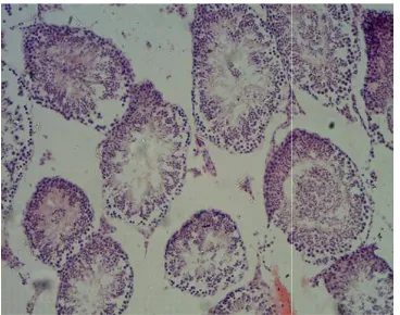
3.2 Histopathological Changes of Testes
The testis tissue in the control negative mice
showing normal seminiferous tubules,
spermatids, and spermatogenic cells at different stages of development Fig. (3). The destructive changes appear more clearly that leads to absence of spermatogenesis with thin of the wall of the seminiferous tube of the testes in first treated dose (2000 mg/kg) Fig. 4. The testes characterize by some seminal duct characterizes by the presence of complete spermatogenesis while some of them show no appearance spermatogenesis do to by generative changes which appear epithelial cells in addition to depletion of the wall of seminal duct for the group treated with (1000 mg/kg) of drug Fig. 5.
3.3 Level of Testosterone
The level of male hormone (Testosterone) directly effects by Zantac drug. The high concentration of drug decreased the testosterone level to (1.07 ng/ml) in compared with control
group (14.07 ng/ml) and the second
(A)
Fig. 1. Sperm head morphological (a)
Fig. 2. Figure represented sperm head abnormality depending on Zantac concentration
Fig. 3. Section from testes of control mice
3.4 Level of Prolactin
The level of prolactin hormone also affected by Zantac drug. In the normal state, the prolactin hormone in the male should be at a low level. The level hormone in control group is (36 while the level in the second group that treated with the drug (2000 mg/kg) is increased to
11.
0 10 20 30 40 50 60
control
p
e
rc
en
ta
ge
s
p
e
rm
h
ea
d
a
b
n
o
rm
al
it
y
(%
)
Sperm Head Abnormality
Oraibi et al.; AJB2T, 5(1): 1-7, 2019; Article no.
(B)
Sperm head morphological (a) normal sperm form (b) abnormal sperm head (100X)
Figure represented sperm head abnormality depending on Zantac concentration
3. Section from testes of control mice
The level of prolactin hormone also affected by Zantac drug. In the normal state, the prolactin hormone in the male should be at a low level. The level hormone in control group is (36 ng/ml) while the level in the second group that treated
mg/kg) is increased to
(115 ng/ml) and another concentration also increase the hormone level (43 ng/ml). The effect of high concentration well be more than the lower one shown in Fig. 7.
4. DISCUSSION
A number of chemicals including various environmental toxicants, eco-bacteria, and even clinically useful drugs can cause cellular damages in different organs of the body through
metabolic activation to highly reactive
substances such as free radicals [8
This study investigates Zantac induced toxicity in testis mice; study results have been shown that Zantac is capable of affecting seminiferous epithelium and altering the spermatogenesis process. The results have been indicated that Zantac induced a significant increase in the
head abnormality associated with severe
histopathological changes [9]. .1
22.8
48.4
control 1000mg\kg 2000mg\kg
treatments
Sperm Head Abnormality
; Article no.AJB2T.48254
sperm head (100X)
Figure represented sperm head abnormality depending on Zantac concentration
ng/ml) and another concentration also ng/ml). The effect of high concentration well be more than the lower
A number of chemicals including various bacteria, and even clinically useful drugs can cause cellular damages in different organs of the body through
metabolic activation to highly reactive
8].
This study investigates Zantac induced toxicity in testis mice; study results have been shown that Zantac is capable of affecting seminiferous epithelium and altering the spermatogenesis process. The results have been indicated that Zantac induced a significant increase in the
Fig. 4. Section of testes tissue of first treated with (2000 mg/kg) of Zantac
The presence of slaughter germinal cells and
their occurrence in the epididymis and
seminiferous tubule luminal are potential due to the loss of contact as consequences of disruption
of cellular junctions between
Spermatogonia are affected by the treatment by which some seminiferous tubules contained spermatogonia but don’t complete and at a low
level. Studies have been reported that
spermatogonia seem to be the most resistant spermatogenic cells to toxicity, however, chronic exposure to the chemotherapeutic drug [1
The successful and complete male germ cell development is depended on the balanced endocrine of hypothalamus, pituitary and the
testis. A gonadotropin-releasing hormone
secreted by the hypothalamus elicits the release of gonadotropins hormones from the pituitary gland [12]. FSH binds with receptors in the Sertoli cells and stimulates spermatogenesis. LH stimulates the production of testosterone in
Fig. 6. Figure representation the concentration of testosterone in
0 2 4 6 8 10 12 14 16 se ru m t es to st e ro n ec o n ce n tr at io n (n g\ m l)
Oraibi et al.; AJB2T, 5(1): 1-7, 2019; Article no.
Section of testes tissue of first group treated with (2000 mg/kg) of Zantac
The presence of slaughter germinal cells and
their occurrence in the epididymis and
seminiferous tubule luminal are potential due to the loss of contact as consequences of disruption
of cellular junctions between cells [10].
Spermatogonia are affected by the treatment by which some seminiferous tubules contained spermatogonia but don’t complete and at a low
level. Studies have been reported that
spermatogonia seem to be the most resistant cells to toxicity, however, chronic exposure to the chemotherapeutic drug [11].
The successful and complete male germ cell development is depended on the balanced endocrine of hypothalamus, pituitary and the
releasing hormone
secreted by the hypothalamus elicits the release of gonadotropins hormones from the pituitary ]. FSH binds with receptors in the Sertoli cells and stimulates spermatogenesis. LH stimulates the production of testosterone in
Leydig cells, which in turn may act on Sertoli cells and stimulates spermatogenesis [1
Fig. 5. Section of testes tissue in second group treated with (1000 mg/kg) of Zantac
The prolactin responsive mechanisms constitute the biological substrates for the pituitary hypothalamo-gonadal feedback system in the male mammals, which relies on the gonadal steroids, testosterone and estradiol for long
and prolactin for short-loop feedback
mechanisms. These mechanisms would operate to contain the adverse effects of reproductive stress and ensure that serum levels of prolactin remain within the physiological range, because even mild-to-moderate hyperprolactinemia, if allowed to become chronic, affects the quality of mature spermatozoa and their fertilizing potential
[14]. Hyperprolactinemia also affects
reproductive behavior in spite of normal testosterone levels. Importantly, whereas the effects of acute hyperprolactinemia appear to be mediated via testosterone inhibition, those due to
moderate hyperprolactinemia would be a
consequence of FSH deficits [15].
Figure representation the concentration of testosterone in Zantac treated albino male mice
14.07
3.42
1.07
control 1000mg\kg 2000mg\kg
treatments
Testosterone
; Article no.AJB2T.48254
g cells, which in turn may act on Sertoli cells and stimulates spermatogenesis [13].
Section of testes tissue in second mg/kg) of Zantac
responsive mechanisms constitute the biological substrates for the
pituitary-gonadal feedback system in the male mammals, which relies on the gonadal steroids, testosterone and estradiol for long-loop,
loop feedback
hanisms. These mechanisms would operate to contain the adverse effects of reproductive stress and ensure that serum levels of prolactin remain within the physiological range, because moderate hyperprolactinemia, if ffects the quality of mature spermatozoa and their fertilizing potential
]. Hyperprolactinemia also affects
reproductive behavior in spite of normal testosterone levels. Importantly, whereas the effects of acute hyperprolactinemia appear to be via testosterone inhibition, those due to
moderate hyperprolactinemia would be a
Oraibi et al.; AJB2T, 5(1): 1-7, 2019; Article no.AJB2T.48254
Fig. 7. Figure representation the concentration of prolactin in Zantac treated albino male mice
5. CONCLUSION
Zantac (Ranitidine) like any others synthetic drugs which have a side effect, and the results of this study found that treatment of mice with zantac negatively effect on the frequency of sperm head abnormality and the level of male sex hormones
COMPETING INTERESTS
Authors have declared that no competing interests exist.
REFERENCES
1. Amit V, Bangia A., Kamath N, Mohan V.
Ranitidine-induced thrombocytopenia:
Arare drug reaction. Indian J Pharmacol. 2011;43(1):76–7.
2. Atanasov AG, Waltenberger B,
Pferschy-Wenzig EM, Linder T, Wawrosch C, Uhrin P, Temml V, Wang L, Schwaiger S, Heiss EH, Rollinger JM, Schuster D, Breuss JM, Bochkov V, Mihovilovic MD, Kopp B, Bauer R, Dirsch VM, Stuppner H. Discovery and resupply of pharmacologically active plant-derived natural products. Biotechnol Adv. 2015;33(8):1582–614.
3. Fedorowicz Z, Zuuren EJ. Histamine
H2-receptor antagonists for urticaria. The
Cochrane Database of Systematic
Reviews. 2012;3:CD008596.
4. Mallow S, Rebuck JA, Osler T, Ahern J,
Healey MA, Rogers FB. Do proton pump
inhibitors increase the incidence of
nosocomial pneumonia and related
infectious complications when compared with histamine-2 receptor antagonists in critically ill trauma patients? Curr Surg. 2004;61(5):452–458.
5. Tuck SP, Francis RM. Testosterone, bone
and osteoporosis Frontiers of Hormone Research. Front Home Research. 2009; 37:123-132.
6. Wyrobek AJ, Bruce WR. Chemical
induction of sperm Abnormalities in mice. Proc. Natl. Acad. Sci. 1975;72:4425-4429.
7. Alwachi SN, Husain DK. Research article
Tamsulosin hydrochloride (flomax) effects
on fertility of albino male mice.
International Journal of Recent Scientific Research. 2014;5(2):326-331.
8. Lemike TL, Williams DH, Foye WO.
Principles of medicinal chemistry. 6th
Edition; 2002.
9. Niran A. Ibraheem, Ruqaya M. Al-Ezzy,
Maysaa CH. Al-Yas, Ebtehal H. Al-Naimy. The effect of lactobacillus acidophilus concentrated filtrate on sperm head abnormality in albino male mice. Journal of Al-Nahrain University Science. 2009;12(4): 151-155.
10. Niran A. Ibrahim, Hanady S. Al-Shmgani,
Ruqaya MI Al-ezzy. Cytarabine induced reproductive histopathological changes in albino male mice. Journal of Biotechnology Research Center. 2017;11(1):6-12.
11. Howell SJ, Shalet SM. Testicular function
following chemotherapy. European society of human reproduction and embryology. Human Reproduction Update. 2001;7(4): 363–369.
12. Lucas BK, Ormandy CJ, Binart N, Bridges
RS, Kelly PA. Null mutation of the prolactin
36 43
115
0 20 40 60 80 100 120 140
control 1000mg\kg 2000mg\kg
se
ru
m
p
ro
la
ct
in
c
o
n
ce
n
tr
at
io
n
(n
g\
m
l)
treatments
Oraibi et al.; AJB2T, 5(1): 1-7, 2019; Article no.AJB2T.48254
receptor gene produces a defect in
maternal behavior. Endocrinology. 1998;
139(10):4102-2107.
13. Al-Hadidyai AAA. Estimation of
testosterone and some biochemical
parameters in infertile women's serum.
Thesis supplement to Microbiology
Department, Medicinal College, Mosul; 2003.
14. Bassil N, Alkaade S, Morley JE. The
benefits and risks of testosterone
replacement therapy. Indian J Urol. 2009; 30(1):2–7.
15. Aleem M, Choudhari J, Padwal V,
Balasinor N, Parte P, Gill-Sharma MK. Hyperprolactinemia affects spermiogenesis in adult male rats. J Endocrinol Invest. 2005;28(1):39-48.
_________________________________________________________________________________
© 2019 Oraibi et al.; This is an Open Access article distributed under the terms of the Creative Commons Attribution License (http://creativecommons.org/licenses/by/4.0), which permits unrestricted use, distribution, and reproduction in any medium, provided the original work is properly cited.
Peer-review history: