The sex Differences in Blood Pressure and Cardiac Autonomic
Modulation during the Menopause Transition at Rest and after Aerobic
Exercise Training
Maycon Jr Ferreira1,2, Rodrigo D Esposti1, Aline O Jarrete1,3, Carlos H G Sponton1,4,
Angelina Zanesco1,5
1Laboratory of Cardiovascular Physiology and Exercise Science, Institute of Biosciences,
UNESP, Rio Claro, Brazil.
2Physiology Exercise Laboratory, Department of Physiology, Federal University of São
Paulo, São Paulo, SP, Brasil.
3Department of Structural and Functional Biology, Institute of Biology,
UNICAMP-University of Campinas, Campinas, SP, Brazil.
4Department of Cell and Tissue Biology, University of California, San Francisco, CA, USA. 5Metropolitan University of Santos (UNIMES), Santos, SP, Brazil.
Running title: Menopausal transition and cardiovascular system
Conflict of Interest/Disclosure: None.
Corresponding author:
Angelina Zanesco, Professor in Physiology Metropolitan University of Santos
Avenida Conselheiro Nébias nº 536 – Encruzilhada Santos/SP
Tel: +55 19 997512416
Email: [email protected]
ABSTRACT
Background: Sex differences in blood pressure (BP) exist during all reproductive life between
women and men whereas a sharper increase in BP occurs after menopause which is associated
with increased prevalence of cardiovascular diseases. This study examined cardiovascular and
biochemical parameters in perimenopausal women (PW) aiming to investigate the influence of sex
on a) office BP and for 24 hours; b) cardiac autonomic modulation; c) redox state by measuring
MDA, SOD, and catalase; d) NOx- concentration. In addition, aerobic exercise training (AET) was
applied for detecting changes in cardiovascular responsiveness during the menopausal transition.
Methods: Thirty-one participants were enrolled, healthy PW and age-matched men.
Cardiovascular and biochemical biomarkerswere evaluatedat baseline and after AET (8 weeks of
exercise on a treadmill, three sessions/week, duration of 30-40 minutes).
Results: At rest, PW presented: a) a lower diastolic BP during daytime; b) a lower absolute and
normalized LF component as well as a higher HF normalized component; d) no sex differences
for redox biomarkers and NOx- concentration. After AET, only PW were responsive in lowering
systolic BP that was accompanied by an increase in NOx- concentration and SOD activity.
Regarding HRV, both groups were responsive to the AET.
Conclusions: During the menopausal transition, systolic BP was similar to men whereas cardiac
autonomic modulation remained unaltered showing the influence of sex steroids on BP. In
Addition, AET was fundamental during the menopause transition by preventing an elevation in
BP, minimizing the effects of aging associated with estrogen deficiency on women's
cardiovascular health.
The existence of sex differences related to blood pressure (BP) levels is well documented.
On the other hand, a sharper increase in BP in women than in men has been reported during midlife
which is associated with increased prevalence of cardiovascular diseases after menopause (Barton
& Meyer, 2009; Benjamin et al., 2018). Estrogen deficiency is considered the main cause of the
deep increase in BP after menopause since estrogen plays a key role in the regulation of a variety
of physiological systems including cardiovascular, endocrine, nervous and immune (Stork et al.,
2004). Nevertheless, most of the data reporting the role of estrogen deficiency on BP levels are
from experimental studies (Reckelhoff, 2018). Therefore, studies with women in different phases
of the climacteric period are fundamental to understand the possible mechanisms as well as
biomarkers that are involved in BP regulation in this particular population. Indeed, many questions
still unanswered related to women and cardiovascular health. Is there an endothelial dysfunction
after menopause? Previous studies have systematically shown that NO/cGMP signaling pathway,
as well as flow-mediated dilation, is not impaired in hypertensive postmenopausal women as
compared with normotensive subjects (Sponton et al., 2014; Puga et al., 2016; Jarrete et al., 2016;
Novais et al., 2017). Additionally, both hypertensive and normotensive postmenopausal women
were responsive to aerobic exercise training (AET) by increasing the endothelial biomarkers
related to NO/cGMP pathway (Sponton et al., 2010; Rezende et al., 2011). Another question in
this scenario that should be raised is: when is the turning point of the increasing BP during the
climacteric period? Thus, the objective of this study was to examine the cardiovascular and
biochemical parameters in perimenopausal women compared with age-matched men. This period
is crucial for women's health since sex hormones fluctuate from month to month and this
postmenopausal women where a complete decline in ovarian function and estrogen levels takes
place. To address this issue, we examine the influence of sex on a) office BP and during 24 hours;
b) autonomic modulation by evaluating heart rate (HR) and its variability (HRV); c) redox state
by measuring biomarkers (MDA, SOD and catalase); d) nitric oxide (NO) production by
measurement of NOx- concentration. In addition, AET was applied as a challenge for detecting
cardiovascular responsiveness during the perimenopausal period.
Materials and methods Participants
This study was approved by the Ethical Committee from University of Sao Paulo State
(Protocol: 908.577/2015). Disclosure and participants recruitment were performed by
advertisement in the surrounding area of the university and local media. Participants were included
according to the following criteria: age between 40 and 59 years old, inactive physically (< 150
minutes of moderate physical activity or < 75 minutes of vigorous physical activity), non-obese ,
nonsmoking, non-prediabetes (fasting blood glucose < 100 mg/dL), being in the perimenopausal
period (exclusive for women), normal renal function (creatinine levels < 135 mmol/L),
non-alcoholic (< 3 cups/day), no menopausal therapy (exclusive for women), no use of antihypertensive
medications, any other diseases that would prevent the practice of physical exercise. After
recruitment, all participants (n=31) underwent a general anamnesis followed by the fulfillment of
questionnaires related to the practice of physical activity (PAR-Q) and quality of life (SF-36).
Participants were invited to sign a free consent term.
The period of evaluation lasted approximately 3 – 4 weeks and 1 – 2 weeks, before and
after of the AET, respectively. At the beginning of the study, all the participants performed a
6 sessions of 20 minutes each. The aims were to teach and to guide them on how to walk in a
treadmill correctly as well as to avoid any stressful situation that would interfere with the
measurements.
Peak oxygen consumption was determined indirectly in a treadmill by performing the
1-mile test according to Kline et al. (1987). Maximal Lactate Steady State (MLSS) test was
performed to determine the optimal walking intensity of each volunteer for AET. After that, the
participants exercised on a treadmill, three sessions/week, totalizing twenty-four sessions of
physical exercise as previously described (Ferreira et al., 2018).
The perimenopause characterization in women was performed through by self-report of
irregular menstrual cycles and the presence of signs and symptoms of this period, as previously
described by Harlow et al. (2012). In addition, follicle-stimulating hormone (FSH), luteinizing
hormone (LH) and 17β-estradiol were measured.
Anthropometric and cardiovascular parameters
Body weight and height of the participants were measured on a digital scale (Toledo® 2096
PP). BMI was calculated by the ratio of body weight/height². Waist circumference measurements
were performed in triplicate between the iliac crest and the last rib.
The measurements of the resting heart rate (HR) and heart rate variability (HRV) were
carried out in a quiet room, noise-free and environment-controlled temperature. The subjects
remained in absolute rest in the sitting position for 15 minutes, comfortably, maintaining a constant
respiratory rate. Values were recorded and stored through of a transmitter belt placed at the level
of the distal third of xiphoid process with a pulse HR monitor (Polar RS800CX, Electro Oy,
Kempele, Finland) as previously described by Barbosa et al. (2016). The participants were
stimulant medication that could interfere in both parameters. In addition, they were instructed not
to engage in any vigorous exercise in the last 48 hours preceding the test. Subsequent to the data
acquisition, the records were transferred to Polar ProTrainer 5™ software. Resting HR was
calculated as the mean value of HR during the recording period.
The HRV measurement was performed through the analysis of the normal intervals RR
(iRR). The selection of the section for analysis was performed by visual inspection of the
distribution of the iRR in the interval of fifteen minutes, in which a central five minutes interval
of greater stability of the signal was selected, containing a sampling frequency of, at least, 256
points. The remaining ten minutes, distributed between initial and final passages, were discarded.
The iRR analysis was performed using Kubios HRV 2.1 software (University of Eastern Finland,
Kuopio, Finland). The HRV measurement was performed using the indices corresponding to the
frequency domain. For this analysis, the indices selected were the power in the low-frequency
range (LF), and in the high-frequency range (HF), (Kleiger et al., 2005). The HF component is
defined as a vagal modulating marker which is synchronized with the respiratory rate . Both powers
were calculated in absolute (ms²) and normalized units (nu). In addition, the variance of all
successive normal iRRs (TP) and the ratio between low- and high-frequency (LF/HF) were used
as an index of sympathovagal balance (Task Force, 1996).
Office BP and Ambulatory BP monitoring (ABPM) were obtained in accordance with
international guidelines as described previously (Sponton et al., 2014).
Biochemical analyses
Blood samples were collected after a 12-hour overnight fast. Samples were centrifuged and
analyses. A specific commercial kit (Cayman Chemical, Ann Arbor, MI, USA) was used for all
the tests.
FSH, LH, and 17-β estradiol: measured by using the sandwich-type immunometric method
(ELISA) (Cayman Chemical, Ann Arbor, MI, USA) in accordance to the manufacturer. The
analysis of 17-β estradiol was performed in a private clinic.
The endogenous production of nitric oxide (NO): quantified indirectly through the nitrite
and nitrate anions (NOx-) measurements, as previously described by Delbin et al. (2012).
Lipid peroxidation: quantified based on the serum concentrations of the compound
malondialdehyde (MDA).
Superoxide dismutase (SOD) activity: determined using a tetrazolium salt for detection of
superoxide radicals generated by xanthine oxidase and hypoxanthine.
Catalase: the method of determining the activity of catalase occurred through its activity
peroxidase.
Statistical analysis
Data are presented as means ± standard deviation (SD). The normality of the data was
verified by the Kolmogorov-Smirnov test. After that, the effects of sex, AET and sex x training
interaction were obtained through the analysis of variance (ANOVA) of repeated measures
adjusted for two factors (sex and training), followed by Bonferroni post-hoc when appropriate.
Differences in the magnitude of behavior ambulatory BP reduction of 24 hours were verified
through analysis of the area under the curve (AUC), using a paired t-test for comparison before
and after AET in each group, and an unpaired t-test for comparison between groups. Analyzes
were performed using SPSS statistical software for Windows (PASW Statistics 18). P<0.05 was
Results
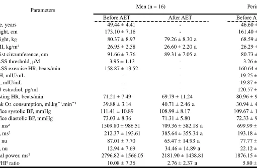
The general characteristics of the participants are shown in Table 1.At the baseline, the
studied population was similar in age and BMI. On the other hand, men showed higher weight
waist circumference as compared with perimenopausal women, ([F(1,29)=11.48; p<0.002] and
[F(1,29)=14.88; p<0.001], respectively). As expected, the VO2 peak values were higher for men
compared with women ([F(1,29)=48.07; p<0.0001]). In contrast, HR was lower in men than women
([F(1,29)=8.93; p<0.007]). Daytime diastolic ABPM ([F(1,29)=12.23; p<0.003]) and 24 hours
diastolic ABPM ([F(1,29)=12.43; p<0.001]) were lower in perimenopausal women compared with
men. In addition, HRV showed a higher absolute LF component in men ([F(1,29)=7.68; p<0.02]). A
higher normalized LF ([F(1,29)=5.74; <0.03) and a lower HF component only before AET,
([F(1,29)=5.66; p<0.03]) were observed in men as compared with women.
Twenty-four sessions of AET promoted a decrease in weight (-1.4%) ([F(1,29)=7.09;
p<0.02]), BMI (-1.3%) ([F(1,29)=6.46; p<0.02]) and waist circumference (-2.6%) ([F(1,29)=9.91;
p<0.005]) only in men. On the other hand, both groups showed an increase in the aerobic capacity
(2.1 and 5% for men and women, respectively) ([F(1,29)=17.25; p<0.001). In addition, no
differences were observed for chronotropic response in both trained groups as measured by mean
resting HR (Table 1). Examining HRV in the frequency domain, we found that AET promoted a
decrease in absolute LF component only in men group (-53.0%) ([F(1,29)=17.32; p<0.001]) whereas
normalized LF component was decreased ([F(1,29)=45.19; p<0.001]) in both groups (24.8 and
-28.6% for men and perimenopausal women, respectively). Similarly, absolute ([F(1,29)=7.53;
p<0.02]) and normalized HF component ([F(1,29)=45.38; p<0.001]) were increased in response to
AET in men (81.6 and 166.3%, respectively) while only normalized HF component was increased
men (-72.6%) and perimenopausal women group (-71.6%) after AET. Data are summarized in
Table 1.
Regarding office BP measurements, only perimenopausal women were responsive to AET,
showing a decrease of -5.6% for systolic office BP ([F(1,29)=17.41; p<0.001]). Additionally, no
difference was found for diastolic office BP in both trained groups (Table 1). In agreement with
that, trained perimenopausal women presented a decrease in systolic BP during nighttime as
measured by ABPM (-4.2%) ([F(1,29)=5.09; p<0.04]) as well as for systolic BP for 24 hours
(approximately -3.0%) ([F(1,29)=5.64; p<0.03]) (Figure 1).
Anova repeated measure showed that there was an interaction for NOX- concentration
([F(1,29)=5.49; p<0.05]). Bonferroni post-hoc showed a significant increase in the NOX-
concentration (38.5%), a biomarker related to NO production, in trained perimenopausal women
but no changes were seen in trained men. Regarding redox status, SOD activity was significantly
increased (55.1%) in response to AET only in perimenopausal women ([F(1,29)=8.67; p<0.007]) but
neither MDA concentrations nor catalase activity was affected by exercise training in both groups.
Discussion
Comparing to age-matched men and prior to the challenge of the AET, our study shows
that perimenopausal women present: a) a lower diastolic BP during daytime ([F(1,29)=12.23;
p<0.003]); b) a lower HR ([F(1,29)=8.93; p<0.007]), absolute ([F(1,29)=7,68; p<0.02]) and
normalized LF component ([F(1,29)=5.74; <0.03]); and a higher HF normalized component
[F(1,29)=5.66; p<0.03]; c) no sex differences for redox biomarkers (MDA levels, SOD and catalase
activity); d) no sex differences in NOx- concentration. After a powerful stimulus to the
cardiovascular system provided by 24 sessions of AET, perimenopausal women were responsive
concentration and SOD activity even though men were more responsive in reducing body weight
and waist circumference. Regarding HRV, both groups were responsive to the AET with a
reduction in LF component and increment in the HF component.
Sex differences at baseline
The perimenopause comprises the period in which the first symptoms of ovarian aging
begin, indicating the approach of the menopause, until the first year after the last menstruation,
occurring on average at forty-seven years of age regardless of the obesity, race/ethnicity, and
smoking (Randolph et al. 2011). In this period occurs several changes in sex hormones with an
increase in FSH and a decrease in estradiol levels (Burger et al., 1999). Estrogen deficiency has
been pointed out as the main cause for the development of cardiovascular diseases after
menopause, mainly arterial hypertension (Wassertheil-Smoller et al., 2000; Stork et al., 2004).
Nevertheless, it is not well clear when the turning point of the increasing BP takes place during
the climacteric period. Our findings revealed that office BP values were similar between the men
and women indicating a change in BP behavior during this particular period of the women's lives.
BP monitoring for 24 hours showed that only diastolic BP (daytime) was lower as compared with
men. These findings show that during the menopausal transition, the sex differences in BP
regulation is altered and neither redox state, measured by MDA and antioxidant enzymes activity,
nor NOx- concentrations were modified. Accordingly, a previous study showed impairment in the
brachial artery flow-mediated dilatation during the perimenopausal transition as compared with
the beginning of this period (early perimenopause), they did not measure any biomarkers though
(Moreau et al., 2012). Another study found morphological alterations in common carotid artery
during the menopausal transition that was associated with a decline in estrogen level (El Khoudary
reduced during the menopausal transition, apparently without a contribution of the vasodilator
agent, NO, measured by its bioavailability. One possibility to explain this change in BP behavior
during menopausal transition could be an unbalance between vasodilation and vasoconstriction
responses in the vasculature. Indeed, a previous study demonstrated that intracoronary infusion of
17β-estradiol to postmenopausal women promoted a decrease in endothelin-1, a potent
vasoconstrictor agent derived from endothelial cells. Moreover, the infusion of 17β-estradiol did
not affect the NOx- concentrations in this population (Webb et al., 2000).
Regarding HRV, our results demonstrate a vagal predominance in perimenopausal women
compared with age-matched men showing a normal sympathovagal balance in this population even
though they are under an intermittent sex hormonal exposure. Previous studies have shown a
reduction in the parasympathetic component in women after menopause (Eaker et al., 1993; Kuo
et al., 1999; Evans et al., 2001). In contrast, other studies show no difference in resting cardiac
autonomic modulation between perimenopausal and postmenopausal women (MyslivecEk et al.,
2002) or any changes in cardiac autonomic activity evaluated by 24-hour indices of HRV after
estrogen administration for 3 months in healthy postmenopausal women (Fernandes et al., 2005).
Accordingly, our study clearly shows that cardiac autonomic modulation is unchanged in
perimenopausal women reinforcing that sex hormone does not affect HRV. Nevertheless, it is
important to emphasize that more studies with a large number of participants would bring more
robust data.
As expected, lower aerobic capacity was observed in perimenopausal women, measured
through the VO2 peak, compared with men, corroborating with previous studies (Fleg et al., 2005;
Jackson et al., 2009; Wang et al., 2010).
Our findings show that the volume and the intensity of the AET program employed in the
study were effective in increasing VO2 peak for both groups. Although the participants were
composed of physically inactive individuals and they exercised at similar relative intensity,
perimenopausal women presented a higher increase in VO2 peak than men. Likely, it is due to the
lower aerobic capacity presented in relation to the men at the baseline, which makes them more
responsive to the same physical stimulus. On the other hand, men were responsive to the AET in
weight loss and in decreasing waist circumference with a consequent reduction in BMI while
perimenopausal women were non-responder for anthropometric parameters to the AET. This lack
of changing in perimenopausal women in response to the AET was expected since a sex difference
has been described related to metabolic rate between men and women which is dependent on the
amount of skeletal muscle mass as well as the density of the adrenergic receptor subtypes present
in fat depot (Thompson et al. 2012).
Regarding cardiovascular parameters, perimenopausal women were responsive in lowering
office (5.6%) and ambulatory (nighttime and 24 hours) systolic BP (approximately 4.2 and
-3.0%, respectively), that was accompanied by increased SOD activity and NOx- levels indicating
a higher NO bioavailability to the cells in trained women. A number of studies has shown the
beneficial effects of the exercise training on cardiovascular system with improvement in the
relaxing responses and reduction in BP either in humans (Maeda et al., 2004; Zaros et al., 2009;
Esposti et al., 2011) or in laboratory animals (Claudino et al., 2011; Delbin et al., 2012). Our
findings show that endothelial cells still reactive to the shear stress induced by exercise training
during menopausal transition promoting a beneficial effect on BP regulation. In contrast, neither
office BP nor ABPM were altered in trained men. Moreover, men were non-responder in changing
Additionally, AET promotes an increase in vagal modulation and a decrease in sympathetic
modulation in both groups. Evidence has systematically shown that physical training promotes an
increase in vagal modulation and a decrease in sympathetic modulation into the heart (Carter et
al., 2003; Melanson & Freedson, 2001; Kiviniemi et al., 2006). On the other hand, studies
examining the effect of the exercise training on cardiac autonomic modulation in women are
scarce, and no studies exist comparing this parameter during menopausal transition and men. An
early study shows a similar response to exercise training, comparing perimenopausal and
postmenopausal women, with a decrease in sympathetic modulation and an increase in
parasympathetic modulation (MyslivecEk et al., 2002).
Conclusions
Taken together, our findings clearly show that during menopausal transition BP levels are
affected by the intermittent hormonal exposure without changes vasodilator bioavailability.
Fortunately, perimenopausal women still responsive to the beneficial effects of AET showing that
endothelial cells are reactive to shear stress induced by physical exercise. In addition, sex
differences in cardiac autonomic modulation remain unaltered with an increase in vagal
modulation and a decrease in sympathetic modulation, showing no interference of sex steroids on
HRV and AET enhanced these parameters. Considering that high BP and physical inactivity are
considered major contributors to death and disability, the relevance of this study is unquestionable
since women live longer than men, as well as, they are more susceptible for cardiovascular diseases
after menopause. Therefore, actions in management BP levels by the practice of regular physical
minimizing the effects of aging associated with estrogen deficiency on women's cardiovascular
Funding information
This research was supported by Sao Paulo Research Foundation (Grant number: 2001/17437-7)
and a scholarship to Maycon Jr Ferreira from Coordination for the Improvement of Higher
Education Personnel (CAPES).
Acknowledgments
The corresponding author, as a principal investigator, had full access to all the data in the
References
Barbosa, M. .P., da Silva, N.T., de Azevedo, F.M., Pastre, C.M. & Vanderlei, L.C. (2016).
Comparison of Polar® RS800G3™ heart rate monitor with Polar® S810i™ and
electrocardiogram to obtain the series of RR intervals and analysis of heart rate variability at
rest. Clinical Physiology and Functional Imaging. 36(2),112-117.
Barton, M. & Meyer, M.R. (2009). Postmenopausal hypertension: Mechanisms and therapy.
Hypertension. 54(1),11–8.
Benjamin, E., Virani, S., Callaway, C., Chamberlain, A., Chang, A.R., Cheng, S….Muntner, P.
(2018). Heart Disease and Stroke Statistics — 2018 Update. A Report from the American
Heart Association. Circulation. 137(12), e67-492.
Burger, H.G., Dudley, E.C., Hopper, J.L., Groome, N., Guthrie, J.R… Dennerstein, L. (1999).
Prospectively measured levels of serum follicle-stimulating hormone, estradiol, and the
dimeric inhibins during the menopausal transition in a population-based cohort of women.
Journal of Clinical Endocrinology & Metabolism. 84(11), 4025–4030.
Carter, J.B., Banister, E.W. & Blaber, A.P. (2003). The effect of age and gender on heart rate
variability after endurance training. Medicine Science Sports & Exercise. 35(8),1333–1340.
Claudino, M.A., Delbin, M.A., Franco-Penteado, C.F., Priviero,, F.B., De Nucci, G…Zanesco,
A. (2011). Exercise training ameliorates the impairment of endothelial and nitrergic corpus
cavernosum responses in diabetic rats. Life Sciences. 88(5–6), 272–277.
Delbin, M.A., Davel, A.P. C., Couto, G.K., de Araújo, G.G., Rossoni, L.V…Zanesco, A. (2012).
Interaction between advanced glycation end products formation and vascular responses in
femoral and coronary arteries from exercised diabetic rats. PLoS One. 7(12), e53318.
Eaker, E.D., Chesebro, J.H., Sacks, F.M., Wenger, N.K., Whisnant, J.P. & Winston, M. (1993).
Cardiovascular disease in women. Circulation. 88(4 (part I)),1999–2009.
El Khoudary, S.R., Wildman, R.P, Matthews, K., Thurston, R.C., Bromberger, J.T. &
Sutton-Tyrrell, K. (2013). Progression rates of carotid intima-media thickness and adventitial
diameter during the menopausal transition. Menopause. 20(1), 8–14.
Esposti, R.D., Sponton, C.H.G., Malagrino, P.A., Carvalho, F.C., Peres, E….Zanesco, A. (2011).
Influence of eNOS gene polymorphism on cardiometabolic parameters in response to
Research. 44(9),855–863.
Evans, J.M., Ziegler, M.G., Patwardhan, A.R., Ott, J.B., Kim, C.S…Knapp, C.F. (2001). Gender
differences in autonomic cardiovascular regulation: spectral, hormonal, and hemodynamic
indexes. Journal of Applied Physiology (Bethesda, Md 1985). 91(6), 2611–2618.
Fernandes, E.O., Moraes, R.S., Ferlin, E.L., Wender, M.C.O. & Ribeiro, J.P. (2005). Hormone
replacement therapy does not affect the 24-hour heart rate variability in postmenopausal
women: results of a randomized, placebo-controlled trial with two regimens. Pacing Clinical
Electrophysiology. 28(Suppl 1), S172–S177.
Ferreira, M. Jr. Jarrete, A.P., Esposti, R.D., Sponton, C.H.G., Anaruma, C.P. & Zanesco, A.
(2018). Evaluation of maximal lactate steady state in middle-aged hypertensive women.
Motriz: Journal of Physical Education. 24 (2),e101896.
Fleg, J.L., Morrell, C.H., Bos, A.G., Brant, L.J., Talbot, L.A... Lakatta, E.G. (2005). Accelerated
longitudinal decline of aerobic capacity in healthy older adults. Circulation. 112(5), 674–
682.
Harlow, S.D., Gass, M., Hall, J.E., Lobo, R., Maki, P… de Villers, T.J. (2012). Executive
summary of the stages of reproductive aging workshop + 10: Addressing the unfinished
agenda of staging reproductive aging. Menopause. 19(4),387–395.
Jackson, A.S., Sui, X., Hébert, J.R., Church, T.S. & Blair, S.N. (2009). Role of lifestyle and aging
on the longitudinal change in cardiorespiratory fitness. Archives of Internal Medicine.
169(19),1781–1787.
Jarrete, A.P., Zanesco, A. & Delbin, M.A. (2016). Assessment of endothelial function by
flow-mediated dilation in diabetic patients: effects of physical exercise. Motriz: Journal of
Physical Education. 22(1), 3–11.
Kiviniemi, A.M., Hautala, A.J., Mäkikallio, T.H., Seppänen, T., Huikuri, H. V. & Tulppo, M.P.
(2006). Cardiac vagal outflow after aerobic training by analysis of high-frequency oscillation
of the R-R interval. European Journal of Applied Physiology. 96(6), 686–692.
Kleiger, R.E., Stein, P.K. & Bigger, Jr J.T. (2005). Heart rate variability: Measurement and
clinical utility. Annual Noninvasive Electrocardiology.10(1),88–101.
Kline, G.M., Porcari, J.P., Hintermeister, R., Freedson, P.S., Ward, A…Rippe, J.M. (1987).
Science Sports & Exercise. 19(3),253–259.
Kuo, T.B., Lin, T., Yang, C.C., Li,C-L., Chen, C-F. & Chou, P. (1999). Effect of aging on gender
differences in neural control of heart rate. American Journal of Physiology. 277(6 (Part 2)),
H2233–H2239.
Maeda, S., Tanabe, T., Otsuki, T. J., Iemitsu, M., Miyauchi, T… Matsuda, M. (2004). Moderate
regular exercise increases basal production of nitric oxide in elderly women. Hypertension
Research. 27(12), 947–953.
Melanson, E.L. & Freedson, P.S. (2001). The effect of endurance training on resting heart rate
variability in sedentary adult males. European Journal of Applied Physiology. 85(5),442–
449.
Moreau, K.L., Hildreth, K.L., Meditz, A.L., Deane, K.D. & Kohrt, W.M. (2012). Endothelial
function is impaired across the stages of the menopause transition in healthy women. Journal
of Clinical Endocrinology & Metabolism. 97(12), 4692–4700.
Myslivecek, P.R., Brown, C.A. & Wolfe, L.A. (2002). Effects of physical conditioning on cardiac
autonomic function in healthy middle-aged women. Journal of Chemical Information and
Modeling.27(1), 1–18.
Novais, I.P., Jarrete, A.P., Puga, G.M., Araujo, H.N., Delbin, M.A. & Zanesco, A. (2017). Effect
of aerobic exercise training on cGMP levels and blood pressure in treated hypertensive
postmenopausal women. Motriz: Journal of Physical Education.23(1), 1–6.
Puga, G.M., Novais, I. P, Katsanos, C.S. & Zanesco, A. (2016). Combined effects of aerobic
exercise and L-arginine ingestion on blood pressure in normotensive postmenopausal
women: A crossover study. Life Sciences. 151, 323–329.
Randolph, J.F., Zheng, H., Sowers, M.R., Crandall, C. Crawford S…Vulga M. (2011). Change in
follicle-stimulating hormone and estradiol across the menopausal transition: Effect of age at
the final menstrual period. Journal of Clinical Endocrinology & Metabolism. 96(3), 746–
754.
Reckelhoff, J.F. (2018). Sex differences in regulation of blood pressure. Advances in
Experimental Medicine & Biology. 1065,139–151.
Rezende, T.M., Sponton, C.H.G., Malagrino, P.A., Bezerra, M.A.C., Penteado, C.F.F. & Zanesco,
A. (2011). Effect of exercise training on the cardiovascular and biochemical parameters in
265–269.
Sponton, C.H.G., Esposti, R.D., Rodovalho, C.M., Ferreira, M.Jr., Jarrete, A.P…Zanesco, A.
(2014). The presence of the NOS3 gene polymorphism for intron 4 mitigates the beneficial
effects of exercise training on ambulatory blood pressure monitoring in adults. American
Journal of Physiology: Heart and Circulatory Physiology. 306(12), H1679–691.
Sponton, C.H.G., Rezende, T.M., Mallagrino, P.A., Franco-Penteado, C. Bezerra, M.A.&
Zanesco A. (2010). Women with TT genotype for eNOS gene are more responsive in
lowering blood pressure in response to exercise. European Journal of Cardiovascular
Prevention Rehabilitation. 17(6), 676–681.
Störk, S., van der Schouw, Y.T., Grobbee, D.E. & Bots, M.L. (2004). Estrogen, inflammation and
cardiovascular risk in women: A critical appraisal. Trends in Endocrinology & Metabolism.
15(2), 66–72.
Task Force of the European Society of Cardiology and the North American Society of Pacing and
Electrophysiology. (1996). Heart rate variability: standards of measurement, physiological
interpretation, and clinical use. Task Force of the European Society of Cardiology and the
North American Society of Pacing and Electrophysiology. European Heart Journal.
17(3),354–381.
Thompson, D., Karpe, F., Lafontan, M. & Frayn, K. (2012). Physical activity and exercise in the
regulation of human adipose tissue physiology. Physiological Review. 92(1),157–191.
Wang, C-YY., Haskell, W.L., Farrell, S.W., Lamonte, M.J., Blair, S.N…Burt, V.L. (2010).
Cardiorespiratory fitness levels among us adults 20-49 years of age: findings from the
1999-2004 National Health and Nutrition Examination Survey. American Journal of
Epidemiology. 171(4), 426–435.
Wassertheil-Smoller, S., Anderson, G., Psaty, B.M., Black, H.R., Manson, J…Lasser, N. (2000).
Hypertension and Its Treatment in Postmenopausal Women Baseline Data from the
Women’s Health Initiative. Hypertension. 36(5),780–789.
Webb, C.M., Ghatei ,M.A., McNeill, J.G. & Collins, P. (2000). 17β-Estradiol decreases
endothelin-1 levels in the coronary circulation of postmenopausal women with coronary
artery disease. Circulation. 102(14), 1617–1622.
Zaros, P.R., Pires, C.E.M.R., Bacci, Jr M., Moraes, C. & Zanesco. A. (2009). Effect of 6-months
Table 1 - The effect of exercise training on anthropometric, cardiovascular and aerobic capacity in perimenopausal women and men
Before AET After AET Before AET After AET
Age, years 49.44 ± 4.41 - 46.60 ± 4.17
-Height, cm 173.10 ± 7.16 - 161.40 ± 3.94
-Weight, kg 80.37 ± 8.97 79.26 ± 8.30 a 68.59 ± 9.75 68.37 ± 10.31
BMI, kg/m² 26.95 ± 2.38 26.60 ± 2.20 a 26.29 ± 3.23 26.19 ± 3.46
Waist circumference, cm 91.66 ± 7.76 89.31 ± 7.05 a 80.73 ± 8.75 79.20 ± 7.50
MLSS threshold, µM 3.95 ± 1.13 - 3.26 ± 1.15
-MLSS exercise HR, beats/min 158.87 ± 13.52 - 160.64 ± 14.47
-FSH, mlU/mL - - 19.25 ± 22.62
-LH, mlU/mL - - 19.87 ± 20.15
-17β-estradiol, pg/ml - - 120.57 ± 113.00
-Resting HR, beats/min 71.21 ± 7.49 69.79 ± 11.24 80.96 ± 9.62 76.83 ± 8.68
Peak O2 consumption, ml.kg ̄ ¹.min ̄ ¹ 39.88 ± 3.14 40.71 ± 2.46 a 30.94 ± 4.02 32.47 ± 4.18 b
Office systolic BP, mmHg 111.41 ± 10.89 108.99 ± 8.17 109.67 ± 12.13 103.49 ± 9.17 b
Office diastolic BP, mmHg 73.03 ± 8.36 71.31 ± 5.80 72.33 ± 9.51 69.93 ± 6.89
LF, ms² 1509.80 ± 986.51 709.36 ± 582.18 a 699.99 ± 764.73 283.46 ± 173.44
HF, ms² 212.37 ± 193.61 385.64 ± 355.34 a 193.18 ± 230.69 316.95 ± 461.41
LF, nu 87.01 ± 7.70 65.47 ± 14.93 a 77.77 ± 13.62 55.54 ± 17.00 b
HF, nu 12.94 ± 7.69 34.46 ± 14.89 a 22.12 ± 13.63 44.31 ± 16.94 b
Total power, ms² 2796.82 ± 1566.05 2181.90 ± 1438.81 1876.15 ± 1365.04 1327.39 ± 995.880
LF/HF ratio 10.08 ± 7.36 2.76 ± 2.37 a 5.80 ± 5.12 1.63 ± 1.39 b
Parameters Men (n = 16) Perimenopausal women (n = 15)