Development of adaptive fuzzy based Image
Filtering techniques for efficient
Noise Reduction in Medical Images
Aneesh Agrawal, Abha Choubey, Kapil Kumar Nagwanshi
1-2 Computer science and Engineering department, ShriShankaracharyaGroup of Institutions, Bhilai, India
3 Member IEEE
Abstract—The existing system available for fuzzy filters for noise
reduction deals with fat-tailed noise like impulse noise and median filter. Only impulse noise reduction uses fuzzy filters. Gaussian noise is not specially concentrated; it does not distinguish local variation due to noise and due to image structure. The proposed system presents a new technique for filtering narrow-tailed and medium narrow-tailed noise by a fuzzy filter. The system first estimates a “fuzzy derivative” in order to be less sensitive to local variations due to image structures such as edges. Second, the membership functions are adapted accordingly to the noise Level to perform “fuzzy smoothing.” A new fuzzy filter is presented for the noise reduction of images corrupted with additive noise. The filter consists of two stages. The first stage computes a fuzzy derivative for eight different directions. The second stage uses these fuzzy derivatives to perform fuzzy smoothing by weighting the contributions of neighboring pixel values. Both stages are based on fuzzy rules which make use of membership functions. The filter can be applied iteratively and effectively reduce heavy noise. In particular, the shape of the membership functions is adapted according to the remaining noise level after each iteration, making use of the distribution of the homogeneity in the image. A statistical model for the noise distribution can be incorporated to relate the homogeneity to the adaptation scheme of the membership functions. Experimental results are obtained to show the feasibility of the proposed approach. These results are also compared to other filters by numerical measures and visual inspection.
Index Terms— Fuzzy smoothing, Wiener Filter, Medical noise,
Gaussian Filter.
I. INTRODUCTION
HEapplication of fuzzy techniques in image processing is a promising research field. Fuzzy techniques have already been applied in several domains of image processing (e.g., filtering, interpolation, and morphology), and have numerous practical applications (e.g., in industrial and medical image processing). In this project, we will focus on fuzzy techniques for image filtering. Already several fuzzy filters for noise reduction have been developed, e.g., the well-known FIRE-filter from, the weighted fuzzy mean FIRE-filter from, and the iterative fuzzy control based filter from. Most fuzzy techniques in image noise reduction mainly deal with fat tailed noise like impulse noise. These fuzzy filters are able to outperform rank-order filter schemes (such as the median filter). Nevertheless, most fuzzy techniques are not specifically designed for Gaussian (-like) noise or do not produce convincing results when applied to handle this type of noise. Therefore, this
project gives a new technique for filtering, narrow-tailed and medium narrow-tailed noise by a fuzzy filter. Two important features are presented: first, the filter estimates a “fuzzy derivative” in order to be less sensitive to local variations due to image Structures such as edges; second, the membership functions are adapted accordingly to the noise level to perform “fuzzy smoothing.” For each pixel that is processed, the first stage computes a fuzzy derivative. Second, a set of 16 fuzzy rules is fired to determine a correction term. These rules make use of the fuzzy derivative as input. Fuzzy sets are employed to represent the properties and while the membership functions are fixed, the membership functions are adapted after each iteration. The adaptation scheme is extensive and can be combined with a statistical model for the noise. The result of this method can be compared with those obtained by other filters [1-2].
II. LITERATURE REVIEW
A novel spatiotemporal fuzzy based algorithm was proposed for noise filtering of image sequences. This algorithm uses adaptive weights based on a triangular membership functions. The proposed algorithm admissibly removes noise without having any knowledge of Salt and Pepper noise density. It is recommended that symmetrical fuzzy algorithm for removing low to medium-density Salt and Pepper noise has some constraint of dissymmetrical implementations[1].
Fuzzy techniques are widely applied in the area of digital image restoration. Main achievements related are presented by four parts, in which the filters based on fuzzy rank selection, fuzzy weighted, fuzzy network (FNN) and soft-switching are systematically analyzed, respectively [2].
A new fuzzy filter is presented for the noise reduction of images corrupted with additive noise. The filter consists of two stages. The first stage computes a fuzzy derivative for eight different directions. The second stage uses these fuzzy derivatives to perform fuzzy smoothing by weighting the contributions of neighboring pixel values. Both stages are based on fuzzy rules which make use of membership functions. The filter can be applied iteratively to effectively reduce heavy noise [3].
intensity in-homogeneities, also known as shading artifacts. We have presented an algorithm for obtaining fuzzy segmentations of images that have been corrupted by intensity in-homogeneities. The algorithm is fully automated, except for the initial specification of some parameters [4].
In this paper, application of artificial neural networks in typical disease diagnosis has been investigated. The real procedure of medical diagnosis which usually is employed by physicians was analyzed and converted to a machine implementable format. There is a problem of advanced training algorithm for neural networks, or an automatic symptoms selection system [5].
In this paper, a filter is introduced which will remove the noise and improve the contrast of the image. To achieve this goal fuzzy-logic-control based approach is used. The filter is tested on the colored images. The contrast is also improved. Visual quality is not satisfactory [6].
The existing system available for fuzzy filters for noise reduction deals with fat-tailed noise like impulse noise and median filter. Only impulse noise reduction uses fuzzy filters. Gaussian noise is not specially concentrated; it does not distinguish local variation due to noise and due to image structure. able to outperform rank-order filter schemes (such as the median filter). Nevertheless, most fuzzy techniques are not specifically designed for Gaussian (-like) noise or do not produce convincing results when applied to handle this type of noise [7].
System (GFIS) in noise image processing. The GFIS is a multi-layer neuro-fuzzy structure which combines both Mamdani model and TS fuzzy model to form a hybrid fuzzy system. The GFIS can not only preserve the interpretability property of the Mamdani model but also keep the robust local stability criteria of the TS model. Simulation results indicate that the proposed model shows a high-quality restoration of filtered images for the noise model than those using median filters or wiener filters, in terms of peak signal-to-noise ratio (PSNR). The image corrupted by different kinds of noises is a frequently encountered problem in image acquisition and transmission. The noise comes from noisy sensors or channel transmission errors. Several kinds of noises are discussed here. The impulse noise (or salt and pepper noise) is caused by sharp, sudden disturbances in the image signal; its appearance is randomly scattered white or black (or both) filtering [8].
The removal of speckle noise in ultrasound medical image is still a challenging one in medical image processing and analysis. Since, there is no common filter for speckle reduction. In this paper, we proposed six nonlinear techniques with combiner approach for image filtering especially for speckle removal task, viz., MNHP Filter, ANH P Filter, TNHP Filter, MMNHP Filter, and AMNHP Filter and TMNHP Filter. The proposed filters are evaluated and compared to achieve using their performance on ultrasound breast cancer images. This procedure is repeated for all non-zero values of {h1}. Similarly, the combiner scans the arrays {h2}, {h3} and {h4} in sequence. For every non-zero element of these arrays, the
combiner performs the removal and replacement of 2N+1 samples in the same way as described above except that it is done along vertical, left diagonal and right diagonal directions respectively [9].
The fuzzy technique is an operator introduced in order to simulate at a mathematical level the compensatory behavior in process of decision making or subjective evaluation. The following paper introduces such operators on hand of computer vision application. In this paper a novel method based on fuzzy logic reasoning strategy is proposed for edge detection in digital images without determining the threshold value. The proposed approach begins by segmenting the images into regions using floating 3x3 binary matrixes. The edge pixels are mapped to a range of values distinct from each other. The robustness of the proposed method results for different captured images is compared to those obtained with the linear Sobel operator. It is gave a permanent effect in the lines smoothness and straightness for the straight lines and good roundness for the curved lines. In the same time the corners get sharper and can be defined easily. Over the last few decades the volume of interest, research, and development of computer vision systems has increased enormously [10].
III. FUZZY BASIS FUNCTIONS AND FUZZY INFERENCE MODELS
Fuzzy systems with singleton fuzzification, product as the fuzzy conjunction operator, addition for fuzzy rule aggregation, and center of average defuzzification, can be expressed as a linear combination of K fuzzy basis functions (FBFs) [11] in the following
∑
∅
(1)whereykis the center of gravity of the output fuzzy set and φk(x) are called fuzzy basis functions and are given by
∅ x ∏ µ
∑ ∏ µ (2)
denotes the membership function of input xjbelonging to the kth rule, μkj: R →[0,1]. Note that Equation 2 is not
well-defined if∑ ∏ 0for some x, which could
network.Specially, if there are n-input, K fuzzy rules and m-output, the kth rule’s activation can be written as
∏ (3)
In fact, rkis the degree (or the firing strength) the input x matches rule computed by the T-norm or product operator. The membership function can have different shapes. For an extensive overview of other membership functions, the reader is referred to [11]. In this study, we choose Gaussian function as the membership function
(4)
The output value of the ith output is ∑
∑ ∑ ∅ (5)
IV. PROBLEM FORMULATION &PROPOSED SOLUTION
A. Problem Identification
Whenever an image is converted from one form to another such as, digitizing, scanning, transmitting, storing, etc., some of the degradation occurs at the output. Hence, the output image has to undergo a process called image enhancement which consists of a collection of techniques that seek to improve the visual appearance of an image. In the image if the local region is somewhat smooth, then the new value of the pixel can be determined by averaging neighboring pixel values. On the other hand, if the local region contains edges a different type of enhancement method should be used. However, it is extremely hard, if not impossible, to set the conditions under which a certain enhancement method should be selected, since the local conditions can be evaluated only vaguely in some portions of an image, therefore, an enhance method needs to be capable of reasoning with vague and uncertain information; this suggests the use of fuzzy logic. In this work a technique is designed by using Fuzzy Inference System in MATLAB 7.5 (is the process of mapping from a given input to an output using fuzzy logic).
B. Objectives
i. To study the existing enhancement methods in spatial, frequency domain and fuzzy domain methods.
ii. To design the technique using FIS (Fuzzy Inference System) to improve the Image contrast .
iii. Test the designed technique using some degraded, low contrasted images.
iv. This designed technique is able to improve
contrast of the image as compare to Thresholding method, Histogram Equalization, Gaussian Low-pass Filter and Spatial Averaging Filter.
v. An algorithm is proposed and implemented to
enhance image using fuzzy Technique. This algorithm is used to convert the image properties into fuzzy data and
vi. Fuzzy data into Defuzzification
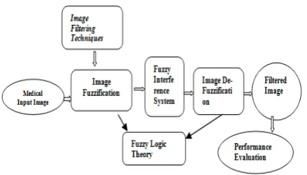
Fig. 1. Flow Chart of the Proposed System
C. Medical Input Image
Medical Images will be taken from renowned hospitals such as District Hospital Durg, BSR Apollo and some of the medical images may be directly taken from open sources. Medical images will include MRI, mammograms, radiographs etc. of various parts of human body. Some preprocessing will be applied over the images taken such as image resizing, image reformatting, image cropping etc.
D. Image Filtering Techniques
Some image filtering techniques will be used such as smoothing filters, sharpening filters and contrast enhancement filters. Smoothing filters will include Gaussian filters. Sharpening filters will be canny edge detectors, Sobel and Prewitt edge detectors. Contrast enhancement technique will use a suitable function such as sigmoid function.
E. Parameter Estimation for Fuzzification and Defuzzification
Image filtering techniques and their mathematical equations will be fuzzified. The fuzzification function will employ some of the membership functions. A set of fuzzified methods will be developed; may be called as Fuzzy interference system. After enhancing the input images by using fuzzified filters, the results will be defuzzified using again the fuzzy membership functions. The goal of parameter estimation is to find the best values for a set of model parameters. Here parameters estimation means taking the value of k- nearest neighboring pixels from all directions (360 degree) and finding the mean and variance and then estimating best intensity pixel value and transform the image visualization according that parameter.The images after enhancement will be stored as filtered images.
Contrast Enhancement: In the current studies, adaptive
attempts to exploit this object coherence by enhancing the contrast within a subrange of the intensity values at the expense of the remaining intensity values. As shown in Fig.2, a subrange or window of the data is chosen and these intensity values are linearly remapped to occupy the entire range of the display device. A single such mapping is applied to the entire image. Intensity values lying above or below the window are mapped to the maximum and minimum possible intensity values, respectively. The result is that the contrast of pixel to see all the information of interest in a clinical image.
While standard windows can be selected to show particular areas of interest for a given anatomical region and imaging modality, manual intervention is usually necessary for the best results. An alternative technique for contrast enhancement which has been widely used is global histogram equalization [2]. In this method, the intensity values in the image are altered such that the resulting image has a constant intensity histogram.
This transformation may be accomplished by the use of the cumulative distribution function of the pixel intensities as the intensity remapping function.Such images utilize the available display levels well, but because the contrast enhancement is based on the statistics of the entire image, some levels will be used for the depiction of parts of the image which are diagnostically unimportant, such as the background site. Selection of the sites was done with the collaboration. The set of normal chest images were chosen from CT scans of five separate patients who were classified as having no pathology in the areas of interest.
The images were obtained on a Technicare 2060 CT scanner; the preliminary selection of images was done by an experienced radiologist. The images were taken from the scanner in digital form. The intensities in each image were calibrated in Hounsfield units, with the CT intensities approximately in the range – 1000 to + 1200; their spatial resolution was 512 X 512 pixels. From about 100 slices in five patients, 32 slices were chosen. Adjacent slices were avoided to maximize anatomical differences between slices and reduce the possibility of memorization of normal variation by the observer. These 32 images were used to generate images for observer training and for the conduct of the actual Lesion Site Selection: In each image, four sites, two sites in the lungs and two in the mediastinum, were chosen for the insertion of artificial lesions.
The criteria for site selection were the presence of appropriate natural anatomy and the prevalence of real lesions at that site in clinical practice. Similar, but not identical, sites were chosen in each base image. Generation of Artificial Lesions. A Gaussian intensity profile was chosen to approximate that which would be generated by a spherical tumor; the widths of the artificial lesions were chosen as appropriate to appear in the given field (lung or mediastinum). The intensity profile for a given lesion is given by Equation 6.
, exp (6)
The variance of the Gaussian determines the width of the lesion in pixels. There are two variances given in the table for each field (lung or mediastinum); the lesions corresponding to these two variances will be referred to as the narrow and wide lesions. These widths correspond visually to a noticeable difference in lesion size. The peak intensity of the lesion is given by A (Q); it depends on the local neighborhood fl of the lesion site, where Q is a square of side 5u, centered at (x, y). The function A (a) must be chosen such that the lesion intensity is not too small, in which case insufficient information will be conveyed to the observer and the observer’s decisions will proceed by guesswork, not too large, in which case the observer will make a correct choice on every trial and no discrimination of the contrast enhancement methods will be possible.
A fixed lesion intensity is unacceptable, since it is well known that the ability of the eye to detect contrast is strongly dependent on both the mean intensity and the presence of structure in the near background of the lesion site. In addition, there is evidence that the far background of the image also exerts influence on the detectability of the artificial lesion. Preliminary tests performed on the images used in this experiment indicated that the appropriate lesion intensities must be chosen on an image-by-image basis. This choice was based on a simple measure of the structural complexity, the average absolute value of the Laplacian of the image intensity in a neighborhood Q of the lesion site. It was assumed that the desired peak intensity of the Gaussian A (Ω) was given by eq.(7)a linear function of the average Laplacian value over the region fl which is a square 5u on a side centered at the position ( x , y ) of the lesion site.
Ω
|
,
|
(7)
V. RESULTS
Contrast Enhancement on the basis of minimum & maximum intensity value of images.In this section we compare the efficiency and performance for grayscale images with other well-known filters for impulse noise reduction.The idea behind these filters is that they try to calculate positive and negative correction terms in order to express the degree of noise for a certain pixel.
A. Fuzzy Derivative Estimation
Estimating derivatives and filtering can be seen as a chicken-and-egg problem; for filtering we want a good indication of the edges, while to find these edges we need filtering.
Calculating fuzzy derivative by applying fuzzy rule for 'small' fuzzy set
for(int t=0;t<8;t++)
fuzzyderiv[t]=SmallFuzzySet.ApplyRule(fofxsmall[0][t],
B. Fuzzy Smoothing
To compute the correction term for the processed pixel value, we use a pair of fuzzy rules for each direction. The idea behind the rules is the following: if no edge is assumed to be present in a certain direction, the (crisp) derivative value in that direction can and will be used to compute the correction term. The first part (edge assumption) can be realized by using
the fuzzy derivative value, for the second part(filtering) we will have to distinguish betweenpositive and negative values.
Fuzzy smoothing calculation membership values for 'positive' and 'negative' fuzzy sets
for(int t=0;t<8;t++)
x( i , j ) = k1*( i , j ) 2B-1 + k2*( i , j ) 2B-2+...+ kB-1*( i , j ) 2 + kB*( i , j )
fofxpositive[t]=PositiveFuzzySet.fofx(simpderiv[0][t],L);
fofxnegative[t]=NegativeFuzzySet.fofx(simpderiv[0][t],L);
C. Defuzzification
Calculating correction term (defuzzification)
delta=0.0;
for(int t=0;t<8;t++)
delta=delta+(positivetruthness[t]-negativetruthness[t]);
delta=((double)L/8.0)*delta;
intoutval=inval+(int)delta;
imgout.setPixel(r,c,outval);
Fig. 2.Original image.
Fig. 3.Fuzzy Enhanced Image.
VI. CONCLUSION
Present approach has implemented a contrast Enhancement technique in which a medical image is given as the input, if the input image is color image then it is converted in to gray scale in order to reduce the processing time the contrast of input image is enhanced.
REFERENCES
[1] Mahmud Saeidi et al., “Noise Reduction in Image Sequences using an Effective Fuzzy Algorithm”, World Academy of Science, Engineering and Technology, page. 351-356, 43, 2008.
[2] PU-YIN LIU et al., “ Fuzzy techniques in image restoration research-a survey”, International Journal of Computational Cognition Volume 2, Number 2, Pages 131–149, June 2004 .
[3] Dimitri Van De Ville et al., “Noise Reduction by Fuzzy Image Filtering”, IEEE Transactions on fuzzy systems, volume. 11, NO. 4, page. 429-436, AUGUST 2003.
[4] Dzung L. Pham et al., “Adaptive Fuzzy Segmentation of Magnetic Resonance Images”, IEEE TRANSACTIONS ON MEDICAL IMAGING, volume. 18, NO. 9, page. 737-752, September 1999.
[5] S. Moein et al., “A Novel Fuzzy-Neural Based Medical Diagnosis System”, World Academy of Science, Engineering and Technology, 37, page. 157-161, 2008.
[6] Harish Kundra et al., “Image Enhancement Based On Fuzzy Logic”, IJCSNS International Journal of Computer Science and Network Security, volume.9 No.10, page. 141-145, October 2009.
[7] Mahesh T R et al., “Noise reduction by using fuzzy image filtering”, Journal of Theoretical and Applied Information Technology, Islamabad PAKISTAN, volume.15, No.2, page. 115-120, 2010.
[8] Nguyen Minh Thanh et al., “Image Denoising Using Adaptive Neuro-Fuzzy System”, IAENG International Journal of Applied Mathematics, page 36:1, IJAM_36_1_11, 2007.
[9] I. Laurence Aroquiaraj et al., “Comparative Analysis of Speckle Filtering Techniques International”, Journal of Recent Trends in Engineering, volume. 2, No. 2, page. 120-123, 2009.
[10] AbdallahA et al., “Edge Detection in Digital Images Using Fuzzy Logic Technique”, World Academy of Science, Engineering and Technology, 53, page. 252-257, 2009.
[11] Kim, H.M., and Mendel, J.M.. ”Fuzzy basis functions: Comparisons with other basis functions.” IEEE Trans. On Fuzzy Systems, 3, 1995, page. 158–168.
[12] Kam H., Hanmandlu M., and Tan W., “An Adaptive Fuzzy System for Smoothing Noisy Images”, IEEE, 2003
[13] A fuzzy filter for images corrupted by impulse noise,” IEEE Signal Processing Lett., volume. 3, page. 168–170,June 1996
[14] E. Kerre and M. Nachtegael, Eds., FuzzyTechniques in Image Processing. New York Springer-Verlag, 2000, volume. 52,Studies in Fuzziness and Soft Computing.
[15] AlirezaBehrad, Ali Shahrokni, Seyed Ahmad Motamedi,