Scholarship@Western
Scholarship@Western
Electronic Thesis and Dissertation Repository
8-14-2013 12:00 AM
Fluorescent Cytidine Analogues
Fluorescent Cytidine Analogues
Kirby J. Chicas
The University of Western Ontario
Supervisor
Robert. H.E. Hudson
The University of Western Ontario Graduate Program in Chemistry
A thesis submitted in partial fulfillment of the requirements for the degree in Master of Science © Kirby J. Chicas 2013
Follow this and additional works at: https://ir.lib.uwo.ca/etd
Part of the Organic Chemistry Commons, and the Other Chemistry Commons
Recommended Citation Recommended Citation
Chicas, Kirby J., "Fluorescent Cytidine Analogues" (2013). Electronic Thesis and Dissertation Repository. 1576.
https://ir.lib.uwo.ca/etd/1576
This Dissertation/Thesis is brought to you for free and open access by Scholarship@Western. It has been accepted for inclusion in Electronic Thesis and Dissertation Repository by an authorized administrator of
(Thesis format: Monograph)
by
Kirby Chicas
Graduate Program in Chemistry
A thesis submitted in partial fulfillment of the requirements for the degree of
Master of Science
The School of Graduate and Postdoctoral Studies The University of Western Ontario
London, Ontario, Canada
ii
Abstract
Two luminescent cytidine analogues have been synthesized in order to perform single
nucleotide polymorphism (SNP) analysis by fluorescence spectroscopy. Herein is described
the synthesis of 6-pyrenylpyrrolocytidine (PypdC), its photophysical characterization, and its
subsequent incorporation into oligodeoxynucleotides (ODNs). The behavior of PypdC in
ODNs is described as well as fluorescence intensity changes with respect to the match and
mismatch cases. In order to obtain a greater understanding of pyrene’s interaction with
pyrrolocytidine, a congener, pyrenyl ethynyl cytidine (PyEtdC) was synthesized. The
congener was photophysically studied and prepared for oligo synthesis
Keywords
Nucleic Acid, Cytidine, Fluorescent Nucleoside Analogue, Single Nucleotide Polymorphism,
Pyrene
iii
Table of Contents
Abstract ... ii
Table of Contents ... iii
List of Tables ... v
List of Figures ... vi
List of Schemes ... viii
List of Abbreviations ... ix
List of Appendices ... xii
Chapter 1 - Introduction ... 1
1 Nucleic Acids ... 1
1.1 Modified Nucleic Acids ... 3
1.1.1 Therapeutics – Small Molecules and Antisense ... 3
1.1.2 Diagnostics – Fluorescent Nucleic Acids ... 5
1.2 Fluorescent C Analogues ... 7
1.3 Fluorescence Characterization of C Analogues ... 11
1.4 DNA Synthesis – Incorporation of Modified Nucleosides ... 14
1.5 Objective ... 16
1.6 References ... 17
Chapter 2 – Pyrenylpyrrolocytidine ... 19
2 Introduction: Precedence and Development ... 19
2.1 General pC Synthesis ... 23
2.2 Results and Discussion ... 25
2.2.1 Towards PypdC ... 25
2.2.2 Synthesis of PypdC ... 27
iv
2.2.4 Nucleoside Fluorescence ... 34
2.2.5 Oligonucleotide Stability ... 38
2.2.6 ODN Fluorescence ... 40
2.3 Conclusions and Future work ... 41
2.4 Experimental ... 43
2.4.1 Synthetic Procedures and Characterization ... 43
2.5 References ... 48
Chapter 3 – Pyrenylethynylcytidine... 50
3 Introduction – Serendipity... 50
3.1 Results and Discussion ... 51
3.1.1 Towards PyEtdC in DNA ... 51
3.1.2 Synthesis of PyEtdC ... 54
3.1.3 PyEtdC Photophysics ... 55
3.1.4 Conclusions and Future Work ... 59
3.2 Experimental ... 60
3.2.1 Synthetic Procedures and Characterization ... 60
4 Summary and Conclusion ... 64
Appendix ... 66
v
List of Tables
Table 1.1 - Et(30) polarity values in comparison to dielectric constants. ... 14
Table 2.1 – Coupling efficiency of the PypdC phosphoramidite... 31
Table 2.2 – Photophysical summary of PypdC ... 36
Table 2.3 – Thermal denaturation of CFTR Mod and control ... 39
Table 2.4 – Photophysical summary of CFTR Mod ... 41
vi
List of Figures
Figure 1.1 – The central dogma of molecular biology... 2
Figure 1.2 – Watson-Crick hybridization of the purines (G / A) and the pyrimidines (C / T) in antiparallel strands. ... 2
Figure 1.3 – Entecavir (left) & Clofarabine (right) ... 3
Figure 1.4 – [bis-o-(aminoethoxy)phenyl]pyrrolocytosine, R = PNA ... 4
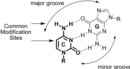
Figure 1.5 – Watson Crick bonding of G and C and common modification sites of C. ... 7
Figure 1.6 – Tricyclic cytosine (tC) ... 8
Figure 1.7 - 5-(fur-2-yl)-2'-deoxycytidine (CFU) ... 9
Figure 1.8 – Thiophen-2-yl pC ... 10
Figure 1.9 – DNA synthesis utilizing phosphoramidite chemistry. ... 15
Figure 1.10 – Pyrene modified C analogues: PypdC (left), PyEtdC (right) ... 16
Figure 2.1 – The pyrimidines, the alkynypyrimidines, and their 5-endo dig products ... 19
Figure 2.2 – Aliphatic vs. aromatic substituted pCs and their quantum yields in ethanol... 20
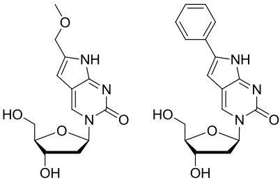
Figure 2.3 - MepdC & phenylpyrrolocytosine-N1-methylene carboxylate ... 20
Figure 2.4 - MmepdC & PhpdC ... 21
Figure 2.5 – Fluorescence intensity changes with respect to complementary base ... 22
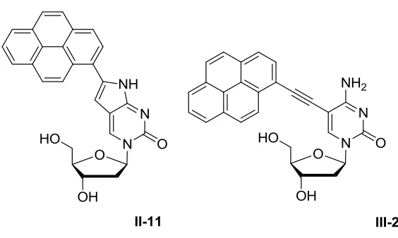
Figure 2.6 – 6-(1-pyrenyl)pyrrolodeoxycytidine (PypdC)... 23
Figure 2.7 – The Watson Crick binding faces of a) furanouracil; b) pC; c) G ... 24
Figure 2.8 – Luminescence profile of PypdC at 2.2 μM in ethanol ... 35
vii
Figure 2.10 – Normalized luminescence of PypdC vs. pyrene. ... 37
Figure 2.11 – Normalized lumincescence of PhpdC and PypdC ... 38
Figure 2.12 – Fluorescence intensity change of CFTR Mod against G,A,C,T and in the ss state at 1.5 μM ... 40
Figure 2.13 – Possible PypdC excimer based probe ... 42
Figure 2.14 – HPLC trace of a failed synthesis towards a new PypdC probe ... 42
Figure 3.1 – PypdC (left) & PyEtdC (right) ... 51
Figure 3.2 – Luminescence profile of PyEtdC in ethanol at 1.1 μM ... 55
Figure 3.3 - Luminescence profile of PyEtdC in water at 1.1 μM normalized to the highest intensity peak in Figure 3.2 ... 55
Figure 3.4 – PyEtdC luminescence profile vs. pyrene. ... 56
Figure 3.5 – Normalized fluorescence of PhpdC and PyEtdC ... 57
viii
List of Schemes
Scheme 2.1 – General methodologies of pC synthesis ... 25
Scheme 2.2 - First attempt towards PypdC ... 26
Scheme 2.3 – Second attempt towards PypdC... 27
Scheme 2.4 – Synthesis of PypdC phosphoramidite ... 29
Scheme 2.5 – The trityl ON / OFF methods of ODN purification ... 33
Scheme 3.1 – Proposed route to PyEtdC phosphoramidite ... 52
ix
List of Abbreviations
A – adenine
Ac – acetyl
AcOH – acetic acid
ALL – acute lymphoblastic leukemia
a.u. – arbitrary unit
BDF – base discriminating fluorophore
boPhpC – [bis-o-(aminoethoxy)phenyl]pyrrolocytosine
Bz - benzoyl
C – cytosine
CFTR – Cystic Fibrosis transmembrane conductance regulator
CPG – controlled-pore glass
DCM – dichloromethane
dmf – dimethylformamidino
DMF - dimethylformamide
DMSO – dimethylsulfoxide
DMT – dimethoxytrityl
DMTCl – dimethoxytrityl chloride
DNA – deoxyribonucleic acid
x
ESI – electrospray ionization
EtOH – ethanol
G – guanine
HBA – Hydrogen bond acceptor
HBD – Hydrogen bond donor
HD – Huntingtin’s disease
HTT - Huntingtin
MepdC – methylpyrrolodeoxycytidine
mM – millimolar
Mme
pdC – methoxymethylpyrrolocytidine
mod - modification
mRNA – messenger ribonucleic acid
MS – mass spectrometer
NBS – N-bromosuccinimide
nm – nanometer
ODN – oligodeoxynucleotide
ORN - oligoribonucleotide
pC – pyrrolocytosine
xi
PNA – peptide nucleic acid
PyEtdC – pyrenylethynyldeoxycytidine
PypdC - pyrenylpyrrolodeoxycytidine
RNA – ribonucleic acid
Rt – retention time
Sn – excited state level n
SNP – single nucleotide polymorphism
T – thymine
tC – tricyclic cytosine
TLC – thin layer chromatography
Tm – thermal melt transition temperature
TMS – trimethylsilyl
U – uracil
UPLC – ultra performance liquid chromatography
UV – ultraviolet
Vis – visible
Ǻ - Angstrom
ε – molar extinction coefficient
xii
Φf – quantum yield of fluorescence
ΦIC – quantum yield of internal conversion
ΦISC – quantum yield of intersystem crossing
°C – degrees celsius
λ – wavelength
λex – excitation maximum
λem – emission maximum
List of Appendices
Chapter 1 - Introduction
1
Nucleic Acids
The importance of nucleic acids is of no secret to us. We became privy when Watson and
Crick famously declared, “[we] discovered the secret of life” on February 28, 1953 at The
Eagle pub in Cambridge, England. Near to their place of work, the lunchtime destination
would become the location in which we were gifted the structure of DNA. The
elucidation however, was not solely the work of Watson and Crick. It was the
culmination of work dating to the mid 19th century.
Swiss chemist Friedrich Miescher isolated a non protein substance from the nuclei of
white blood cells in 1869. He would call this substance nuclein 1. He would isolate
nuclein from white blood cells and later from salmon sperm. Despite his efforts he found
the isolated substance tainted with protein. It wasn’t until 1889 that Richard Altmann
would isolate a protein free sample and call it nucleic acid 1.
Identification of the sugar, phosphate, and nitrogenous base components of nucleic acid
came by way of Levene and Jacobs in 1909 1. Female scientist, Rosalind Franklin from
Kings College at The University of London would later provide a key component to the
mystery of nucleic acid. It was her work with Maurice Wilkins that would produce the
X-ray diffraction pattern needed to lead Watson and Crick towards the double helix. The
elucidation of DNA structure was recognized with a Nobel Prize in 1962 and shared
amongst James Watson, Francis Crick, and Maurice Wilkins.
Francis Crick would frame the gravity of DNA through the central dogma of molecular
biology. It explained in a simplified manner the flow of genetic information: transcription
of DNA coding for phenotypic traits to messenger RNA (mRNA) in preparation for
translation into proteins and expression of those coded phenotypes (Figure 1.1). By this
model and still in line with our understanding, DNA is the blueprint defining our very
Figure 1.1 – The central dogma of molecular biology
We are by now well aware of the antiparallel nature of the duplex (Figure 1.2). We know
that nucleic acids are composed of a sugar phosphate backbone and of a nitrogenous base
whose strand direction has been defined by its ribose moiety. Much progress has been
made since 1953 and in 2003 the Human Genome Project was declared complete - the
complete sequencing of the approximately 20 500 genes or three billion chemical units
coding for the human being.
Figure 1.2 – Watson-Crick hybridization of the purines (G / A) and the pyrimidines (C /
1.1
Modified Nucleic Acids
The important and complex nature of DNA and RNA has generated a great demand for
tools and techniques to examine and treat nucleic acids. Modified nucleosides have been
successfully applied to both therapeutic and diagnostic technologies in nucleic acids.
1.1.1
Therapeutics – Small Molecules and Antisense
Small molecule nucleosides have attracted attention due to their use in the treatment of
cancer and viral diseases. These small molecules fall into one or both of the categories
typically used to identify modified nucleosides: (1) ribose modified and / or (2) base
modified nucleosides2. Research has led to the development of Entecavir for the
treatment of chronic hepatitis B virus infection and Clofarabine (Figure 1.3) for the
treatment of refractory acute lymphoblastic leukemia (ALL) in children.
Figure 1.3 – Entecavir (left) & Clofarabine (right)
Modified nucleosides have also found success in the world of antisense therapy.
Antisense therapy is a treatment for genetic disorders or infections. If a gene is known to
be responsible for a particular disease state, nucleic acids capable of binding
the messenger RNA (mRNA) of the gene can be synthesized in order to prevent
translation into protein. Binding of the exogenous strand with mRNA effectively silences
the gene and turns it "off". The off response is brought about by the requirement for
mRNA to be in single stranded state for translation3.
The synthetic nucleic acid is known as the "anti-sense" strand. The term antisense
describes the nucleic acids’ complementary base sequence to the gene's messenger RNA
Zamecnik and Stephenson4 first proposed the concept of antisense therapeutics in 1978.
Their seminal publication detailed the synthesis of a 13 mer oligoribonucleotide (ORN),
complementary to a sequence in the respiratory syncytial virus genome. They suggested
that the ORN could be stabilized by terminal modifications and showed evidence of
antiviral activity4.
The Hudson group has forayed into antisense technologies through the incorporation of
boPhpC (Figure 1.4) into peptide nucleic acids with the help of the Corey Group at The
University of Texas Dallas Southwestern Medical School. They planned to use PNA
oligomers for the selective inhibition of mutant Huntington (HTT) protein. Mutant HTT
is responsible for Huntington’s disease (HD), an incurable neurological disorder. The
Corey group reasoned that silencing of the mutant HTT protein would be a useful
strategy for the treatment of Huntingtin’s disease. When oligos containing the boPhpC moiety were added at 1 μM concentration, selective HTT inhibition was observed5
. The
introduction of one or two boPhpC substitutions did not greatly increase the potency of
inhibition of mutant HTT or improve selectivity. Alternatively, introduction of three or
four boPhpC bases significantly eroded selectivity5. The boPhpC insert included the
added advantage of intrinsic fluorescence which permitted visualization and localization
of intracellular PNAs without the need for an appended fluorophore. Confocal
fluorescence microscopy showed punctate intracellular PNA distribution consistent with
known uptake / distribution mechanisms for PNA5.
1.1.2
Diagnostics – Fluorescent Nucleic Acids
Of the many diagnostic tools available for nucleic acids, none may find the ease of use
and potential for broad application as readily as fluorescent nucleoside analogues.
Possessing no analytically exploitable fluorescence 6, natural nucleosides are readily
given fluorescence properties enabling them to be used for spectroscopic studies. The
combination of hybridization specificity and fluorescence enables them to fulfill a
number of requirements needed for an ideal detection device or assay.
Ideal detection devices and assays must offer great selectivity, excellent sensitivity, ease
of use and low cost of production 7. Nucleic acids have long been admired for their
genetic coding properties but have recently emerged as important materials for molecular
diagnostic technologies. Nucleic acids satisfy ideal detection device and assay
requirements by a number of assets such as specific Watson-Crick base pairing, high
stability (DNA), low cost of synthesis and excellent adaptability to modifications 7.
Nucleic acids are often used in combination with fluorescence spectroscopy for a number
of reasons: (1) a large selection of fluorophores for nucleic acid conjugation exists; (2)
there are minimal health risks associated with fluorophore handling; (3) there is
instrumentation capable of detection at ultralow concentrations; (4) portability of
instrumentation for on-site detection; (5) the relatively long shelf life of fluorophores 7.
Fluorescent nucleoside analogues have shown utility in a wide range of applications
including but not limited to single nucleotide polymorphism (SNP) detection 8a - e, nucleic
acid structure and function, and microenvironmental studies. Structure and function
experiments have allowed for the resolution of hybridization events 9, folding 10,
conformational change 11, and enzyme action 12. Microenvironmental probing studies
with fluorescent nucleosides have shown nucleobase damage 13,
depurination/depyrimidation 14, and base flipping 10. To this day new uses for fluorescent
nucleoside analogues continue to emerge broadening the utility of the technique and
driving development in the field.
Numerous modifications have been explored to introduce favourable fluorescent
their congeners such as the Alexa dyes have been appended to oligonucleotides by linkers
to the sugar phosphate backbone or have been tethered to nucleobases. Modifications
have also been conjugated to the base or utilize the base as the fluorophore itself. Due to
the seemingly limitless possibilities for modification, numerous classifications have been
assigned to the fluorescent nucleoside field. Tor has divided the field into five categories;
(1) chromophoric base analogues; (2) pteridines; (3) expanded nucleobases; (4) extended
nucleobases and (5) isomorphic bases15. Wilhelmson has segmented the field in terms of
internal (base) and external (sugar or phosphate) modifications 16. Further classifications
have been offered by Asseline 17 and terms such as base discriminating fluorophore
(BDF) have been introduced by Saito 18.
The Hudson group focuses upon modifications of cytosine for the fluorescent probing of
nucleic acids. Typical modifications fall into two fields capable of canonical base
pairing: base analogues possessing pendant fluorophores (extrinsic) and intrinsically
fluorescent nucleoside analogues 19. These terms are used frequently within the Hudson
group to classify C modifications.
The pendant class often has an advantage in that higher overall brightness (defined as
× ) is obtained by the attachment of a well characterized fluorophore. The high
brightness is due to the combination of efficient luminescence (high quantum yield, )
and large molar absorptivity coefficients (). Despite the usually favourable
photophysical properties, this class suffers from a number of drawbacks. The use of a
fluorophore covalently bound by a tether to a nucleobase can allow for the independent
movement of the fluorophore making interpretation difficult. Furthermore, due to the
distance of the fluorophore from the base-pairing moiety, fluorescence does not directly
report on the bases’ environment. Thus, observed fluorescence does not reflect
hybridization at a specific base, protonation or other electronic changes at the site of
interest 19.
Intrinsically fluorescent base analogues are those in which fluorescence is observed from
the nucleobase itself and not from an appended (extrinsic) moiety. Intrinsically
microenvironmental changes, thus enabling one to elucidate events occurring in close
proximity to the nucleobase. Modest chemical modifications can also yield stunning
photophysical properties allowing them to be competitive with traditional fluorescent
probes. Historically, intrinsically fluorescent base analogues have been able to
communicate hybridization change.
The Hudson group continues to work towards new fluorescent C analogues for
applications in both antisense and diagnostic systems.
1.2
Fluorescent C Analogues
Described by Albrecht Kossel in 1903 20, the pyrimidine cytosine enjoys a three hydrogen
bond Watson-Crick face for complementary pairing with guanine and shows good
tolerance for chemical modifications (Figure 1.5).
Figure 1.5 – Watson Crick bonding of G and C and common modification sites of C.
Cytosine is readily halogenated at the 5-position, providing a handle for well established
carbon-carbon bond forming chemistry. Modification of the exocyclic amine also
provides entry into fluorescent C analogues. Modifications of the C scaffold either by
manipulation at the 5-position or of the amine generally produces a C analogue where
modifications tend to be nonperturbing in nucleic acids and in many cases increase
duplex stability by favourable base stacking or additional hydrogen bond engagement of
G. Modification at the 6-position is generally regarded to be detrimental due to the steric
interaction of the sugar moiety in the anti-glycosidic conformer 21.
A wide variety of fluorescent C analogues have been developed over the years, each with
their own unique characteristics and applications. The following is a brief window into
the scope of fluorescent C analogues.
Tricyclic cytosine (tC, Figure 1.6) was first reported in 1995 for use in antisense
systems.
Figure 1.6 – Tricyclic cytosine (tC)
Studies on tC showed melt temperature increases with respect to 5-methylcytidine 22 and
in 2001, spectrophotometric studies were undertaken 23. Wilhelmsson showed that tC
could be selectively excited over DNA (λabs = 260 nm) at a wavelength of 375 nm with
corresponding emission at 505 nm 23. Wilhelmsson reported increased melt temperatures
24
as was also reported by Matteucci in 1995. Quantum yield determinations by Albinsson
et al. showed moderate quantum yield values for the free nucleoside (Φ = 0.13) in water
25
. Incorporation of tC into ODNs produced slightly greater quantum yields than that of
the free nucleoside. Values ranged from 0.17 – 0.24 in the ss form and 0.16 – 0.21 in the
ds form. Very little change in quantum yield was observed with respect to flanking bases
or incorporation into dsDNA making it a relatively insensitive analogue to
RNA 28 polymerase experiments in their respective ribo and deoxy forms, and in DNA /
protein interaction experiments 29.
The Tor group at UCSD investigated a number of conjugated five member ring systems
based upon 2-phenylfuran. 2-phenylfuran is a fluorophore (λex = 280 nm; λem. = 340 nm)
with a desirably high molar extinction coefficient of 20 000 M-1cm-1 and good quantum
yield (Φ = 0.4) 30
. By the conjugation of a furan moiety to the six member aromatic
system of cytosine (Figure 1.6), Tor hoped to emulate the favourable properties
2-phenylfuran. Following a previously reported synthesis 31, Tor produced the furan labeled
C analogue from 5-iodo-2-deoxyuridine.
Figure 1.7 - 5-(fur-2-yl)-2'-deoxycytidine (CFU)
Unfortunately the quantum yield of the CFU nucleoside proved to be less than ideal with a
value of 0.02 in water. Although possessing a low efficiency, the CFU base could still be
selectively excited over DNA at ~ 310 nm with emission at ~ 440 nm. The Tor group
proceeded to investigate the fluorescent C analogue as a potential candidate for the
detection of 8-oxoG. CFU proved sensitive to 8-oxo-G and relatively insensitive to
unmodified G 32. Anticipating transverse mutation, fluorescence response with respect to
T was also measured and determined to provide the greatest fluorescence intensity.
Moreover, the emission wavelength was observed to change with complementary base.
From these observations it was proposed that CFU could facilitate rapid and
non-destructive real time fluorescence based methods for the in vitro monitoring of oxidative
stress 32.
The Tor group has since sought ways to increase the efficiency of their C analogues. In
the group turned their attention to the pC core which had been shown to possess
favourable fluorescence characteristics in other systems.
Figure 1.8 – Thiophen-2-yl pC
The group synthesized both the deoxy and ribose variants of the thiophen-2-yl pC base.
Foregoing acylation of the exocyclic amino group (Discussed in Chapter 2, Scheme 2.1)
it was observed that the intermediate nucleoside resisted annulation by copper and
formation of the pyrrole ring. The intermediate nucleoside was screened against metal
catalysts to induce cyclization. It was found that sodium tetrachloroaurate(III) dihydrate
produced the desired cyclized product in low to moderate yield 33. Photophysical
characterization of the deoxy and ribose thiophen-2-yl pC nucleosides yielded little to no
difference in their photophysical properties. It was found that they underwent excitation
at ~ 370 nm and emission at ~ 471 nm in water with an efficiency of ~ 0.42. A greater
efficiency was observed in dioxane (~ 0.48) 33 with red shifted absorption and blue
shifted emission corresponding to decreased Stokes shifts with respect to those in water.
The extinction coefficients when taken into consideration with quantum yield led to
brightness factors that were approximately 14 – 24 times brighter than those of the MepC
or MepdC.
Characteristics of C analogues range from the insensitive (tC) to the sensitive and from
the highly fluorescent to nominally fluorescent. Our work develops the scope of C
analogues in the hopes of obtaining new favourable fluorescence properties or improved
phenylpyrrolocytosine developed from pyrrolocytosine and is further discussed in
Chapter 2.
1.3
Fluorescence Characterization of C Analogues
Traditionally the Hudson group has obtained a particular set of values for photophysical
characterization of C analogues. The values obtained allow for the comparison of
fluorescent C analogues and the development or improvement of nucleic acid probes.
Typically photophysical characterization includes: excitation and emission spectra in
ethanol and water; fluorescence quantum yield determination in ethanol & water; molar
absorptivity determinations in ethanol & water. In addition to the above mentioned
values, the Hudson group has recently added the Et(30) method of polarity sensitivity
measurement.
Excitation and emission spectra provide insight into the excitation and relaxation
pathways used in the molecule of interest. The excitation spectra in conjunction with
UV/Vis, details the excited states involved upon photon absorption. Peaks of defined
structure or shoulders become clear indicators of excitement of ground state electrons
into a multitude of Sn levels. Mirror image emission spectra detail a relaxation process
which participates in the release of a photon from the excited Sn state. More often than
not, a mirror image is not obtained as the rate of internal conversion tends to be much
greater than the rate of fluorescence emission. This rate difference manifests itself as a
radiationless relaxation from Sn S1 followed by photon emission and relaxation from
S1 S0 which we observe as a single fluorescence peak lacking significant structure.
From the excitation and emission spectra we further derive Stokes shift thus determining
the energy difference between the S0 and S1 states.
Fluorescence quantum yield (Φf) provides a measure of the fluorescence efficiency for a
molecule. Φf is defined as the ratio of the numbers of photons emitted over the number of
photons absorbed, it is a process that competes with internal conversion (IC); inter system
crossing (ISC) and other functions of excited state decay. Excited state decay can be
expressed as the sum of the possible pathways or, 1 = Φf + ΦIC + ΦISC + Φn. The higher
photonic emission. Vice versa, the lower the fluorescence quantum yield, the more the
molecule dissipates energy by non fluorescent processes. Quantum yield (a measure of
fluorescence efficiency) cannot be directly ported or compared to other molecules as it is
a concentration independent value and provides little information on the luminosity of
molecules at varied concentrations.
Molar absorptivity also known as the molar extinction coefficient (ε) is expressed by the
Beer-Lambert law, A = εlC, where A = absorption; ε = molar extinction coefficient; l =
path length; C = concentration. It relates the concentration of a molecule to its
absorptivity making it a valuable measure for concentration determinations. More
specifically, ε is a measure of the probability, favourability, or likelihood of a transition
(absorption) at a particular wavelength. The product of epsilon taken in conjunction with
fluorescence quantum yield (ε x Φf) is a value deemed brightness. The value can be
conceptualized as a measure of fluorescence efficiency per unit volume and allows for
the direct comparison of two molecules that exhibit fluorescence.
Recently, the Hudson group has begun polarity sensitivity measurements in the hopes of
probing duplexes and biological pockets. Microenvironmental understanding is key since
intra and intermolecular forces are dependent on their immediate surroundings.In the
past many methods of polarity measurement have been used. Unfortunately many of
these studies utilize the dielectric constant - a bulk solvent property expressed in units of
Debye (D). The value represents a molecule’s ability to attenuate an electric field
generated between electrodes relative to vacuum 34. It can be thought of as the ability of
a group of molecules to respond to the applied field and reorganize to minimize the
generated potential. A solvent such as water is highly capable of attenuating the
generated field and would therefore have a high dielectric constant. Conversely,
hydrocarbons have little or no ability to respond to an applied field and attain low
dielectric constants. These bulk values do not represent the first or second solvation
spheres surrounding a molecule and do not represent the environment within a small
biological cavity 35. Microenvironmental analysis became plausible when spectroscopic
studies were approached. Recently the Et(30) scale developed by Dimroth and Reichardt
transfer band of a pyridinium betaine dye in a solvent or mixture of solvents. It is thought
that the ET(30) value then reflects the polarity of the immediate environment surrounding
the dye as it is the first or second solvation spheres that cause changes in the measured
transfer band.
The polarity sensitivity of a new molecule can be determined by plotting the stokes shift
of the new molecule in varied mixtures of dioxane / water against the predetermined
Et(30) value for the same mixture. It has been shown that the linear relation typically
produced is of greater reliability than other spectroscopic methods and the dielectric
methods for polarity determinations 23. The slope of the resultant line is termed the
polarity sensitivity of the molecule. The greater the slope of the line, or the greater the
change in stokes shift with respect to change in solvent composition, the greater the
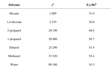
polarity sensitivity. Table 1.1 contains ET(30) values for common solvents displaying the
Table 1.1 - Et(30) polarity values in comparison to dielectric constants.
Solvents εa ET(30)b
Hexane 1.889 31.0
1,4-dioxane 2.219 36.0
2-propanol 20.190 48.4
1-propanol 20.800 50.7
Ethanol 25.290 51.9
Methanol 33.520 55.4
Water 80.180 `63.1
a
in units of Debye (D), b kcal mol-1
1.4
DNA Synthesis – Incorporation of Modified
Nucleosides
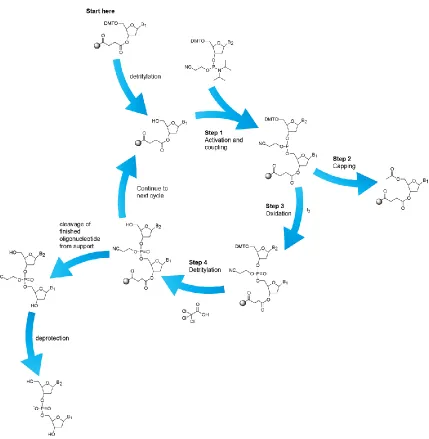
Currently the most popular method of oligodeoxynucleotide (ODN) synthesis is the
phosphoramidite approach (Figure 1.7) 36. Construction of the DNA biopolymer follows
sequential additions of the phosphoramidite monomer to an already coupled base tethered
Figure 1.9 – DNA synthesis utilizing phosphoramidite chemistry.
The already present base undergoes detritylation by treatment with low percentage DCA
or TCA in DCM - typically at percentages below 3% m/v. Detritylation affords the free
5` hydroxyl which reacts readily with a monomer activated by treatment with tetrazole.
To ensure easy purification, a capping step is involved to minimize the ODN products by
“sealing” unreacted 5’ hydroxyls through acetylation. Capping is followed by oxidation
of the phosphite triester to create the cyanoethyl protected phosphate. The cycle is
support and deprotection of the phosphate backbone is undertaken by heating in the
presence of ammonia.
1.5
Objective
The aim of the research described herein is to expand the scope of fluorescent analogues
by the synthesis and characterization of new fluorescent C analogues. We aim to test the
limits of base discriminating fluorescence and duplex stability of these new analogues by
the conjugation of the large and bright fluorophore pyrene.
We will describe the development of new synthetic procedures towards pyrene modified
C analogues (Figure 1.7) as well as their incorporation or progress towards synthetic
ODNs. We will then outline their ability to act as sensors in nucleic acids by the means of
fluorescence spectroscopy.
Figure 1.10 – Pyrene modified C analogues: PypdC (left), PyEtdC (right)
The analogues will have their photophysical properties studied in the nucleoside form.
We will gain insight into the interactions of substituent and base electronics and the
consequences that these electronics may have on the analogues as fluorescence sensors.
Specifically we aim to answer whether or not the fluorescence we observe from these
pyrene modified C analogues is attributed to the pyrene substituent, the base, or some
1.6
References
1. Lagerkvist, U. DNA Pioneers and their legacy. Yale University Press, New Haven and London, 1998.
2. Zhang, Li He; Xi, Zhen; Chattophaya, Jyoti; Medicinal Chemistry of Nucleic Acids. John Wiley & Sons, Hoboken, N.J., 2011.
3. Crooke, Stanley T.; Antisense Drug Technology: Principles, Strategies, and Applications. CRC Press, Boca Raton, 2008.
4. Zamecnik, P.C.; Stephenson, T.C., Proc. Natl. Acad. Sci. USA. 1978, 75(1), 280 – 284
5. Hua, J.X.; Dodd, David, D.W.; Hudson, R.H.E.; Corey, D.R., Bioorg. Med. Chem. Lett. 2009, 19 (21), 6181 – 6184.
6. Vaya, I.; Gustavsson, T.; Mianny, F.A.; Douky, T.; Markovitsi, D.J.; J. Am. Chem. Soc. 2010, 132 (34), 11834 – 11835.
7. Mayer, G.; The Chemical Biology of Nucleic Acids. 2010.
8. a) Okamoto, A.; Tainaka, K.; Saito, I., Chem. Lett. 2003,32 (8), 684-685. b)Okamoto, A.; Tanaka, K.; Fukuta, T.; Saito, I., J. Am. Chem. Soc. 2003,125 (31), 9296-9297. c) Hattori, M.; Ohki, T.; Yanase, E.; Ueno, Y., Bioorg. Med. Chem. Lett. 2012,22 (1), 253-257. d) Yoshida, Y.; Niwa, K.; Yamada, K.;
Tokeshi, M.; Baba, Y.; Saito, Y.; Okamoto, A.; Saito, I., Chem. Lett. 2010,39 (2), 116-117. e) Saito, Y.; Motegi, K.; Bag, S. S.; Saito, I., Bioorg. Med. Chem. 2008, 16 (1), 107-113.
9. Tinsley, R. A.; Walter, N. G., RNA-Publ. RNA Soc. 2006,12 (3), 522-529.
10.Sinkeldam, R. W.; Greco, N. J.; Tor, Y., Chem. Rev. 2010,110 (5), 2579-2619.
11.Zhang, C. M.; Liu, C. P.; Christian, T.; Gamper, H.; Rozenski, J.; Pan, D. L.; Randolph, J. B.; Wickstrom, E.; Cooperman, B. S.; Hou, Y. M., RNA-Publ. RNA Soc. 2008,14 (10), 2245-2253.
12.Wahba, A. S.; Esmaeili, A.; Damha, M. J.; Hudson, R. H. E., Nucleic Acids Res. 2010,38 (3), 1048-1056.
13.Greco, N. J.; Sinkeldam, R. W.; Tor, Y., Org. Lett. 2009,11 (5), 1115-1118.
14.Tanpure, A. A.; Srivatsan, S. G., Chem.-Eur. J. 2011,17 (45), 12820-12827.
15.Greco, N.J.; Tor, Y., Tetrahedron2007, 63 (17), 3515 – 3527.
16.Wilhelmsson, L.M., Q. Rev. Biophys. 2010, 43 (2), 159 – 183.
17.Asseline, U., Curr. Org. Chem.2006, 10 (4), 491 – 518.
18.Okamato, A; Saito, Y; Saito, I., J. Photochem. Photobiol. C-photochem. Rev.2005, 6 (2-3), 108 – 122
19.Dodd, D.W.; Hudson, R.H.E., Mini-Rev. Org. Chem. 2009, 6 (4), 378 – 391.
21.Schweize, M.P.; Kreishma, G.P., J. Magn. Reson. 1973, 9 (2), 334 – 337.
22.Lin, K. Y.; Jones, R. J.; Matteucci, M., J. Am. Chem. Soc. 1995,117 (13), 3873-3874.
23.Wilhelmsson, L. M.; Holmen, A.; Lincoln, P.; Nielson, P. E.; Norden, B., J. Am. Chem. Soc. 2001,123 (10), 2434-2435.
24.Engman, K. C.; Sandin, P.; Osborne, S.; Brown, T.; Billeter, M.; Lincoln, P.; Norden, B.; Albinsson, B.; Wilhelmsson, L. M., Nucleic Acids Res. 2004,32 (17), 5087-5095.
25.Tahmassebi, D. C.; Millar, D. P., Biochem. Biophys. Res. Commun. 2009,380 (2), 277-280.
26.Stengel, G.; Gill, J. P.; Sandin, P.; Wilhelmsson, L. M.; Albinsson, B.; Norden, B.; Millar, D., Biochemistry 2007,46 (43), 12289-12297.
27.Stengel, G.; Urban, M.; Purse, B. W.; Kuchta, R. D., Anal. Chem. 2010,82 (3), 1082-1089.
28.Stengel, G.; Purse, B. W.; Wilhelmsson, L. M.; Urban, M.; Kuchta, R. D., Biochemistry 2009,48 (31), 7547-7555.
29.Greco, N. J.; Tor, Y., Tetrahedron 2007,63 (17), 3515-3527.
30.Wigerinck, P.; Pannecouque, C.; Snoeck, R.; Claes, P.; Declercq, E.; Herdewijn, P., J. Med. Chem. 1991,34 (8), 2383-2389.
31.Greco, N. J.; Sinkeldam, R. W.; Tor, Y., Org. Lett. 2009,11 (5), 1115-1118.
32.Noe, M. S.; Rios, A. C.; Tor, Y., Org. Lett. 2012,14 (12), 3150-3153.
33.Sinkeldam, R.W.; Yitzhak, T., Org. Biomol.Chem. 2007, 5, 2523 – 2528.
34.Kosower, E.M., An Introduction to Physical Organic Chemistry. JohnWiley & Sons, Inc., New York, 1968, 259 – 264.
Chapter 2 – Pyrenylpyrrolocytidine
2
Introduction: Precedence and Development
Initially, with the intent of synthesizing 5-alkynylpyrimidines to determine their affect on
the biophysical properties of PNA oligomers 1, Hudson and coworkers “rediscovered” the
cyclization of N4-acyl-protected cytosine to pyrrolcytosine - an observation first made by
Ohtsuka that had gone largely underappreciated 2. During these studies, conditions were
defined for the synthesis of the simple cross-coupled products versus the annulated
furanouracil and pyrrolocytosine (Figure 2.1). It was found that the structurally simple
5-alkynylpyrimidine derivatives were luminescent 3; however, the similarly substituted
pCs were determined to be better fluorophores 4.
Figure 2.1 – The pyrimidines, the alkynypyrimidines, and their 5-endo dig products
Pyrrolocytosines possessing aromatic substitutions were discovered to be remarkably
good fluorophores; better than those with aliphatic substitution (Figure 2.2). The pCs
shown in Figure 2.2 are blue fluorophores ( em ~ 450 nm) except for the para-(N,N
-dimethylamino)phenyl which is bathochromically shifted (em ~ 500 nm) and the para
-nitrophenyl which displays weak orange fluorescence (em ~ 575 nm). Interestingly the p
-nitrophenyl exhibits an absorbance band that overlaps the emission of
6-phenylpyrrolocytosine (PhpC). Good luminescence is maintained in water as PhpC
displays a ~ 0.35, approximately 10-fold greater than that of the well established
Figure 2.2 – Aliphatic vs. aromatic substituted pCs and their quantum yields in ethanol
6-Methylpyrrolo-dC (MepdC), a fluorescent C analogue dating back to the late 1980s 2
was a relatively unexploited modification until the turn of the century. In the early 2000s,
methylpyrrolo-dC experienced a surge 5 in use and has since become one of the more
popular fluorescent C analogues. The popularity is likely due in part to its commercial
availability 6. Methylpyrrolo-dC has been shown to act similarly to C in terms of
hybridization selectivity and stability 7. MepdC has been used for the characterization of
the transcription bubble of T7 RNA polymerase 5, the kinetics of DNA repair by a human
alkyl transferase 8, and in investigations of the HIV-1-polypurine tract 9. With
methylpyrrolo-dC proving its utility in a number of experiments with considerably lesser
fluorescence than PhpC, it is not surprising that PhpC garnered much excitement within
the Hudson group
Hudson continued his investigation by converting the PhpC acetate to the Fmoc PNA
monomer followed by incorporation into PNA and DNA duplexes. It was found that the
PhpC containing PNA bound complementary DNA with stability on par to the
corresponding unmodified PNA. The PNA oligomers also displayed excellent sequence
discrimination for a complementary G versus mismatch that rival or best C 4.
The first pC studies in DNA carried out by Hudson included the synthesis and
incorporation of 6-methoxymethyl pyrrolodeoxycytidine (MmepdC) into ODNs for the
purpose of selective fluorimetric detection of guanosine-containing sequences. Prior to
the synthesis of the MmepdC analogue, it had been well known from PNA systems that
aromatic substitutions provided the most dramatic fluorescence response and that
interesting characteristics would be observed from a phenylpyrrolodeoxycytidine
(PhpdC) (Figure 2.4). The analogue was synthesized by the deoxyuridine route (vide
infra). It was found that the PhpdC phosphoramidite could be synthesized in three steps
with good overall yield 10.
Figure 2.4 - MmepdC & PhpdC
Thermal denaturation experiments of a centrally located PhpdC yielded a moderate
increase in duplex stability (+3.3 °C) 11 relative to dC while maintaining excellent
mismatch discrimination that was equal to or better than that of MepdC. Fluorescence
emission from the PhpdC ODN was 18 times greater than that of MepdC 11. Additional
fluorescence studies showed the ability of PhpdC to communicate the identity of the
It was proposed that modification of pyrrolocytosine with the large polycyclic aromatic
hydrocarbon, pyrene, would yield interesting fluorescence properties as it is a well known
fluorophore with a desirably high quantum yield (0.65) in ethanol. It was also thought
that the substitution would yield insight into a number of questions important to pC
understanding. We aimed to explore: (1) does conjugating highly fluorescent moieties to
pC overwhelm fluorescence identification of the complementary base? (2) How large can
a substitution become before it compromises duplex stability?
In regard to question (1), 6-(1-pyrenyl)pyrrolodeoxycytidine (PypdC) was suggested to
behave in one of two ways. (A) It would act like PhpC as a sensitive reporter group in
nucleic acid studies, or (B) the pyrene substituent would overwhelm any fluorophore-like
contribution from the nucleobase. If (B) were to be true it was imagined that the
hybridization dependent fluorescence would be lost. 0
200000 400000 600000
350 400 450 500 550 600 650
intens ity (au) wavelength (nm) SS G (match) A (mismatch) C (mismatch) T (mismatch)
Figure 2.6 – 6-(1-pyrenyl)pyrrolodeoxycytidine (PypdC)
In regard to question (2), duplex stability was expected to change in one of two ways:
either the duplex stability would increase through greater base stacking interactions or the
duplex stability would decrease due to the sheer size of the modified base. In a wild type
vs. mutant case as in boPhpC (Chapter 1), these stability changes could possibly manifest
themselves as such. In the case of a net stabilization for the match and mismatch cases,
little to no selectivity would be observed. However if fluorescence changes were
observed then a fluorimetric SNP probe would be possible. A reduction in duplex
stability would either increase match strand selectivity or in the case of severe
destabilization impair the target as a fluorimetric probe for G containing sequences and
SNP associated disorders. In the case of increased selectivity, an antisense ODN could be
developed.
2.1
General pC Synthesis
Synthetic routes towards the pC nucleoside typically follow one of two methodologies.
The general features of pC synthesis are illustrated in Scheme 2.1. The chemistry
utilized depends on the substitution desired at the 6 position of the pyrrole and the nucleic
acid required. For peptide nucleic acid (PNA), the most convenient starting point is the
cytosine or uracil nucleobase. For DNA / RNA structures, the deoxy- or ribonucleosides
are most commonly used.
The substrates are prepared for derivatization by halogenation at the C5 position, most
fused bicyclic structure is achieved by the intramolecular cyclization (5-endo dig
annulation) of the 5-alkynyl pyrimidine. The annulation reaction (Scheme 2.1, Approach
1) usually occurs under mild conditions for the uracil derivative, although there is some
substrate dependence. For instance, electron rich alkynes cyclize more rapidly than those
that are electron deficient. The cyclization reaction is metal 12a-f, base 13a-b or electrophile
14
catalyzed and is facilitated by conventional heating or microwave irradiation 15. Lewis
acidic, alkynophilic metals have proven to be effective for this type of reaction (M+ =
Cu(I) 2, Zn(II) 12c, Au(III) 12a-b, Hg(II) 12a, Pd(0) 12f). Utilizing approach one, the final
transformation is the atom exchange reaction to convert the furanouracil nucleobase to
pyrrolcytosine by treatment with ammonia 16. This step is critical, of course, because
furanouracil no longer pairs with any of the natural nucleobases. Complementarity to
guanine is manifested once the furanouracil is converted to the pyrrolocytosine (Figure
2.7).
Figure 2.7 – The Watson Crick binding faces of a) furanouracil; b) pC; c) G
The second approach starts with cytosine or cytidine in preparation for
cross-coupling by iodination of the base. In approach 2 (Scheme 2.1), partitioning of the
products between the simple cross-coupled 5-alkynylcytosine and the annulated
pyrrocytosine is controlled by the substrate. If the nucleobase is unprotected (pathway a),
and the conditions are not forcing, then the 5-alkynylcytosine derivatives are achieved in
good yield 17. For the intramolecular cyclization to occur (pathway b), acylation of the
N4 must be undertaken prior to Sonogashira chemistry. Using this substrate and elevated
occurs during which the benzoyl group is cleaved.The N-benzoylpyrrolocytosine is not
fluorescent and not stable to silica gel column chromatography. Thus, the benzoyl group
is removed to facilitate isolation and purification of the desired pyrrolcytosine.
Foregoing benzolyation of the exocyclic amine, cyclization has been reported using a
gold catalyst providing low to moderate yields 18.
Scheme 2.1 – General methodologies of pC synthesis
The best synthetic route towards a pC analogue is case specific and must be chosen
appropriately as each modification to the pC scaffold will generate its own synthetic
challenges.
2.2 Results and Discussion
2.2.1
Towards PypdC
Our work towards a pyrene modified pC can be divided in terms of the two synthetic
schemes (vide supra) generally utilized to synthesize pC analogues.
Initial attempts towards the target utilized the 2’-deoxy-5-iodouridine (II-1) starting
material. The iodinated nucleoside readily underwent tritylation under standard
conditions to produce the 2’-deoxy-5’-O-(4,4-dimethoxytrityl)-5-iodouridine (II-2) in
furanouracil intermediate (II-3) that would allow ready entry into the trityl protected
nucleoside via ammoniolysis. Attempts at a one pot conversion of the cross coupled
product via Cu(I) and Ag(I) catalysis after Sonogashira conditions with 1-ethynyl pyrene
failed to produce the furanouracil intermediate (Figure 2.8). Prolonged exposure to
catalytic amounts of the metals at elevated temperatures produced intractable mixtures
defiant of characterization
Scheme 2.2 - First attempt towards PypdC
The 4,4-dimethoxytrityl (DMT) protecting group, was thought to be the culprit for
degradation as our group has known it to become labile under prolonged exposure to
elevated temperatures. In order to circumvent decomposition related to protecting group
loss, 5-iodouridine (II-1) was acetylated to afford the 3’,5’-acetyl protected iodouridine
(II-4). Treatment of the nucleoside with 1-ethynylpyrene under Sonogashira conditions
acetyl protected cross coupled product and treatment under the aforementioned Lewis
acidic conditions that the furanoruracil was observed in low impure yields.
Ammoniolysis of the furanouracil afforded only intractable mixtures of non fluorescent
products. This deoxyuridine route was left in favour of the deoxycytidine scheme.
Scheme 2.3 – Second attempt towards PypdC
2.2.2
Synthesis of PypdC
It was unfortunate that at the time of PypdC synthesis the deoxycytidine route had fallen
out of favour within the Hudson group as it would prove to be the most successful and
portable method of pC synthesis. The issue of contention lay within the reproducibility of
the benzoylation of the exocyclic amine contained within C. Seemingly attempts at
benzoylation of the amine led to uncontrollable bis-benzoylation and a product unsuited
for the required following transformations. With failure observed via the deoxyuridine
route, the deoxycytidine methodology was undertaken (Scheme 2.4).
Deoxycytidine was treated under acidic conditions with acetyl chloride to generate the
3’,5’-acetyl protected cytidine (II-7) in quantitative yield. The protected nucleoside
proved highly amenable to iodination (II-8) with subsequent purification by column
chromatography made possible by its increased lipophilicity.
To realize the monobenzoyl intermediate (II-9), microwave conditions were utilized. The
reaction mixture was subjected to 120 °C at 45 s intervals and checked by TLC until
be applied and the lack of constant access to a microwave lead to the exploration of
thermal techniques to reach the intermediate. Treatment of (II-8) with benzoic anhydride
in dry pyridine at 90 °C under nitrogen allowed for the selective production of
monobenzoylated C (II-9) with little to no bis benzoylated product observed. Despite a
lower yield and increased reaction time, we were able to circumvent the need for
microwave chemistry and produce (II-9) on the gram scale.
With the selective protection and therefore access to the aforementioned acyl protected
N4 cytidine possible (II-9), the application of the Sonogashira tandem annulation
reaction was attempted. While a number of conditions were screened, only one provided
the desired result in acceptable yield. An oven dried flask charged with 1-ethynyl pyrene
and (II-9) in dry deoxygenated DMF with subsequent addition of the metal catalysts and
triethylamine in appreciable excess would allow for the eventual isolation of the acyl
protected PypdC. Purification of the reaction mixture proved problematic. Initial attempts
followed conditions outlined within the group led to the capture of impure acetylated
PypdC. The impurities were due to excessive band broadening and rapid coelution of
products. The purification process was revised. The crude reaction mixture was washed
against EDTA and brine as opposed to the suggested removal of DMF by rotary
evaporation. Column chromatography was then performed under a number of conditions,
however, the most successful conditions were determined to be the use of
toluene/methanol with gravity controlled flow. The desired fractions were taken to
dryness, dissolved in DCM and added by slow drop wise addition to stirring hexanes. The
slow addition was pivotal as rapid addition yielded inferior product with severe
To reach oligonucleotide synthesis and to obtain nucleoside photophysical
characterization, a deacetylation procedure was required. While conceptually simple,
preliminary attempts proved only mildly fruitful. The conditions chosen called for
potassium carbonate in alcoholic solvent. The conditions were based upon the idea of
easy clean up (filtration of the insoluble base) as column chromatography of the
unprotected nucleoside was an unattractive proposition considering the difficulty in
processing the protected nucleoside. First attempts at deacetylation proved only mildly
fruitful as reaction progress was difficult to obtain reliably. This was likely due to the
production of a molecule that on one end contained a hydrophilic carbohydrate and on the
other a hydrophobic pyrene “head”. This functionality led to difficulty in the monitoring
of reaction progress by TLC as extreme streaking was observed. To take advantage of the
potential clean reaction, UPLC ESI/MS was employed to follow reaction progress. UPLC
allowed resolution between the three possible materials whose identities were confirmed
by the in tandem mass spectrometer. Monitoring the reaction by this method allowed for
complete conversion of (II-10) to the PypdC nucleoside (II-11) in high yield. First
success was observed in methanol, however the reaction times proved far too long to be
suitable. The alcoholic solvent was changed to ethanol and a reduction in reaction time
from 4 days to 18 hours was obtained.
In order to obtain selectivity for the carbohydrate hydroxyls in preparation for
phosphoramidite ODN synthesis, the bulky 4,4-dimethoxytrityl protecting group is used
to protect the primary alcohol of the carbohydrate. A notoriously finicky reaction, the
tritylation reaction was attempted under stringent dry conditions with firm thermal
controls to allow the combination of the requisite materials under cool temperatures and
reaction at room temperature. Utilizing oven dried flasks and anhydrous pyridine treated
with 4 Ǻ molecular sieves, the nucleoside (II-11) was dried by rotary evaporation with
dry pyridine a number of times and placed under high vacuum overnight to ensure an
absence of water. Both the dried nucleoside and protecting group chloride were gently
dissolved in their respective dry flasks under nitrogen with pyridine at 0 °C by dropwise
dissolution. Rapid dissolution had been observed to reduce yields and even cause no
reactions for otherwise simple substrates. Mixing of the solutions at 0 °C under nitrogen
initially expected to be an unpleasant affair, mirrored the acetyl protected PypdC and was
undramatic.
The phosphoramidite monomer for oligo synthesis contains not only the trityl protected
primary hydroxyl but the phosphitylated secondary hydroxyl of the carbohydrate
allowing for orthogonal deprotection and coupling steps. Entry into the PypdC monomer
followed conditions outlined by Dr. Ghorbani Choghamarani 11 and while allowing for
facile synthesis of the monomer provided a phosphorous based impurity. The impurity
carried through aqueous work up, chromatography and even precipitation from
DCM/hexanes. Distillation of the phosphitylating agent removed the impurity from the
reaction mixture and isolated material.
2.2.3
Incorporation into DNA
The PypdC phosphoramdite was incorporated into synthetic oligodeoxynucleotides
(ODNs) utilizing the standard phoshphoramadite cycle and purified by the trityl ON
method (Scheme 2.5). All sequences synthesized utilized a T-resin. Three sequences
containing PypdC were synthesized, Mano 1 Mod, Mano 2 Mod, and CFTR Mod.
Incorporation of the modifications was a facile process and proceeded by acceptable yield
without the modification of standard coupling time (Table 2.1).
Table 2.1 – Coupling efficiency of the PypdC phosphoramidite
Sequence Name Sequence (5’ 3’) Coupling Yield (%)
Mano 1 Mod (II-14) GTA GAT X ACT 89.2
Mano 2 Mod (II-15) GTA GAT CXC T 94.6
CFTR Mod (II-16) CTT TCC TXC CAC TGT 93.5
X = PypdC
Mano 1 Mod and Mano 2 Mod were chosen as synthetic targets as they have a rich
“g-clamp” 19. Use of the known sequences is an attempt by the Hudson group for the
standardization of initial sequences for comparison of newly synthesized analogues.
The CFTR Mod sequence was chosen as a practical application model. CFTR, an
acronym for Cystic Fibrosis Transmembrane Conductance Regulator describes a
particular gene susceptible to mutation leading to the disease state of Cystic Fibrosis (CF)
20
. CF is the most frequent recessive autosomal disease in the Caucasian population and
can be caused by a number of point mutations in the CFTR gene. One of these point
mutations (SNP) known as W1282X 20 is thought to be the most common CF mutation
in the Askenzai Jewish population where it may be present in up to 50 – 60 % of CF
chromosomes 20.
We aimed to develop a fluorescent probe capable of seeking out and identifying the
W1282X mutation. We implemented the PypdC modification in the hopes of observing
fluorescence changes communicating the SNP condition.
The modified oligos were cleaved from the T-resin by treatment with ammonia in water
at 50 °C overnight in small screw cap vials. After cleavage the vessels were left at the
elevated temperature sans cap to remove any ammonia in preparation for HPLC
Scheme 2.5 – The trityl ON / OFF methods of ODN purification
Filtration of the cleaved oligo from the glass resin typically uses a 0.2 μM pore filter
fed by 1 mL syringe. However, due to the viscosity of the solution and small pore size of
the filter, filtration using the syringe proved impossible as excessive force was required
filtration losses, the pore size was increased to 0.45 μM. The increased pore size of the
filter proved sufficient for the separation of the solvated oligo from the resin.
In order to avoid preparative scale HPLC and expedite the purification process, the
solvated oligos were treated by lyophilization and taken to dryness. They were
re-dissolved in 250 μL pH 7 buffer (triethylammonium acetate) in preparation for
concentrated injections upon the analytical scale HPLC column. Most of the residual
white solid dissolved readily, however, a fine white cloud was observed and 10 μL
injections provided UV-Vis spectra with absorptions at ~ 4 minutes; a retention time too
fast to be an olignucleotide. The absorption proved so dominant that any present oligo
was not observed by UV-Vis trace. Despite suspension in mixtures of up to 25% DMSO
and 75% buffer, dissolution was not observed and the ODNs could not be detected. The
suspensions were therefore treated by centrifugation for purification of only the mother
liquor was undertaken. Removal of the mother liquor from the resulting white pellet and
observation by HPLC yielded both the desired DNA and truncated sequences produced
by the DNA synthesizer. The trityl ON sequences were then separated from the truncated
sequences. The trityl ON sequences were deprotected by treatment with 80% acetic acid
in water for 45 minutes and purified as above.
2.2.4
Nucleoside Fluorescence
Pertinent to our understanding of the interaction of the substituent pyrene with the
scaffold pdC, is the fluorescence characterization of the naked nucleoside. Observation of
these properties helps to explain the electronics of the molecule when incorporated into
oligonucleotides or when used as a standalone probe.
Fluorescence characterization began with obtaining the normalized excitation and
Figure 2.8 – Luminescence profile of PypdC at 2.2 μM in ethanol
Figure 2.9 - Luminescence profile of PypdC at 2.2 μM in water normalized to Figure
2.8.
It is evident that equimolar concentrations of PypdC behave very differently in ethanol
than in water. Likely due to polarity difference, one observes λex = 388 nm and λem = 443
nm in ethanol in contrast to λex = 377 nm and λem = 485 nm in water. A considerable
0.00 0.25 0.50 0.75 1.00
200 300 400 500 600
N o rm al ize d In te n si ty Wavelength (nm) Emission Excitation 0.0000 0.0025 0.0050 0.0075 0.0100 0.0125 0.0150 0.0175
200 300 400 500 600
change in Stokes shift of 53 nm occurs when traversing between the two solvents (Table
2.2). Concurrent with Stokes shift change is marked decrease in fluorescence intensity.
The increased Stokes shift in water points towards a destabilization of the excited state in
comparison to ethanol. The energy required to cause transition becomes larger in the
more polar solvent or conversely smaller in the less polar solvent. The change in energy
between the So and S1 may even go as far as to explain the decrease in fluorescence
intensity. As the energy gap becomes larger (greater stokes shift), then the likelihood of
non radiative processes such as internal conversion becomes more favourable. The
consequence of this is that as ΦIC increases, Φf decreases. This would manifest itself as
decreased fluorescence intensity at equimolar concentration and as a decrease in quantum
yield (Table 2.2)
Table 2.2 – Photophysical summary of PypdC
Compound Solvent λexa λema εb Φf
Stokes
Shifta Brightness
Polarity
Sensitivity
PypdC ethanol 388 443 16.0 0.6 55 9.6
34
water 377 485 - 0.02 108 -
a
nm, b103 M-1cm-1,ccm-1/(kcal∙mol-1)
With such a drastic change in behaviour observed in fluorescence intensity (an
approximate 30 fold increase in ethanol vs. water) and near doubling of stokes shift from
ethanol to water, one becomes tempted to label PypdC as a solvatchromatic fluorophore.
Such a fluorophore could be envisioned as a polarity reporter of enzymatic active sites, a
reporter of duplex polarity, or more classically as a base discriminating fluorophore.
To test the possibility of PypdC as a microenvironmental polarity reporter, the Et(30)
method was applied. Unfortunately, the polarity sensitivity determined proved to be of
lesser value and less dramatic than literature molecules. The use of PypdC as a polarity
Normalized luminescence plots of PypdC and pyrene (Figure 2.10) were used to
understand pyrene’s interaction with pdC. We observe a red shift in tandem with the
retention of the S3, S2, and S1 (left to right respectively – Figure 2.10) excited states
indicative of pyrene in the plot of PypdC. While we see excitation structure similar to that
of pyrene, we see very little in the emission that is reminiscent of the moiety. The lack of
fine structure points towards a high rate of internal conversion from upper energy states
to the S1 followed by relaxation to the S0 with the release of light. The broadening of the
excitation and emission curves of PypdC with respect to that of pyrene indicates a mix of
electronic character between that of pdC and pyrene and that the fluorescence emission
may not be wholly attributed to the pyrene moiety itself.
Figure 2.10 – Normalized luminescence of PypdC vs. pyrene.
0.00 0.25 0.50 0.75 1.00
200 300 400 500 600
Figure 2.11 – Normalized lumincescence of PhpdC and PypdC
Further bolstering the mixed electronic character hypothesis of PypdC is the comparison
of normalized excitation and emission plots of PhpdC with that of PypdC (Figure 2.11).
Both the excitation and emission plots of PypdC closely resemble those of PhpdC in
structure and even wavelength emission maxima indicating that PypdC electronics more
closely resemble that of PhpdC than that of pyrene.
2.2.5
Oligonucleotide Stability
When introducing a modification into DNA, one needs to consider the effect that the
modification has upon the hybridization of the duplex it is forming. Hence, “how large
can the substitution become before it comprises duplex stability?” is of great importance
to practical application in biological samples and with respect to the tuning of
fluorescence properties.
We set out to understand the effect that the large poly aromatic hydrocarbon
substituted pdC might have on hybridization by performing UV-Vis thermal denaturation
experiments. Of the three oligonucleotides synthesized only a partial data set of CFTR
Mod could be obtained due unfortunate material shortages and instrumental errors. The
CTFR Mod oligo was compared to its natural C counterpart and it was found that a
0.00 0.20 0.40 0.60 0.80 1.00
225 325 425 525 625
stabilization effect of + 6 °C was observed for the PypdC modified oligo vs. its C
counterpart (Table 2.3).
Table 2.3 – Thermal denaturation of CFTR Mod and control
Tm (°C)
Target strand (5’3’)
ACA GTG GXA GGA AAG
DNA Sequence
(5’3’) X = G X = A X = C X = T
CTT TCC T PypdC C CAC TGT (II-16) 51 ± 1 42 ± 1 46 ± 1 45 ± 1
CTT TCC TCC CAC TGT 45 ± 1 - - -
100 mM NaCl, 10 mM Na2HPO4, 0.1 mM EDTA, pH 7
The stabilization effect is reminiscent of a tricyclic cytosine analogue which was
designed as a helix stabilizing modification for antisense applications in 1995 by
Matteucci and coworkers 21. Matteuci planned to use an extended aromatic face to engage
greater pi stacking with neighbouring bases in order to produce irreversible binding for
gene knockdown. Their studies similarly showed melt temperature increases with respect
to the control in addition to good discrimination between guanine and adenine 21.
While an incomplete data set for the CFTR Mod oligo makes interpretation difficult, if
not impossible, one is able to recognize the unusually high and yet consistent Tm values
for the mismatch cases. One would expect melt temperatures for the mismatch cases with
respect to the natural C to be greatly reduced from the match case by up to 10 °C. One
could postulate mismatch melts to be in the 30 – 40 °C range, which is yet lower than the
temperatures observed for the mismatch cases of the CFTR Mod oligo. These early melt
studies indicate that a net destabilization should not be observed for the modification but
rather a net stabilization will be observed likely due to the extended aromatic face of

![Figure 1.4 – [bis-o-(aminoethoxy)phenyl]pyrrolocytosine, R = PNA](https://thumb-us.123doks.com/thumbv2/123dok_us/7789240.1289922/17.612.266.378.507.611/figure-bis-o-aminoethoxy-phenyl-pyrrolocytosine-r-pna.webp)