International Journal of Nanomedicine
Dovepress
R e v I e w
open access to scientific and medical research
Open Access Full Text Article
value of phagocyte function screening
for immunotoxicity of nanoparticles in vivo
eleonore Fröhlich
Center for Medical Research, Medical University of Graz, Graz, Austria
Abstract: Nanoparticles (NPs) present in the environment and in consumer products can cause immunotoxic effects. The immune system is very complex, and in vivo studies are the gold standard for evaluation. Due to the increased amount of NPs that are being developed, cellular screening assays to decrease the amount of NPs that have to be tested in vivo are highly needed. Effects on the unspecific immune system, such as effects on phagocytes, might be suitable for screening for immunotoxicity because these cells mediate unspecific and specific immune responses. They are present at epithelial barriers, in the blood, and in almost all organs. This review summarizes the effects of carbon, metal, and metal oxide NPs used in consumer and medical applications (gold, silver, titanium dioxide, silica dioxide, zinc oxide, and carbon nanotubes) and polystyrene NPs on the immune system. Effects in animal exposures through different routes are compared to the effects on isolated phagocytes. In addition, general problems in the testing of NPs, such as unknown exposure doses, as well as interference with assays are mentioned. NPs appear to induce a specific immunotoxic pattern consisting of the induction of inflammation in normal animals and aggravation of pathologies in disease models. The evalua-tion of particle acevalua-tion on several phagocyte funcevalua-tions in vitro may provide an indicaevalua-tion on the potency of the particles to induce immunotoxicity in vivo. In combination with information on realistic exposure levels, in vitro studies on phagocytes may provide useful information on the health risks of NPs.
Keywords: immunotoxicity, phagocytes, cytokines, respiratory burst, nitric oxide generation, phagocytosis
Introduction
Nanoparticles (NPs) are used in many industrial applications and consumer products,
and they are also being developed for targeted drug delivery, imaging, and implants
in the medical sector. In addition to cytotoxicity, NPs can act on the immune system.
Potential immunotoxic effects of NPs are relevant for human health because the immune
system is present at all potential portals of entry of NPs and a variety of
immuno-modulatory actions of NPs has been proposed.
1The immunmodulatory action of a
compound usually describes a desired change in the immune system – for instance, for
therapeutic intervention – while “immunotoxicity” is used for adverse
immunomodu-lation indicating nondesired effects on the immune system. Immunotoxicity includes
interactions with blood (hemolysis, coagulation, and protein binding), accumulation
in the mononuclear phagocyte system (MPS), adjuvant properties, binding of haptens,
interference with phagocytosis, and modulation of the Th2/Th1 response to antigens.
Epidemiological studies in regions with increased concentrations of ultrafine particles
suggested that NPs could influence the immune system. High levels of airborne
par-ticles caused worsening of asthma and pneumonia in exposed individuals.
2–5Ultrafine
particles in the atmosphere do not meet the size requirements of NPs because their
Correspondence: eleonore Fröhlich Center for Medical Research, Medical University of Graz, Stiftingtalstr 24, 8010 Graz, Austria
Tel +43 316 3857 3011 Fax +43 316 3857 3009
email [email protected]
Journal name: International Journal of Nanomedicine Article Designation: Review
Year: 2015 Volume: 10
Running head verso: Fröhlich
Running head recto: Nanoparticle effects on phagocytes DOI: http://dx.doi.org/10.2147/IJN.S83068
International Journal of Nanomedicine downloaded from https://www.dovepress.com/ by 118.70.13.36 on 23-Aug-2020
For personal use only.
This article was published in the following Dove Press journal: International Journal of Nanomedicine
26 May 2015
Dovepress
Fröhlich
upper size limit is usually 2.5
µ
m, but the reports stimulated
further studies on size-related particle effects and raised the
awareness that the large surface area of NPs was the reason
for their high biological reactivity and toxicity.
6In contrast to cytotoxicity, the role of in vitro
immuno-toxicity testing is not well established. This is firstly due to
general problems in simulating the complexity of the
immu-nological system in vitro, as well as in the extrapolation of
in vitro and animal data to human reactions and, secondly, to
NP-specific problems. The immune system is redundant and
has the capacity to compensate for minor
immunotoxicologi-cal effects. High interindividual variations of the immune
system further complicate the identification of a link between
NP exposure and immunotoxicity in humans. Due to the high
proliferation rate and compensation capacity of the immune
system, only extreme alterations will result in clinical
symp-toms. On the other hand, decreased immunosurveillance
may have long-term consequences, which cannot be directly
linked to immunotoxicity. One example of such effects is the
three- to fourfold increase in cancer incidence by
immuno-suppression with cyclosporine A for 5 years.
7Engineered NPs, to which humans might be exposed,
comprise titanium dioxide (TiO
2) and zinc oxide (ZnO) NPs
in consumer products, silver (Ag) NPs in clothing, and silica
(SiO
2) NPs in food. For medical products, gold (Au), carbon
nanotubes (CNTs), and iron oxide are likely candidates.
The main exposure routes are dermal for NPs in consumer
products and oral for NPs in food and intravenous for NPs
in medical use. The exposure of humans to engineered NPs,
due to the different use of these products, is expected to be
highly variable. Site-specific composition and reaction of
the immune system (lung, skin, blood, etc) affords
exposure-specific models because the same NPs might cause no immune
effects when applied by the oral and dermal route, but they
may induce sensitization after intradermal injection.
8This
cre-ates a high number of different testing scenarios and renders
the testing of all variations in vivo ethically and financially
problematic. In this situation, prescreening by in vitro assays,
similar to cytotoxicity screening for systemic toxicity, would
be helpful. Of course, in vitro testing has the limitation that
only one or a few cell types can be evaluated. Data produced
after exposure to high doses for a short period are not
rep-resentative for the exposure to most NPs.
9Furthermore, the
protective mechanisms of the body – for instance, mucociliary
clearance in the lung and radical scavenging by glutathione in
the blood – will mitigate the toxic effect observed in vitro.
According to the Agence Française de Sécurité Sanitaire
des Produits de Santé (AFSSAPS), immunotoxicity testing
of NPs should focus on macrophages, granulocytes, and
dendritic cells (DCs), and the testing should use cytokines
as readout parameters.
10Since phagocytes are involved in
the unspecific defense, as well as in the specific immune
response, impairment of phagocyte function can indicate
a decreased reserve of the immune system in NP-exposed
individuals.
Therefore, phagocytes appear to be suitable for
discrimi-nating between NPs interfering or not interfering with the
immune system. Several studies report interference with
phagocyte function by iron oxide particles, but the iron oxide
NPs, which have been approved for medical use (such as
Ferumoxtran-10 [Sinerem
]), did not influence the different
aspects of phagocyte function. The secretion of
proinflamma-tory cytokines, oxidative burst, phagocytosis, and chemotaxis
was not affected by the exposure to the particles in vitro.
11The
few studies in which the same NPs were assessed by animal
exposure and by exposure of cells to Ag and SiO
2NPs show
that impairment of phagocytes function in vitro accords with
immune inflammation in vivo.
12,13Proinflammatory action
was seen in vivo as well as in macrophages isolated from
animals exposed to TiO
2and ZnO NPs.
14,15This review is focused on plain (not pegylated or
for-mulated) metal and metal oxide NPs, such as SiO
2, iron
oxide, Ag, Au, TiO
2, and ZnO NPs, and single-walled CNTs
(SWCNTs) and multiwalled CNTs (MWCNTs). These NPs
are relevant for humans because they are used in a variety
of consumer products and as imaging reagents in medicine.
Their classification as non- or low biodegradable NPs is often
used to differentiate these particles from the enzymatically
degradable NPs, such as liposomes, poly(lactic-
co
-glycolic
acid), dendrimers, and so on, which can cause additional
effects by their degradation products. However, it should
not be forgotten that metal and metal oxide release ions
which can interact with proteins and induce inflammation.
16Nevertheless, the NPs mentioned in this review form a more
homogeneous group than nanocarriers for drug delivery,
which consist of different materials and possess different
surface charges and functionalization. Polystyrene (PS)
particles are included in this review because they are often
used as model particles for nonbiodegradable NPs.
17Role of phagocytes in the immune
system
Professional phagocytes are a group of immune cells that
share the feature that they can ingest 0.5–10
µ
m sized particles
better than epithelial cells. Since they are key players in the
immune defense, they are represented in almost all organs.
18,19International Journal of Nanomedicine downloaded from https://www.dovepress.com/ by 118.70.13.36 on 23-Aug-2020
Dovepress Nanoparticle effects on phagocytes
Mononuclear phagocytes are derived from myeloid
progeni-tor cells in bone marrow and develop into granulocytes and
monocytes. Monocytes circulate in the blood and differentiate
into macrophages (M
φ
) in the tissue, where they reside as
peri-toneal M
φ
, alveolar M
φ
, mesangial phagocytes of the kidney,
synovial type A cells, bone marrow stromal M
φ
, splenic red
pulp and splenic white pulp M
φ
, osteoclasts in the bone,
histio-cytes in the connective tissue, and as microglia in the brain.
20DCs are a specific lineage of monocytic phagocytes and are
mainly present as myeloid and plasmacytoid DCs in the blood,
as interstitial DCs in many organs, and as interdigitating DCs
in the lymphatic organs. Based on the history of their
discov-ery, some of them received specific names, such as the DCs in
the epidermis (Langerhans cells) and M
φ
s in the liver (Kupffer
cells). Phagocytes express different surface markers and differ
in their optimum size of phagocytosis. Peritoneal macrophages
and monocytes in the peripheral blood optimally phagocytose
0.3–1.1
µ
m particles. The optimal size for phagocytosis by
alveolar macrophages is 3–6
µ
m particles.
21–23Granulocytes
are classified into neutrophilic, eosinophilic, and basophilic
granulocytes. The phagocytosis of invading pathogens is the
main role of neutrophilic granulocytes. After self-destruction,
they are the main component of pus. Compared to neutrophilic
granulocytes, eosinophilic and basophilic granulocytes have
only a low potential for phagocytosis and act mainly against
pathogens by the release of enzymes, as well as toxic and
proinflammatory substances.
Macrophages possess a variety of receptors for the binding
of bacterial constituents (Figure 1). Complement C3b and the
Fc fragment of immunoglobulin (Ig)G enable better uptake of
opsonized particles. Distinct adhesion molecules, intercellular
adhesion molecule (ICAM)-1 (CD54), ICAM-2 (CD102),
lymphocyte function-associated antigen (LFA)-1 (CD11a),
and LFA-3 (CD58), together with costimulatory molecules
B7.1 (CD80), B7.2 (CD86), or CD40, and processed
cytoso-lic proteins presented by major histocompatibility complex
(MHC) I or extracellular proteins presented by MHC II,
molecules activate T-cells.
24Cytokines such as tumor necrosis
factor-alpha (TNF-
α
) and interferon (IFN)-gamma (IFN-
γ
),
as well as their interaction with lipopolysaccharide
(LPS)-binding protein, activate macrophages. Phagocytes ingest a
variety of pathogens, such as bacteria, mycobacteria, virus,
fungi, and nonpathogenic particles (for instance, dyes and
dust) in an unspecific way. On the other hand, they fulfill a
Figure 1 Receptors linked to main functions of phagocytes.
Note: Activation of these receptors regulates macrophage function, which can be evaluated by a panel of in vitro assays.
Abbreviations: CD14, lipopolysaccharide-binding protein receptor; LPS, lipopolysaccharide; IFN, interferon; TNF, tumor necrosis factor; MHC, major histocompatibility
complex; LFA, lymphocyte function-associated antigen; ICAM, intercellular adhesion molecule; Ig, immunoglobulin; NO, nitric oxide.
%DFWHULDODGKHVLRQ
&'/36UHFHSWRU 6FDYHQJHUUHFHSWRU *O\FDQUHFHSWRU 0DQQRVHUHFHSWRU
&HOODFWLYDWLRQ
,)1γUHFHSWRU 71)αUHFHSWRU 7ROOOLNHUHFHSWRU
$VVD\V
&KHPRWD[LV
&\WRNLQHVHFUHWLRQ
3KDJRF\WRVLV 5HVSLUDWRU\EXUVW
12SURGXFWLRQ
)DFLOLWDWHGXSWDNH
&RPSOHPHQW&EUHFHSWRU ,J*)FUHFHSWRU$JSUHVHQWDWLRQ
&' %% 0+&,0+&,, /)$/)$ ,&$0,&$0
International Journal of Nanomedicine downloaded from https://www.dovepress.com/ by 118.70.13.36 on 23-Aug-2020
Dovepress
Fröhlich
definite function as antigen-presenting cells for the correct
function of the specific immune system.
In vitro assays to study phagocyte
function
A panel of in vitro assays of different complexities can
assess phagocyte function. Cytokine secretion, chemotaxis,
phagocytosis, and respiratory burst can be measured in all
phagocytes. Nitric oxide generation is used only for
mono-cytes and macrophages, whereas the detection of the release
of myeloperoxidase and elastase is specific for neutrophilic
granulocytes.
25The evaluation of DC function is more
complex because it requires interactions with T-cells. Cell
isolation, cell exposure, and the detection platform for the
performance of the respective assays are described in the core
publication and in the supplements of
Current Protocols in
Immunology
.
26Cytokine secretion
A wide spectrum of cytokines is being used in
immunotoxic-ity studies, and phagocytes isolated from exposed animals
or cultures of primary cells, and cell lines are equally
suit-able for these analyses.
27In the presence or absence of the
test substance, the release of cytokines/chemokines can be
analyzed by linked immunosorbant assays,
enzyme-linked immunosorbent spot assays, antibody array assays, and
bead-based assays. To identify proinflammation responses,
interleukin (IL)-1, IL-6, IL-8, and TNF-
α
are routinely
used.
28The classification of allergic responses is based on
the type of lymphocyte helper cells that are activated. IL-4
and IL-5 identify T
H2 responses, while marker cytokines for
T
H1 responses are IFN-
γ
and TNF-
β
.
Chemotaxis
The migration of leukocytes from an upper chamber across a
membrane to a lower chamber containing a chemoattractant
is termed chemotaxis. Human serum-derived complement
5a, human lymphocyte-derived chemotactic factor,
mono-cyte chemoattractant protein 1, or
N
-formyl-methionyl-leucyl-phenylalanine are commonly used attractants.
29All
leukocytes are able for chemotaxis, but monocytes, either
as primary cells or as cell lines, are used most frequently. In
conventional assays, membrane-containing inserts
separat-ing the upper from the lower chamber are used. The amount
of cells that passed the membrane and reached the lower
chamber is counted or quantified by viability assays.
Alter-natively, an impedance-based system (eg, xCELLigence and
ECIS/Taxis) can be used.
30,31Phagocytosis
The phagocytosis assay evaluates the phagocytic activity
of fluorescein-labeled bacteria (
Staphylococcus aureus,
Escherichia coli
) in macrophages, monocytes, and
polymor-phonuclear neutrophils exposed to the test compound.
32Respiratory burst (reactive oxygen
production)
This assay can be performed in macrophages, monocytes, and
polymorphonuclear neutrophils by the detection of reactive
oxygen species (ROS), which is produced upon
phagocyto-sis. For the assays, mostly unlabeled
E. coli
is used as the
phagocytic stimulus. Either chemiluminescent detection by
lucigenin or the oxidation of dyes to fluorescent products
(eg, rhodamine 123) can be employed for the quantification
of the produced oxygen species.
33Nitric oxide (NO) generation
Murine macrophages are routinely used because, when
compared to human monocytes, they possess a much higher
production of NO.
34An additional advantage of their use is
that, in contrast to human macrophages, they do not need
a differentiation step. Differentiation of monocytes with
the commonly used phorbol 12-myristate 13-acetate or
vitamin D3 cannot reproduce the phenotype of human
mac-rophages in vivo, and it introduces additional variations in
the assay.
35The common and very reliable detection method
of NO uses the Griess reagent.
36Release of elastase and myeloperoxidase
These enzymes are used as indicators for neutrophilic
granu-locyte activation.
37Assays are performed in whole blood or
in neutrophilic granulocytes isolated from peripheral blood.
These cells only rarely show direct effects to conventional
chemicals, but they are activated by particles.
38,39The
rel-evance of granulocyte activation for immunotoxicity in vivo,
however, is currently unclear.
Function of dendritic cells
DCs for testing cannot be obtained directly from the blood in
sufficient amounts, but they require differentiation in vitro.
CD14
+mononuclear cells isolated from peripheral blood
mononuclear cells (PBMCs) are treated with recombinant
(rh) granulocyte macrophage colony-stimulating factor and
IL-4 for 7 days. Maturation to DCs induced by LPS in the
presence and absence of the test compound is verified by
the surface expression of CD80, CD83, CD86, and human
leukocyte antigen-DR, and by the secretion of IL-12.
International Journal of Nanomedicine downloaded from https://www.dovepress.com/ by 118.70.13.36 on 23-Aug-2020
Dovepress Nanoparticle effects on phagocytes
DC function requires a mixed lymphocyte culture,
which analyzes the ability of T-cells to recognize
allo-genic cells as not belonging to the organism (nonself) as a
result of the presence and proliferation of different MHC
class II antigens on their surface. This assay is used to
identify sensitizing agents. A DC to T-cell ratio of 1:100
is sufficient to initiate vigorous and optimal responses.
40Splenocytes or lymph node cells from treated animals
(responder cells) with genetically dissimilar cells (stimulator
cells) are cocultured. The assay is usually performed in mice,
where cells from another strain can be used as stimulators.
41Stimulator cells are inactivated by irradiation or treatment
with a DNA intercalating agent such as mitomycin C. After
incubation for several days, proliferation of the responder
cells is measured using
3H-thymidine uptake.
42The reaction
can also be performed using human PBMC-derived DCs
mixed with allogenic lymphocytes,
43and the proliferation of
the responder T-cells after contact with allogenic
lympho-cytes is assessed using a viability (formazan bioreduction)
assay. Human myeloid leukemia-derived MUTZ-3 cells
have the ability to differentiate into DCs,
44and this assay is
in the process of validation as an alternative to the in vivo
identification of sensitizing agents.
45Specific issues in the assessment
of NPs
The specific nature of NPs, mainly linked to their high surface
reactivity, complicates their assessment by in vitro assays.
The adsorption of molecules (either bacterial proteins or
macromolecules from the body to the particle surface) holds
importance for the in vivo and in vitro testing of phagocyte
function.
In vivo and in vitro – binding of endotoxin
NPs may bind endotoxin, an LPS and pyrogenic compound
of the wall of Gram-negative bacteria. Endotoxin is a strong
stimulant of the immune response and causes a pyrogenic
reaction in the human body.
46Endotoxin contamination
of metal and metal oxide NPs and CNTs is less expected
because synthesis often includes steps that kill bacteria.
However, contamination is often difficult to exclude
because endotoxin can be present in distilled water.
47Due to the strong stimulation of endotoxin, its presence
in the sample does not allow for the identification of NP
effects. The detection of endotoxin is usually achieved by
evaluation in the limulus amebocyte lysate assay, one of
the accepted alternatives to the in vivo endotoxin detection
assays.
48This assay can be performed in different formats,
generally as clotting tests and by colorimetric detection.
49Unfortunately, several NPs interfere with this assay. While
for some NPs (TiO
2, Ag, CaCO
3, SiO
2NPs), interference
with the gel-clotting assay was more prominent,
50for other
particles (Au NPs), interference with the colorimetric
limulus amebocyte lysate assay has been reported.
51The
release of inflammatory cytokines (IL-6, IL-8, IL-1) from
PBMCs produced variable results and it has been suggested
that NPs and endotoxin compete against each other in the
induction of cytokines.
52In vivo and in vitro – protein corona
High surface activity leads to the binding of macromolecules
to the particle surface once they get into contact with
physi-ological solutions. This coating consists mainly of proteins and
has been termed “protein corona”.
53It is hypothesized that the
composition of the protein corona determines the trafficking
and biological effects of NPs. For a description of the
composi-tion and variability of the protein corona, the reader is referred
to reviews focusing on this topic.
54,55The physicochemical
parameters of the NPs and the composition of the biological
fluid are the main factors determining the composition of the
protein corona. As a general rule, hydrophobic particles bind
more proteins than do hydrophilic particles,
56and abundant
proteins in the incubation solution are bound faster on the
NP surface than the low abundant proteins.
57Dependence on
size and shape, as well as surface charge, has been reported
in the following way: Au and SiO
2NPs
10 nm bound more
proteins than particles
10 nm; more proteins were attached to
TiO
2nanospheres than to nanorods and nanotubes; and
bind-ing to positively charged Au, PS, and carbon black particles
was higher than to particles without charged groups.
58–62While
the composition of the inner coating (hard corona) appeared
to be more stable, the composition of the outer part (soft
corona) was dynamic and changed in its composition when
the particle was transferred from one medium to the other.
63The passage through various media left a fingerprint of the
protein composition of the previous media on the NP.
64NPs
retained the protein corona during endocytosis; the coat was
subsequently removed in lysosomes.
65The role of the protein corona composition for biological
effects is still not entirely clear. The reduction of toxic effects,
such as cytotoxicity and hemolysis, by protein coating of
NPs has been observed in several studies of nonphagocytic
cells.
66–70This decreased effect was linked to reduced cellular
uptake. Bovine serum albumin (BSA) bound to the surface of
carboxyl-functionalized PS, quantum dot (Qdot), and Au NPs
decreased cell uptake. The opposite was observed for BSA
International Journal of Nanomedicine downloaded from https://www.dovepress.com/ by 118.70.13.36 on 23-Aug-2020
Dovepress
Fröhlich
bound to these types of NPs when they were functionalized
with amine groups instead of carboxyl groups.
71All
BSA-coated NPs displayed the same effective surface charge, but
apparently the BSA structure was influenced by the binding in
such a way that different groups were visible for the cells. As
a result, BSA-coated carboxylated NPs bound to the albumin
receptor, while BSA-coated amine-functionalized NPs were
ingested after binding to the cellular scavenger receptor.
Protein-coated NPs are expected to produce more
pro-nounced immunological effects because coating with serum
increased the uptake by phagocytes.
72The secretion of
proin-flammatory cytokines by DCs was higher for spherical-than
sheet-shaped ZnO NPs, which also bound more proteins
on their surface.
73While increased protein binding might
have caused the higher secretion of cytokines, the opposite
behavior has also been observed: coating of SiO
2NPs with
serum decreased cytokine secretion of murine macrophages.
74The presence of complement in the protein corona plays a
specific role because the binding of complement C3b and IgG
increases uptake by phagocytes by binding to the complement
and Fc-receptors. Responses to complement binding were
variable; firstly, complement proteins could be activated or
inactivated by the binding, and secondly, increased uptake
could lead to the activation or inhibition of phagocytes.
75,76Changes in protein conformation appear to be the reason
for the different effects; binding of fibrinogen to negatively
charged poly(acrylic acid)-conjugated Au NPs induced
acti-vation of the Mac-1 receptor on THP-1 monocytes, resulting
in a proinflammatory response.
77While these studies support
a specific role of the bound proteins, other studies do not
support the hypothesis of a protein corona-specific effect
because the composition of the protein corona did not
cor-relate with hemocompatibility.
78In vitro – cellular doses
Dose-dependent effects are more difficult to identify for NPs
than for conventional compounds because cellular uptake is
influenced by the diffusion and sedimentation of the single
NPs and agglomerates of the NPs. Several mathematical
mod-els have been developed to calculate the deposition of particles
suspended in liquids on adherent cells.
79,80Particle-dependent
minimal deposition was seen between 50–200 nm, while
larger and smaller particles deposited at higher rates.
79Small
changes in the dispersion factor caused considerable
varia-tions in the deposited dose.
80The differences are due to the
formation of agglomerates, but the extent of agglomeration
and its effect on deposition are difficult to quantify by
math-ematical models. The measured deposition of 50–1,000 nm
plain PS particles on macrophages increased over time and
showed a minimum for 100 nm particles.
17Carboxyl PS
par-ticles of 20–1,000 nm showed the cellular uptake of 25%–40%
in macrophages with a minimum at 100 nm.
81The cellular
dose of the same type of particles with sizes of 20–500 nm in
endothelial cells increased from 4.6% to 28.4%,
demonstrat-ing higher particle uptake by phagocytic cells, as compared to
nonphagocytic cells, in general.
82When adherent cells were
cultured upside-down, they ingested much less NPs than the
cells cultured in the standard orientation.
83Further
complica-tions arise when cells are exposed to aerosolized NPs because
cell contact is dependent on the used exposure system, as well
as on the variations in the size and concentration of the
aero-sol; great variations in deposition rates between 0.037% and
30% of the applied dose per well for different particles have
been reported.
84–88Furthermore, the influence on flow has to
be considered when assessing NP uptake from the systemic
blood circulation.
89Endothelial cells best ingested Qdots and
SiO
2NPs at a shear stress of 0.05 Pa, which corresponds to
postcapillary venules and peripheral arteries.
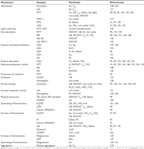
89In vitro – assay interference
The interference of NPs with several assay systems can
strongly influence the results (Table 1). The absorbance of NPs
could lead to false-negative results (absence of cytotoxicity is
detected, although the NP is cytotoxic) because the metabolic
activity (according to absorbance) is estimated to be higher
than it actually is.
90Enzyme inhibition by NPs could also
cause false-negative results. Lactate dehydrogenase (LDH) is
released into the supernatant of cells when the plasma
mem-brane integrity is lost. Its enzymatic activity correlates to the
amount of damaged cells. If LDH activity is inhibited by NPs,
a lower degree of cell damage will be determined.
False-positive results (cytotoxicity is detected although
the NP is not toxic) are detected when the fluorescent signals
of dihydrofluorescein (the detection of oxidative stress) or
of propidium iodide (the disruption of membrane integrity)
are enhanced by NPs.
91–95Depending on the assays used, the
masking of toxic effects and the identification of nonexistent
toxicity by NPs can occur simultaneously. Increased
absor-bance by colored NPs will result in a higher signal of LDH
(indicating more dead cells) and in the MTT assay
(indicat-ing more viable cells). The use of multiple assays, therefore,
helps to reveal assay interference. The addition of protein,
mostly BSA, could prevent interference, but it also could
increase it. While false-negative results by the inhibition of
LDH activity by Si, Au, and CdSe NPs
96was avoided, the
addition of BSA caused false-negative effects in protein
International Journal of Nanomedicine downloaded from https://www.dovepress.com/ by 118.70.13.36 on 23-Aug-2020
Dovepress Nanoparticle effects on phagocytes
Table 1 Mechanisms of interference between nonbiodegradable NPs and in vitro assays
Mechanism Assay(s) Particle(s) Reference(s)
Absorbance Hemolysis Au, C60 128–130
LAL Au, C60 131
MTT Au, CB, C60, Qdots, Ag, AgO, iron oxide, SwCNT
90, 92, 93, 129, 132–136
wST-1 Iron oxide 137
MTS Al, Qdots 51, 97, 138
LDH Au, SiO2, iron oxide, CeO2 51, 98, 135, 139
Light scattering MTT, ATP Carbon nanodiamonds 140
Dye absorption MTT SwCNT, CB, Al, iron oxide 95, 141–144
NR CB, SwCNT, C60, Si, TiO2 90, 134, 141, 145–148
AB, AK SwCNT 146
CB, wST-1 SwCNT 145
enzyme inactivation/inhibition LDH Cu, Ag 149, 150
LDH ZnO 135
LDH Si, Au, Qdots 96
LDH Au 151
AK PS 105
enzyme adsorption LDH Cu, Qdots, TiO2 93, 95, 133, 149, 152, 153
Reduction/enzymatic activity MTT Si, SwCNT, C60, TiO2 91, 95, 129, 145, 148, 152, 154, 155
AB Si 148
AK SwCNT 146
Prevention of reduction MTT Zn 93
Oxidation H2DCF CB 135
Hemoglobin Iron oxide 128
Protein binding Cytokines CB, SwCNT, iron oxide, Cu, SiO2, Al2O3, CeO2, NiO2, TiO2
93, 135, 141, 146, 156–159
Increase enzymatic activity LAL Iron oxide 131
Hemoglobin PS, SwCNT 129, 146
Physical interaction TB, calcein AM, live/dead SwCNT, C60, CB, Qdots 90
COMeT Ge 160
Quenching of fluorescence H2DCF CB, SiO2, SiO2-iron 161, 162
LDH CB, SwCNT, C60, Qdots 90
Calcein AM/ethD-1 CB, SwCNT 90
Increase of fluorescence H2DCF Au, iron oxide, TiO2, C60, SiO2, CB, SwCNT
91–95
PI Qdots, PS 95
Calcein AM/ethD-1 CB, iron oxide 91
AB CB, SwCNT, TiO2, Qdots 90, 97, 145
Resazurin CoO 51
COMeT TiO2, CuO 163
Increase of luminescence Phagocytosis Qdots 130
ATP SiO2 139
Quenching of luminescence Phagocytosis Au 129, 131
Aggregation Platelet aggregation Au, C60 129
Abbreviations: NPs, nanoparticles; Au, gold; C60, C60 fullerenes; LAL, limulus amebocyte lysate; CB, carbon black; Qdots, CdSe quantum dots; Ag, silver; SwCNT,
single-walled carbon nanotube; wST-1, water soluble tetrazolium salt; MTS, 3-(4,5-dimethylthiazol-2-yl)-5-(3-carboxyphenyl)-2-(4-sulfophenyl)-2H-tetrazolium); Al, aluminum; LDH, lactate dehydrogenase; SiO2, silica; CeO2, cerium oxide; ATP, adenosine triphosphate; NR, neutral red; TiO2, titanium dioxide; AB, alamarBlue; AK, adenylate kinase; Cu, copper; ZnO, zinc oxide; PS, polystyrene; Zn, zinc; H2DCF, dihydrodichlorofluorescein; Al2O2, aluminum oxide; NiO2, nickel oxide; TB, trypan blue; AM, acetoxymethylester; Ge, germanium; ethD-1, ethidium homodimer 1; PI, propidium iodide; CoO, cobalt oxide; CuO, copper (II) oxide.
detection via the Bradford reagent.
97For the identification
of potential assay interference, the incubation of NPs with
the assay compounds alone (in the absence of cells) and
with cells alone (in the absence of assay compounds) can be
used. These controls are, however, only useful when the NPs
interact with assay compounds and with the readout; assay
interactions by Au NPs, which increased the detected amount
of dead cells by shuttling the indicator dye, propidium iodide,
into the cells, would not have been revealed.
98,99Similarly,
the more global effects of NPs on cultured cells, such as the
depletion of nutrients by SWCNTs,
100would go unnoticed.
Interference can show dose dependency; dye (acridine)
fluorescence is increased by low concentrations of Ag NPs
and quenched at high concentrations of NPs.
101International Journal of Nanomedicine downloaded from https://www.dovepress.com/ by 118.70.13.36 on 23-Aug-2020
Dovepress
Fröhlich
Some general rules may help identify and prevent the
false interpretation of results. The use of low NP
concentra-tions reduces the problem of interference, but the removal
of NPs by centrifugation is generally not recommended
because analytes adsorbed to the particles might be removed.
Assay interference of the colored CNTs, carbon black, C
60fullerenes, and Au NPs, and of the fluorescent Qdots, may
occur more frequently than interference with noncolored
Si, SiO
2, TiO
2, and ZnO NPs. Testing of NPs with several
assays based on different detection methods can reduce the
risk of misinterpretation.
90,102In this regard, immunotoxicity
testing poses more problems than cytotoxicity testing because
a lower number of assays for a given immunological
func-tion are usually available. On the other hand, compared to
cytotoxicity testing, NPs are usually studied at much lower
NP concentrations, reducing the risk for interference.
Immunotoxicological data from NP
exposure
In vivo exposure includes voluntary inhalation and oral
application, forced inhalation (intranasal and intratracheal
instillation, oropharyngeal administration), forced oral
(intragastric/gavage) application as well as noninvasive
der-mal and invasive (intraderder-mal injection) derder-mal applications.
Parenteral applications include intravenous and
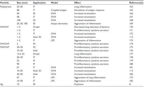
intraperito-neal injection. Table 2 shows the general reaction pattern of
the immune system after in vivo exposure to NPs.
Systemic immune effects
Effects in the respiratory tract with only a thin epithelium
were more pronounced than effects after dermal or oral
inges-tion exposure, where a horny layer or a thick mucus layer
separated NPs from epithelial and immune cells.
103Inflammation in the lung is one of the most frequently
reported effects of respiratory exposure to NPs.
104Since
cytokines are produced by several cell types, it is not clear
whether the reported increases in cytokine secretion and
subsequent inflammation were due to specific activation of
immune cells, or if they were a consequence of cytotoxic
action on alveolar epithelial cells. Heavy metal-containing
NPs reacted in a similar manner as PS particles.
105,106Given
that heavy metal-containing NPs show ROS generation,
and since they are expected to have greater cytotoxicity, the
similarity of the reaction does not support the hypothesis of
cell death (induced by more cytotoxic heavy metal-containing
NPs) as a main inductor of inflammation.
Only a few studies have reported the absence of
immu-nological effects, which could be due to restricted access to
Table 2 effects of NPs after inhal, IN, IG, IP, Iv, oral, oroph, and SC application, and ID and IT in normal animals and in animal models (Model)
Particle Size (nm) Application Model Effect Reference(s)
Polystyrene 25–50 IT Lung inflammation 164
68 IT Coupled antigen Stimulation of antigen response 165
40 ID OvA Increased sensitization 166
58 IT OvA Increased sensitization 167
100 SC OvA Increased sensitization 168
20, 50, 100 ID Atopic dermatitis Aggravation of inflammation 169
SwCNT 1–2 Oroph Decreased lung clearance of bacteria 170
1–2 IT Proinflammatory cytokine secretion 171
1–2 IT OvA Increased sensitization 172
1–2 Inhal, SC OvA Increased sensitization 113
1–2 IT LPS Aggravation of inflammation 173
DwCNT 2 IN Proinflammatory cytokine secretion 174
MwCNT 20–30 SC Proinflammatory cytokine secretion 175
10–20 Inhal Proinflammatory cytokine secretion 176
12.5–25 Oroph Lung inflammation 177
20–30 IT Proinflammatory cytokine secretion 178
25 Iv Proinflammatory cytokine secretion 179
90 IP Proinflammatory cytokine secretion 180
67 IT OvA Increased sensitization 181
10–50 Inhal, SC OvA Increased sensitization 113
30–50 Inhal OvA Increased sensitization 182
67 IT LPS Aggravation of lung inflammation 173
10–50 IT LPS Aggravation of inflammation 183
Ag 10 ID erythema 8
(Continued)
International Journal of Nanomedicine downloaded from https://www.dovepress.com/ by 118.70.13.36 on 23-Aug-2020
Dovepress Nanoparticle effects on phagocytes
Table 2 (Continued)
Particle Size (nm) Application Model Effect Reference(s)
18 Inhal Lung inflammation 184
18 Inhal Lung inflammation 185
20 Iv Suppressed immune response to KLH immunization 187
22, 42, 71 Oral Increased TGF-β levels 186
52 IT Proinflammatory cytokine secretion 12
33 IN OvA Increased sensitization 188
Au 50 IT Lung inflammation 189
50, not 10 IP Proinflammatory cytokine secretion 190
21 IP Anti-inflammatory action in adipose tissue 108
15 Oroph TDI Aggravation of asthma 191
5, 15 IP IL-1β inflammation Decrease of inflammation 109
Iron oxide 5.3 IT Lung inflammation and allergic response 193
20 Iv Proinflammatory cytokine secretion 194
36 IT Lung inflammation and cytokine secretion 192
58 Iv Decreased OVA-specific antigen production 195, 196
43 IT OvA Increased sensitization 197, 198
35 IT OvA Increased sensitization 199
SiO2 10 IT Lung inflammation 200
12 IP Proinflammatory cytokine secretion 13
30, 70 IP Proinflammatory cytokine secretion 201
15 Iv Proinflammatory cytokine secretion 202
70 Iv Proinflammatory cytokine secretion 203
30, 70, 100 ID Atopic dermatitis Aggravation of inflammation 204
10–20 IT OvA Increased sensitization 205
TiO2 2–5 Inhal Lung inflammation 207
5 IP Proinflammatory cytokine secretion 215
5.5 IG Infiltration of immune cells in spleen 213
8–10 IN Lung inflammation 206
20 ID, not oral Immune activation 8
20 Inhal Lung inflammation 208
20 IT Lung inflammation 209
25 IT Proinflammatory cytokine secretion 210
25 IT Lung inflammation 211
15, 28 IT Lung inflammation 212
30–40 IT Lung inflammation 200
66 Oral Proinflammatory cytokine secretion 14
20 IG Proinflammatory and allergic cytokine secretion 214
14, 29 Inhal OvA Increased sensitization 217
15 Oroph TDI Aggravation of asthma 191
20 IP LPS Aggravation of lung inflammation 218
28 Inhal OvA Increased sensitization 216
ZnO 10 Inhal Lung inflammation 219
10 IT Lung inflammation 200
21 Oral No effect on oral tolerance to OvA 107
21 IP OvA Increased sensitization 114
55 IP OvA Increased sensitization 115
Abbreviations: NPs, nanoparticles; inhal, inhalation; IN, intranasal; IG, intragastral; IP, intraperitoneal; Iv, intravenous; oroph, oropharyngeal; SC, subcutaneous; ID,
intradermal; IT, intratracheal instillation; OvA, ovalbumin; SwCNT, single-walled carbon nanotube; LPS, lipopolysaccharide; DwCNT, double-walled carbon nanotube; MwCNT, multiwalled carbon nanotube; Ag, silver; TGF, transforming growth factor; KLH, keyhole limpet hemocyanin; Au, gold; TDI, toluene diisocyanate; IL, interleukin; SiO2, silica; TiO2, titanium dioxide; ZnO, zinc oxide.
immune cells.
107The absence of immune effects after the oral
ingestion of and exposure to ZnO and TiO
2NPs could be
explained by the hindered assessment of the particles to the
cells by mucus.
8,107On the other hand, the low reactivity of
intraperitoneally applied Au NPs appears to be due to their
high biocompatibility given that few studies have reported
on the adverse cellular effects of Au NPs.
108,109This statement
is supported by a lack of immunological interference in the
cellular assays showing no increased cytokine secretion,
110,111and no effect on DC maturation and activation.
94,112When NPs were applied to diseased animals, the
pathol-ogy of the disease was aggravated. This aggravation was seen
International Journal of Nanomedicine downloaded from https://www.dovepress.com/ by 118.70.13.36 on 23-Aug-2020
Dovepress
Fröhlich
in asthma models, as well as in atopic dermatitis (Table 2).
Aggravation of asthma is unlikely to be caused by
cytotoxic-ity of the NPs because exposure by the respiratory tract and
by other routes (subcutaneous, intraperitoneal), where no
direct contact with the alveolar epithelium occurred, caused
the same effects.
113–115The mechanisms for amplifying
pre-existing pathologies have been proposed through the
following mechanisms:
116pre-existing inflammation in the
respiratory tubes could be amplified by enhancing the levels
of inflammatory factors or humoral immunity. Second, NPs
within the size range of
100 nm were able to stimulate
and enhance hypersensitivity, which is primarily mediated
by Th2 cells.
116In vitro and ex vivo effects
Phagocyte function after in vitro (cells exposed in wells) and
ex vivo (cells harvested from exposed animals) exposure is
summarized in Table 3. To evaluate the potential of
screen-ing in phagocytes, first, data obtained from ex vivo and
in vitro studies have to be compared. Second, the similarity
of ex vivo and in vitro exposures to in vivo exposure has to
be tested. In vitro data on cytokine secretion and chemotaxis
corresponded to the respective ex vivo data (Table 3). NPs
showed a similar pattern of interference with phagocyte
functions; proinflammatory cytokine secretion (mostly IL-6,
IL-1
β
, and TNF-
α
) and respiratory burst increased, while
phagocytosis and chemotaxis decreased. The
degranula-tion of neutrophilic granulocytes has been shown for a few
particles.
81,117The influence on DC maturation and function
varied markedly between the particles. MWCNTs inhibited
maturation, Au and iron oxide showed no prominent effect,
and SiO
2and TiO
2activated DCs.
94,112,118,119The different
results could be due to the use of different readouts
(matura-tion and activa(matura-tion).
Table 3 Immune effects in isolated phagocytes, either after in vivo treatment with nanoparticles (ex vivo) or by in vitro treatment Particle Effects
Ex vivo In vitro
Polystyrene Proinflammatory cytokine secretion81
Increased respiratory burst81 Neutrophilic granulocyte activation81
MwCNT Proinflammatory cytokine secretion220
Inhibition of DC maturation118
SwCNT Decreased chemotaxis221
Decreased phagocytosis221
Ag Proinflammatory cytokine secretion after IT application8 Proinflammatory cytokine secretion222–224 Proinflammatory cytokine secretion after inhalation225 Decreased phagocytosis12
Proinflammatory cytokine secretion after oropharyngeal application226 Increased respiratory burst227 Proinflammatory cytokine secretion after oral application186 Decreased NO production228
Neutrophilic granulocyte activation117
Au Proinflammatory cytokine secretion112,229,230
No increased cytokine secretion110,111 No effect on DC maturation, no activation94,112 Iron oxide Proinflammatory cytokine secretion after IT application199,231 Proinflammatory cytokine secretion232
Upon LPS challenge, decreased cytokine secretion after IT application233 Decreased phagocytosis234
Increased NO production with and without LPS challenge233,234
No effect on DC maturation94
SiO2 Increased NO production after IT application235 Proinflammatory cytokine secretion13,236,237 Activation of DC119
TiO2 Proinflammatory cytokine secretion after IT application235,238,239 Proinflammatory cytokine secretion240 Proinflammatory cytokine secretion after IG application10 Decreased chemotaxis241
Decreased phagocytosis12 Increased NO production after IT application235,239 Increased respiratory burst242 Decreased chemotaxis after IT application235,239 Activation of DC119
ZnO Decrease of cytokine secretion after oral application116 Proinflammatory cytokine secretion243–245 Proinflammatory cytokine secretion after IT application199 Decreased chemotaxis241
Proinflammatory cytokine secretion after inhalation11,219 Decreased phagocytosis12 Increased respiratory burst246
Abbreviations: MwCNT, multiwalled carbon nanotube; DC, dendritic cells; SwCNT, single-walled carbon nanotube; Ag, silver; IT, intratracheal instillation; NO, nitric
oxide; Au, gold; LPS, lipopolysaccharide, SiO2, silica; TiO2, titanium dioxide; IG, intragastral; ZnO, zinc oxide.
International Journal of Nanomedicine downloaded from https://www.dovepress.com/ by 118.70.13.36 on 23-Aug-2020
Dovepress Nanoparticle effects on phagocytes
The secretion of proinflammatory cytokines was
increased by all NPs when applied by in vitro exposure, and
after the ex vivo respiratory exposure, to NPs. The lower
sensitivity of phagocytes by the oral route was confirmed in
an ex vivo study.
120Uptake of NPs by phagocytes
When NPs are coated with proteins in biological fluids, they
are well ingested by phagocytes.
121Phagocytosis of NPs
by primary cells, cell lines, macrophages, monocytes, and
monocyte-derived macrophages indicated accumulation in
the MPS and showed a good correlation to the accumulation
of particles in the MPS of the spleen and liver in vivo.
122Due
to the crucial function of macrophages and DCs in the
spe-cific immune response, the accumulation of NPs in the MPS
could result in immunotoxicity. The indication of uptake by
the MPS or accumulation in lymphatic organs, however,
was not correlated to adverse effects on the immune system
in vivo or in vitro.
81,123Accumulation in the spleen was only
observed for 30 nm Au particles, while adverse effects on
the immune system according to increases in relative spleen
weight and immune cell numbers were seen for 5 nm, 10 nm,
and 60 nm Au particles.
123Small carboxyl PS particles were
ingested in much higher numbers than 1,000 nm particles by
macrophages.
81While the 1,000 nm large particles induced
oxidative burst and cell damage, particles in the size range
between 40 nm and 500 nm were taken up without obvious
interference with cell viability and function. Taken together,
these data suggest that the uptake of NPs may not result in
impaired phagocyte function.
Guidelines for sample preparation
and exposure
Physiologically relevant testing is based on sample
prepara-tion, as well as on the use of dispersant and intended exposure
routes. Most NPs form stable solutions in distilled water,
which cannot be used for in vitro studies. The presence of
ions and protein in the physiological solution leads to NP
agglomerates, which may increase in size, but they may
also disintegrate. The surface coating of NPs determines
their penetration of barriers, cellular uptake, and immune
response.
124The Office of Economic Co-operation and
Development (OECD) guidelines for sample preparation and
dosimetry had advised that dose should be indicated in terms
of mass, surface area, and particle number at a minimum.
125To get information on the stability of the dispersion, repeated
measurements are recommended with the documentation
of agglomeration and ion release. The dispersants should
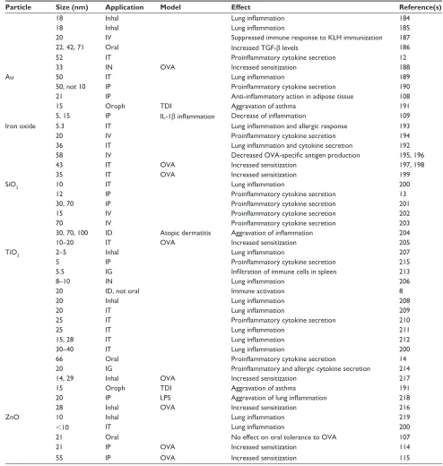
Table 4Overview of nanoparticle actions on phagocyte functions
Particles
Cytokine secretion
Chemotaxis
Phagocytosis
Respiratory burst
NO generation
Degranulation
DC maturation
Ex vivo
In vitro
Ex vivo
In vitro
Ex vivo
In vitro
Ex vivo
In vitro
Ex vivo
In vitro
Ex vivo
In vitro
Ex vivo
In vitro
Polystyrene
↑
↑
↑
M
w
CNT
↑
↓
Sw
CNT
↓
↓
Ag
↑
↑
↓
↑
↓
↑
Au
↑
-–
Iron oxide
↑
↑
↓
↑
–
SiO
2
↑
↑
↑
TiO
2
↑
↓
↓
↓
↑
↑
↑
ZnO
↑
↓
↑
↓
↓
↑
Notes:
Red arrow: increase; black arrow: decrease; –: no change.
Abbreviations:
NO, nitric oxide; DC, dendritic cell; M
w
CNT, multiwalled carbon nanotube; S
w
CNT, single-walled carbon nanotube; Ag, silver; Au, gold; SiO
2
, silica; TiO
2
, titanium dioxide; ZnO, zinc oxide.
International Journal of Nanomedicine downloaded from https://www.dovepress.com/ by 118.70.13.36 on 23-Aug-2020
Dovepress
Fröhlich
Due to the specific composition of the immune system
at different portals of entry, exposure-specific coculture
models including immune cells could serve as a possibility
to assess immunotoxicants in vitro. Alveolar epithelial cells
and alveolar macrophages in cocultures released
inflamma-tory cytokines at lower concentrations of TiO
2NPs than did
the respective monocultures.
127At the expense of greater
complexity, these systems could increase the sensitivity of
immunotoxicity in vitro screening and enable
exposure-specific testing. However, until a correlation of these findings
in these systems to data obtained in humans has been shown,
their value remains elusive.
Acknowledgments
Support by the European integrated project
NMP4-CT-2006-026723 and by the Austrian Science Fund grant P22576-B18
is gratefully acknowledged.
Disclosure
The author reports no conflicts of interest in this work.
References
1. Dobrovolskaia MA, McNeil SE. Immunological properties of engi-neered nanomaterials. Nat Nanotechnol. 2007;2(8):469–478. 2. D’Amato G. Outdoor air pollution in urban areas and allergic respiratory
diseases. Monaldi Arch Chest Dis. 1999;54(6):470–474.
3. Delfino RJ, Zeiger RS, Seltzer JM, Street DH, McLaren CE. Associa-tion of asthma symptoms with peak particulate air polluAssocia-tion and effect modification by anti-inflammatory medication use. Environ Health Perspect. 2002;110(10):A607–A617.
4. Li N, Xia T, Nel AE. The role of oxidative stress in ambient particu-late matter-induced lung diseases and its implications in the toxicity of engineered nanoparticles. Free Radic Biol Med. 2008;44(9): 1689–1699.
5. Penttinen P, Timonen KL, Tiittanen P, Mirme A, Ruuskanen J, Pekkanen J. Ultrafine particles in urban air and respiratory health among adult asthmatics. Eur Respir J. 2001;17(3):428–435.
6. Buzea C, Pacheco II, Robbie K. Nanomaterials and nanoparticles: sources and toxicity. Biointerphases. 2007;2(4):MR17–71.
7. Sodemann U, Bistrup C, Marckmann P. Cancer rates after kidney transplantation. Dan Med Bull. 2011;58(12):A4342.
8. Auttachoat W, McLoughlin CE, White KL Jr, Smith MJ. Route-dependent systemic and local immune effects following exposure to solutions prepared from titanium dioxide nanoparticles. J Immunotoxi-col. 2014;11(3):273–282.
9. Warheit DB, Sayes CM, Reed KL, Swain KA. Health effects related to nanoparticle exposures: environmental, health and safety consider-ations for assessing hazards and risks. Pharmacol Ther. 2008;120(1): 35–42.
10. Claude JR, Domenjoud L, Fattal E, et al. Recommendations for Toxico-logical Evaluation of Nanoparticle Medicinal Products. Paris, France: Agence Française de Sécurité Sanitaire des Produits de Santé; 2011. Available from: http://ansm.sante.fr/var/ansm_site/storage/original/ap plication/2968a90b774b563b03405379b7d4f4e6.pdf. Accessed April 24, 2014.
11. Müller K, Skepper JN, Posfai M, et al. Effect of ultrasmall superpara-magnetic iron oxide nanoparticles (Ferumoxtran-10) on human mono-cyte-macrophages in vitro. Biomaterials. 2007;28(9):1629–1642.
preferentially contain macromolecules that are present in
the target tissue. For exposure with aerosols, and in
addi-tion to the NP parameters, the mass median aerodynamic
diameter and aerosol concentration should be determined.
Guidelines for sample preparation for nanoscale TiO
2are
already available,
126and existing guidelines for exposure by
spontaneous inhalation, oral gavage, and dermal application
are applicable for NP exposure. Moreover, the additional
effects of intravenous exposure (for instance, behavior in the
syringe) have to be considered.
Freshly prepared solutions from stock solutions
pre-pared in water, diluted in cell culture medium, and treated
by sonification should be added to the cells. In the case that
no route-specific surfactants, such as 1,2 dipalmitoyl-
sn
-glycero-3-phosphocholine for pulmonary exposure, are used,
BSA appears to be a good choice because this zwitterionic
molecule prevents the binding of protein from the solution.
Conclusion
Due to the complexity of the immune system, in vivo testing will
remain the gold standard. However, intraindividual variations
in the immune system, as well as its compensatory abilities,
are major limitations. As has been observed in environmental
studies of airborne particles, individuals with impaired immune
function were affected by small particle doses, while no effects
were observed in the healthy population.
2–5This overview on a variety of carbon, metal, and metal
oxide NPs shows that these particles caused relatively similar
patterns of immunotoxicity in vivo, which involved
inflam-mation and immunosuppression in healthy animals and
aggravation of the pathology in animals with pre-existing
diseases. This suggests that the classification of particles
as more or less immunotoxic by in vitro screening might
be helpful. The extent to which such screening could lead
to valid results was studied by comparing data obtained
by in vivo exposure, in vitro testing and in vitro data
(Table 4). This analysis showed that the results obtained
in cells isolated from NP-exposed animals were similar to
the data obtained of cells, which were exposed to NPs
in vitro. Secondly, NPs that inhibited phagocyte functions
in vitro reacted in an immunotoxic manner in vivo (Tables 2
and 4). The data suggest that the in vitro testing of phagocytes
might predict the typical immunotoxicity pattern of NPs in
vivo. Cellular assays may also be suitable to identify
disease-related alterations in the immune reaction to NPs because
comparison between reactions of PBMCs from healthy and
allergic donors showed that the cells exhibited disease-related
differences upon challenge.
118International Journal of Nanomedicine downloaded from https://www.dovepress.com/ by 118.70.13.36 on 23-Aug-2020
Dovepress Nanoparticle effects on phagocytes
12. Liu H, Yang D, Yang H, et al. Comparative study of respiratory tract immune toxicity induced by three sterilisation nanoparticles: silver, zinc oxide and titanium dioxide. J Hazard Mater. 2013;248–249:478–486. 13. Park EJ, Park K. Oxidative stress and pro-inflammatory responses
induced by silica nanoparticles in vivo and in vitro. Toxicol Lett. 2009; 184(1):18–25.
14. Nogueira CM, de Azevedo WM, Dagli ML, et al. Titanium dioxide induced inflammation in the small intestine. World J Gastroenterol. 2012;18(34):4729–4735.
15. Chen JK, Ho CC, Chang H, et al. Particulate nature of inhaled zinc oxide nanoparticles determines systemic effects and mechanisms of pulmonary inflammation in mice. Nanotoxicology. 2015;9:43–53. 16. Karlsson HL, Cronholm P, Hedberg Y, Tornberg M, De Battice L,
Svedhem S, Wallinder IO. Cell membrane damage and protein inter-action induced by copper containing nanoparticles–importance of the metal release process. Toxicology. 2013;313(1):59–69.
17. Ahmad Khanbeigi R, Kumar A, Sadouki F, et al. The delivered dose: Applying particokinetics to in vitro investigations of nanoparticle inter-nalization by macrophages. J Control Release. 2012;162(2):259–266. 18. Roitt I, Delves P, editors. Roitt’s Essential Immunology. 10th ed. Oxford,
UK: Wiley; 2001.
19. Janeway C Jr, Travers P, Walport M, Shlomchik M, editors. Immuno-biology: The Immune System in Health and Disease. 6th ed. New York, NY: Garland Science Pubishing; 2005.
20. Gordon S, Pluddemann A, Martinez Estrada F. Macrophage hetero-geneity in tissues: phenotypic diversity and functions. Immunol Rev. 2014;262(1):36–55.
21. Hirota K, Hasegawa T, Hinata H, et al. Optimum conditions for efficient phagocytosis of rifampicin-loaded PLGA microspheres by alveolar macrophages. J Control Release. 2007;119(1):69–76.
22. Kawaguchi H, Koiwai N, Ohtsuka Y, Miyamoto M, Sasakawa S. Phagocy-tosis of latex particles by leucocytes. I. Dependence of phagocyPhagocy-tosis on the size and surface potential of particles. Biomaterials. 1986;7(1):61–66. 23. Seymour L, Schacht E, Duncan R. The effect of size of polystyrene
particles on their retention within the rat peritoneal compartment, and on their interaction with rat peritoneal macrophages in vitro. Cell Biol Int Rep. 1991;15(4):277–286.
24. Abbas AK, Lichtman H, Pallai S, editors. Cellular and Molecular Immunology. 6th ed. Philadelphia, PA: Saunders; 2007.
25. Dale DC, Boxer L, Liles WC. The phagocytes: neutrophils and mono-cytes. Blood. 2008;112(4):935–945.
26. Coligan JE, Bierer B, Margulies D, Shevach E, Strober W, Kruisbeek A. Current Protocols in Immunology. New York, NY: John Wiley and Sons; 1991.
27. Krebs FC, Miller SR, Catalone BJ, Fichorova R, Anderson D, Malamud D, Howett MK, Wigdahl B. Comparative in vitro sensitivities of human immune cell lines, vaginal and cervical epithelial cell lines, and primary cells to candidate microbicides nonoxynol 9, C31G, and sodium dodecyl sulfate. Antimicrob Agents Chemother. 2002;46(7):2292–2298. 28. Borish LC, Steinke JW. 2. Cytokines and chemokines. J Allergy Clin
Immunol. 2003;111(2 Suppl):S460–S475.
29. Falk W, Goodwin RH Jr, Leonard EJ. A 48-well micro chemotaxis assembly for rapid and accurate measurement of leukocyte migration. J Immunol Methods. 1980;33(3):239–247.
30. Pietrosimone KM, Yin X, Knecht DA, Lynes MA. Measurement of cellular chemotaxis with ECIS/Taxis. J Vis Exp. 2012;(62). pii: 3840. 31. Iqbal AJ, Regan-Komito D, Christou I, et al. A real time chemotaxis
assay unveils unique migratory profiles amongst different primary murine macrophages. PLoS One. 2013;8(3):e58744.
32. Gille C, Spring B, Tewes L, Poets CF, Orlikowsky T. A new method to quantify phagocytosis and intracellular degradation using green fluorescent protein-labeled Escherichia coli: comparison of cord blood macrophages and peripheral blood macrophages of healthy adults. Cytometry A. 2006;69(3):152–154.
33. Elsner J, Kapp A. Reactive Oxygen Release. In: Proudfoot A, Wells T, Power C, editors. Methods in Molecular Biology: Chemokine Protocols. Human Press: Totowa; 2000:153–157.
34. Schneemann M, Schoeden G. Macrophage biology and immunology: man is not a mouse. J Leukoc Biol. 2007;81(3):579; discussion 580. 35. Daigneault M, Preston JA, Marriott HM, Whyte MK, Dockrell DH.
The identification of markers of macrophage differentiation in PMA-stimulated THP-1 cells and monocyte-derived macrophages. PLoS One. 2010;5(1):e8668.
36. Mosser DM, Edwards JP. Exploring the full spectrum of macrophage activation. Nat Rev Immunol. 2008;8(12):958–969.
37. Mann BS, Chung KF. Blood neutrophil activation markers in severe asthma: lack of inhibition by prednisolone therapy. Respir Res. 2006;7:59.
38. Jovanović B, Anastasova L, Rowe EW, Palić D. Hydroxylated fullerenes inhibit neutrophil function in fathead minnow (Pime-phales promelas Rafinesque, 1820). Aquat Toxicol. 2011;101(2): 474–482.
39. Vesnina LÉ, Mamontova TV, Mikitiuk MV, et al. [Effect of fullerene C60 on functional activity of phagocytic cells]. Eksp Klin Farmakol. 2011;74(6):26–29. Russian.
40. Steinman RM, Witmer MD. Lymphoid dendritic cells are potent stimu-lators of the primary mixed leukocyte reaction in mice. Proc Natl Acad Sci U S A. 1978;75(10):5132–5136.
41. Kang HG, Lee JE, Yang SH, et al. Donor-strain-derived immature den-dritic cell pre-treatment induced hyporesponsiveness against allogeneic antigens. Immunology. 2010;129(4):567–577.
42. House RV, Thomas PT, Bhargava HN. In vitro evaluation of fenta-nyl and meperidine for immunomodulatory activity. Immunol Lett. 1995;46(1–2):117–124.
43. Li Y, Li X, Li Z, Gao H. Surface-structure-regulated penetration of nanoparticles across a cell membrane. Nanoscale. 2012;4(12): 3768–3775.
44. Masterson AJ, Sombroek CC, De Gruijl TD, et al. MUTZ-3, a human cell line model for the cytokine-induced differentiation of dendritic cells from CD34+ precursors. Blood. 2002;100(2):701–703.
45. Nelissen I, Selderslaghs I, Heuvel RV, Witters H, Verheyen GR, Schoeters G. MUTZ-3-derived dendritic cells as an in vitro alternative model to CD34+ progenitor-derived dendritic cells for testing of chemical sensitizers. Toxicol In Vitro. 2009;23(8): 1477–1481.
46. Magalhães PO, Lopes AM, Mazzola PG, Rangel-Yagui C, Penna TC, Pessoa A Jr. Methods of endotoxin removal from biological prepara-tions: a review. J Pharm Pharm Sci. 2007;10(3):388–404.
47. Anderson WB, Huck PM, Dixon DG, Mayfield CI. Endotoxin inac-tivation in water by using medium-pressure UV lamps. Appl Environ Microbiol. 2003;69(5):3002–3004.
48. Tsuji K, Steindler KA, Harrison SJ. Limulus amoebocyte lysate assay for detection and quantitation of endotoxin in a small-volume parenteral product. Appl Environ Microbiol. 1980;40(3):533–538.
49. Hofman J. Bacterial endotoxins. In: Shier W, Mebs D, editors. Handbook of Toxicology. New York, NY: Marcel Dekker Inc.; 1990: 655–682.
50. Smulders S, Kaiser JP, Zuin S, et al. Contamination of nanoparticles by endotoxin: evaluation of different test methods. Part Fibre Toxicol. 2012;9:41.
51. Oostingh GJ, Casals E, Italiani P, et al. Problems and challenges in the development and validation of human cell-based assays to determine nanoparticle-induced immunomodulatory effects. Part Fibre Toxicol. 2011;8(1):8.
52. Dobrovolskaia MA, McNeil SE. Endotoxin and Engineered Nano-materials. In: Dobrovolskaia MA, McNeil SE, editors. Handbook of Immunological Properties of Engineered Nanomaterials. Republic of Singapore: World Scientific Pub Co Inc; 2012:77–116.
53. Cedervall T, Lynch I, Foy M, et al. Detailed identification of plasma proteins adsorbed on copolymer nanoparticles. Angew Chem Int Ed Engl. 2007;46(30):5754–5756.
54. Monopoli MP, Aberg C, Salvati A, Dawson KA. Biomolecular coronas provide the biological identity of nanosized materials. Nat Nanotechnol. 2012;7(12):779–786.
International Journal of Nanomedicine downloaded from https://www.dovepress.com/ by 118.70.13.36 on 23-Aug-2020