1
Cycling Exercise Training Enhances Mitochondrial Bioenergetics of Platelets in Patients with Peripheral Arterial Disease: A Randomized Controlled Trial
Ming-Lu Lin, PhD1; Tieh-Cheng Fu, MD, PhD2+; Chih-Chin Hsu, MD, PhD2; Shu-Chun
Huang, MD, PhD3; Yu-Ting Lin, PhD1; Jong-Shyan Wang, PhD1, 2, 4 *
1 Healthy Aging Research Center, Graduate Institute of Rehabilitation Science, Medical
Collage, Chang Gung University, Tao-Yuan, Taiwan
2 Department of Physical Medicine and Rehabilitation, Chang Gung Memorial Hospital,
Keelung, Taiwan
3 Department of Physical Medicine and Rehabilitation, Chang Gung Memorial Hospital,
Tao-Yuan, Taiwan
4 Research Center for Chinese Herbal Medicine, College of Human Ecology, Chang Gung
University of Science and Technology, Tao-Yuan, Taiwan
+ Equal contribution as the first author
Short title: Exercise effect on platelet mitochondrial function in PAD patients * Please send correspondence to:
Professor, Jong-Shyan Wang
Graduate Institute of Rehabilitation Science Chang Gung University
259 Wen-Hwa 1st Road, Kwei-Shan, Tao-Yuan, 333, Taiwan Fax # 886-3-2118700
E-mail address: [email protected]
2
Abstract
Exercise training influences the risk of vascular thrombosis in patients with peripheral arterial disease (PAD). Mitochondrial functionalities in platelets involve the cellular bioenergetics and thrombogenesis. This study aimed to elucidate the effect of cycling exercise training (CET) on platelet mitochondrial bioenergetics in PAD patients. Forty randomly selected patients with PAD engaged in general rehabilitation (GR) with CET (i.e., cycling exercise at ventilation threshold for 30 min/day, 3 days/week) (GR+CET, n=20) or to a control group that only received GR course (n=20) for 12 weeks. Systemic aerobic capacity and platelet mitochondrial bioenergetics that included oxidative phosphorylation (OXPHOS) and electron transport system (ETS) were measured using automatic gas analysis and high-resolution respirometry, respectively. The experimental results demonstrated that GR+CET for 12 weeks significantly (i) elevated VO2peak and lowered VE-VCO2 slope, (ii) raised resting ankle-brachial
index and enhanced cardiac output response to exercise, (iii) increased the distance in 6-minute walk test and raised the Short Form-36 physical/mental component scores, and (iv) enhanced capacities of mitochondrial OXPHOS and ETS in platelets by activating FADH2 (Complex II)-dependent pathway. Moreover, changes in VO2peak levels were positively associated with
changes in platelet OXPHOS and ETS capacities. However, no significant changes in systemic aerobic capacity, platelet mitochondrial bioenergetics, and health-related quality of life (HRQoL) occurred following GR alone. Hence, we conclude that CET effectively increases the capacities of platelet mitochondrial bioenergetics by enhancing Complex II activity in patients with PAD. Moreover, the exercise regimen also enhanced functional exercise capacity, consequently improving HRQoL in PAD patients.
3
1. Introduction
Peripheral arterial disease (PAD) is a manifestation of atherosclerosis or thrombosis that causes chronic narrowing of peripheral arteries, consequently reducing the capacity of blood flow to contracting muscles in legs [1]. Moreover, reduced exercise capacity negatively affects the ability of PAD patients to perform the activities required for daily life, further decreasing their independence and quality of life [2,3]. However, exercise training may not only improve physical performance but also reduce vascular thrombotic risk in patients with PAD [2,3].
Platelets play a critical role in thrombogenesis of PAD patients [4,5]. Platelet mitochondrial functionality are mainly involved in the cellular redox balance and activation, thereby modulating thrombogenesis [6-8]. Cycling exercise training (CET) improves aerobic fitness, concurrence with reducing the risk of major vascular thrombotic events in patients with circulatory disorders [9,10]. According to our early studies, moderate-intensity exercise training on a bicycle ergometer depressed platelet adhesion/aggregation at rest and attenuated the enhancement of platelet reactivity caused by acute strenuous exercise [11,12]. Recently, our investigation further demonstrated that CET improved the capacity for platelet mitochondrial bioenergetics [13,14] and reduced platelet-induced thrombin generation [13] in healthy sedentary people [13] or heart failure (HF) patients [14]. However, the effects of CET on platelet mitochondrial bioenergetics in PAD patients have not yet been established. The electron transport complexes in mitochondrion are interconnected in mitochondrial inner membrane and turn into respiratory supercomplexes [15]. Mitochondrial structure and function are disrupted as isolating independent organelles from whole cells [16]. A high-resolution respirometry can measure mitochondrial functionality by the addition of exogenous substrates and inhibitor to the permeablized cells [17]. Recently, we have established two novel reference protocols of the substrate-uncoupler-inhibitor titration (SUIT-RPs), those were employed to determine the capacities of mitochondrial oxidative phosphorylation (OXPHOS) and electron transport system (ETS) in platelets, respectively [18]. Therefore, the present study further evaluated how CET effects systemic aerobic capacity and mitochondrial OXPHOS and ETS activities of platelets in PAD patients.
4 2. Methods
2.1. Participants
Fifty-two patients diagnosed with PAD have been surveyed for the interventions from April 1, 2018 to December 31, 2019 at the Chang Gung Memorial Hospital, Keelung, Taiwan. Inclusion criteria in this investigation were listed as follows: (i) > 20 years old, (ii) PAD for more than 2 weeks; (iii) ankle-brachial index (ABI) < 0.9, and (iii) active voluntary exercise. Exclusion criteria were listed as follows: (i) less than 20 years old; (ii) unstable angina; (iiii) systolic blood pressure (SBP) at rest is greater than 200 mm Hg or diastolic blood pressure (DBP) greater than 110 mm Hg; (iv) the orthostatic blood pressure drop is greater than 20 mm Hg with symptoms; (v) severe aortic stenosis with a peak systolic pressure gradient greater than 50 mm Hg, with an aortic valve opening area less than 0.75 cm2; (vi) acute discomfort or
fever; (vii) uncontrolled atrial or ventricular dysrhythmias; (viii) uncontrolled sinus rhythm tachycardia (more than 120 per minute); (ix) uncompensated congestive heart failure; (x) third degree atrioventricular block; (xi) acute pericarditis or myocarditis; (xii) recent embolism; (xiii) thrombophlebitis; (xiv) ST segment displacement is more than 2 mm at rest; and (xv) uncontrolled diabetes (glycemic blood glucose greater than 300 mg/dL or greater than 250 mg/dL with ketone bodies). Afterwards, forty eligible PAD patients were randomly divided into general rehabilitation (GR) with CET (GR+CET, n=20) and only received GR (GR, n=20) groups (Fig. S1). All subjects provided informed consent after the experimental procedures were explained. This study was performed in accordance with the tenets of the Declaration of Helsinki and approved by the Institutional Review Board of Chang Gung Memorial Hospital, Taiwan (ClinicalTrials.gov Identifier: NCT03965520).
2.2. Ankle-brachial index (ABI) measurement
Doppler measurements were performed in accordance with AHA guidelines for ABI measurement [19,20]. A digital vascular doppler HUNTLEIGH Dopplex DMX (Huntleigh Healthcare, United Kingdom) with an 8 MHz probe was used to measure the individual systolic pressures. An appropriately sized pneumatic cuff was applied to the right upper arm, inflated to suprasystolic pressure and deflated slowly until a Doppler flow signal was detected. The process was repeated for right leg and values for both dorsal pedal and anterior tibial arteries were measured, followed by left leg and left arm. ABI was subsequently calculated for each lower limb separately using the value of pressure from the respective arm as a denominator. Moreover, we used duplex ultrasound scanning as the gold standard. DUS was performed using Vivid S6 Ultrasound System (GE Healthcare, USA) equipped with 8L-RS (a 5-13 MHz linear transducer) and 4C-RS (1.8-6 MHz curvilinear transducer).
2.3. Grade exercise test
5 or GR subjects to assess their cardiopulmonary parameters 2 days before and 2 days after 12-week intervention. Moreover, the data collector was isolated from the data analytic specialist. Each participant was instructed to fast for at least 8 h and to refrain from exercise for at least 24 h before the test. All participants arrived at the testing center at 9:00 A.M. to eliminate diurnal effects. The subject first collected two minutes of resting parameters on the stationary bicycle. Then, the subjects performed on the unloaded free pedal for two minutes, after which the load was increased by 1 watt every 6 seconds (10 watts per minute) until the subjects were exhausted (i.e., progressive exercise to peak oxygen consumption, VO2peak). During the process,
the bicycle speed was required to be maintained at 60 rpm. Minute ventilation (VE), VO2, and
carbon dioxide production (VCO2) were measured on a breath-by-breath basis, using a
computer-based system (Master Screen CPX, Cardinal-health Germany) [14]. Heart rate (HR) was determined from the R-R interval on a 12 lead electrocardiogram, mean arterial pressure (MAP) was measured using an automatic blood pressure system (Tango, SunTech Medical, UK), and arterial O2 saturation was monitored through finger pulse-oximetry (model 9500,
Nonin Onyx, Plymouth, MN, USA) [14]. VO2peak was defined by the following criteria: VO2
increased by <mL/kg/min over at least 2 min, HR ≥85% of its predicted upper threshold, respiratory exchange ratio ≥ 1.10, or some other symptom/sign limitations in accordance with the guidelines of the American College of Sports Medicine for exercise testing [21]. Additionally, the ventilation threshold (VT) was determined when VE/VO2 increased without
a corresponding increase in the VE-to-VCO2 ratio, end-tidal PO2 increased without a decrease
in end-tidal PCO2, or there was a departure from linearity for VE [21].
Ventilation and VCO2 responses, acquired from the initiation of exercise to the peak values,
were used to calculate the VE-VCO2 slope, using least-squares linear regression (y = m · x + b,
m = slope) [22]. Additionally, the 6-minute walk test (6 MWT) was used to assess functional capacity and exercise endurance in patients with PAD [23]. The distance covered over a time of 6 minutes was used as the outcome by which to compare changes in performance capacity [23].
2.4. Hemodynamic measurements
6 2.5. Cycling exercise training (CET) protocol
In addition to the daily PAD rehabilitation course, the GR+CET subjects also performed supervised hospital-based training on a bicycle ergometer (Ergoselect 150P, Germany), completing 3 weekly sessions for 12 weeks. The CET protocol comprised a warm-up at 30% of VO2peak for 3 min, followed by continuous work-rate at VT for 30 min, then a cool-down at
30% of VO2peak for 3 min. All subjects used an HR monitor (Tango, SunTech Medical, UK) to
obtain the assigned intensity of exercise. Borg 6-to-20 scale was used to assess the rate of perceived exertion during and after each exercise session. The work-rate of bicycle ergometer was adjusted continuously to ensure that the intensity of exercise matched the target HR throughout the training period. Patients were instructed to immediately stop exercise training if they had leg pain or other signs/symptoms of circulatory disorders. The GR subjects only engaged in general PAD rehabilitation course, as instructed by their rehabilitation physicians. The rates of compliance with the GR+CET and GR subjects were 100% and 100%, respectively. 2.6. Platelet isolation
Before the graded exercise test at the beginning of the present study and 12 weeks later in various groups, 20 ml of blood was sampled from each subject's antecubital vein within 1 min by venipuncture (20-gauge needle). Blood samples (20 mL) were collected in polypropylene tubes containing sodium citrate (3.8 g/dL, 1–9 vol. blood). Platelet rich plasma (PRP) was prepared through centrifugation at 300 g for 10 min at approximately 20°C. Platelets were sedimented through centrifugation of the PRP at 1500 g for 10 min at approximately 20 °C and then washed once with phosphate buffered saline (PBS) containing ethylenediaminetetraacetic acid (EDTA, final concentration, 4 mM) (Sigma) to inhibit platelet activation [13,14]. The number of platelets was adjusted to 2 × 10⁸ cells/mL with mitochondrial respiration medium
(MiR05, containing sucrose 110 mM, HEPES 20 mM, taurine 20 mM, K-lactobionate 60 mM, MgCl2 3 mM, KH2PO4 10 mM, EGTA 0.5 mM, BSA 1 g/L, pH 7.1). Blood analysis was
repeated twice to ensure reproducibility of the results. All platelet fractions were analyzed within 2 h after cell purification. Blood cells were enumerated using a Sysmax SF-3000 cell counter (GMI, Inc., Ramsey, MN, USA).
2.7. High-resolution respirometry
Platelet mitochondrial respiration was measured in 2 mL glass chambers of a high-resolution respirometry (Oxygraph-2K, Oroboros Instrument, Austria) with a stirrer speed of 750 rpm at a constant temperature of 37 °C. Data was acquired and recorded every 2 s by DatLab software version 6 (Oroboros Instrument, Austria). The O2 sensors and instrument
background O2 consumption had been already calibrated before experiments following the
manufacturer's instruction [13, 14]. Fixed number of platelets (2 x 10⁸ cells/mL) were added in
7 experiment for quality control. O2 concentration was automatically calculated from barometric
pressure and solubility factor was 0.92 for MiR05. 2.8. Mitochondrial ETS and OXPHOS in platelets
A documented protocol, the substrate-uncoupler-inhibitor titration reference protocol (SUIT-RP), was applied, consisting of two harmonized mitochondrial substrate-controlled experiments (RP1 and RP2) to acquire most information (Figs. S2A-S2D) [18]. All chemicals were purchased from Sigma-Aldrich (St. Louis, MO), if not stated otherwise.
RP1 is the SUIT experiment to measure mainly the capacity of mitochondrial ETS (Figs. S2A and S2B). Routine respirations of platelets were obtained after the cells stabilized in the O2K chamber. The respirations of intact cells were driven by endogenous substrates in this state. To access the mitochondrial respiration with saturating exogenous substrates, the cell membrane was then permeabilized with digitonin [18]. The concentrations of digitonin titration were 50 μg/4×108 platelets. After cell permeabilization, concomitantly added NADH-linked
(N-linked) substrates malate (2 mM) and pyruvate (5 mM). Because of absent in ADP, the oxygen consumption was only driven by uncoupling proton leakage (LEAK state, PML). The
oxidative phosphorylation (OXPHOS) capacity (PMP) was then evaluated by ADP
(Calbiochem, USA) titration (1mM). A cytochrome C (10 μM) test was applied to check if any disruption of the mitochondrial outer membrane and loss of cytochrome c during the process of cell permeabilization. The ETS capacity driven by malate and pyruvate (PME) was
subsequently obtained by mitochondrial protonophore carbonyl cyanide-p-trifluoromethoxyphenylhydrazone (FCCP) titration (0.5 μM/step) until no further respiration increase. Glutamate (10 mM) was added to fulfill the N-linked substrates, accessing the maximal ETS capacity driven by NADH or complex I (CI)-related resources (MPGE). 10 mM
succinate was added to complete the convergent input of CI and complex II (CII) (MPGSE),
followed by an octanoyl-carnitine titration (0.5 mM) to check the additional effect of fatty acid oxidation (MPGSOctE). Mitochondrial CI inhibitor, rotenone (0.5 μM), was added to block the
respiration from N-linked substrates dependent and fatty acid oxidation pathways (SE). The
additional contribution of the mitochondrial complex of glycerophosphate dehydrogenase (CGpDH) was evaluated with 10 mM glycerophosphate addition (SGpE). At last, the
mitochondrial respiration was inhibited by antimycin A (2.5 μM), the mitochondrial complex III inhibitor.
The capacities of mitochondrial ETS in permeabilized platelets were calculating from the results of RP1 protocol using the following equations:
ETSCI (PMGE) = Pyruvate + Malate + ADP + FCCP + Glutamate ………. [1]
ETSCI+CII (PMGSE) = Pyruvate + Malate + ADP + FCCP + Glutamate + Succinate ... [2]
ETSCI+CII+FAO (PMGSOctE) = Pyruvate + Malate + ADP + FCCP + Glutamate + Succinate
8 ETSCII (SE) = Pyruvate + Malate + ADP + FCCP + Glutamate + Succinate +
Octanoyl-Carnitine + Rotenone ………... [4] ETSCII+Gp (SGpE) = Pyruvate + Malate + ADP + FCCP + Glutamate + Succinate +
Octanoyl-Carnitine + Rotenone + Glycerophosphate ………...………...…. [5] RP2 is another SUIT experiment to measure mainly the OXPHOS state (ATP synthase dependent) of mitochondrial respiration (Figs. S2C and S2D). Following routine respiration and digitonin permeabilization, 1 mM ADP was titrated to accelerate the depletion of residual endogenous substrates. Afterwards, octanoyl-carnitine (0.5 mM) and malate (0.1 mM) were added to evaluated the fatty acid oxidation. High malate concentration (2 mM) was required to reduce the flux from succinate dehydrogenase, but could also activate the mitochondrial malic enzyme and led to higher O2 consumption (OctMP). Cytochrome C test was applied after high
malate titration, to ensure the quality and intactness of mitochondria. 5 mM pyruvate (OctMPP)
and 10 mM glutamate (OctMPGP) were sequentially titrated to acquire the N-linked respiration.
A consecutive titration of succinate (10 mM) and glycerophosphate (10 mM) stimulated the respiration to maximal through CII (OctMPGSP) and CGpDH (OctMPGSGpP) input. To
evaluate the limitation of OXPHOS system, FCCP (0.5 μM/ step) is titrated to access the maximum ETS capacity (OctMPGSGpE). 0.5 μM rotenone was added to reach the identical
SGpE state in RP1 and RP2, and linked these two protocols for harmonization. Antimycin A
(2.5 μM) was added in final to block the whole respiration.
The capacity of mitochondrial OXPHOS in permeabilized platelets were calculating from the results of RP2 protocol using the following equations:
OXPHOSFAO (OctP) = ADP + Octanoyl-Carnitine…………...…………....…………... [1]
OXPHOSFAO+CI (OctMPGP) = ADP + Octanoyl-Carnitine + Malate + Pyruvate +
Glutamate ………. [2] OXPHOSFAO+CI+CII (OctMPGSP) = ADP + Octanoyl-Carnitine + Malate + Pyruvate +
Glutamate + Succinate ……….………...….. [3] This study defines the maximal capacities of OXPHOS and ETS in platelets as the OctMPGSGpP and OctMPGSGpE states in RP2 protocol, respectively (Fig. S2B).
OXPHOSFAO+CI+CII+Gp (OctMPGSGpP) = ADP + Octanoyl-Carnitine + Malate + Pyruvate
+ Glutamate + Succinate + Glycerophosphate …….……….…... [1] ETSFAO+CI+CII+Gp (OctMPGSGpE) = ADP + Octanoyl-carnitine + Malate + Pyruvate +
Glutamate + Succinate + Glycerophosphate + FCCP ………...… [2] 2.9. Health-related quality of life
9 2.10. Statistical analysis
Data were expressed as mean±SEM and were analyzed using the statistical software package StatView. Experimental results were analyzed by 2 (groups) × 2 (time sample points;
i.e., pre- and post-interventions) repeated measures ANOVA with Bonferroni's post hoc test to compare cardiopulmonary fitness and capacities of mitochondrial OXPHOS and ETS in platelets at the beginning of this study and after 12 weeks in the GR+CET and GR groups.
Pearson correlation analysis was used to determine the association between aerobic capacity (i.e., VO2peak) and platelet mitochondrial OXPHOS and ETS in patients with PAD. The criterion
10
3. Results
3.1. Cardiopulmonary fitness and HRQoL
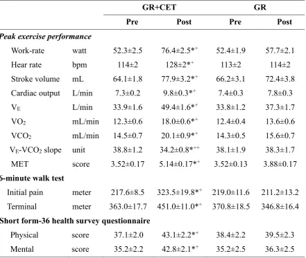
Fifty-two patients diagnosed with PAD have been surveyed for the interventions. A total of 40 eligible subjects completed the study in the GR+CET (n=20) and GR (n=20) groups. No adverse vascular or thrombotic event occurred in the two groups throughout the periods of investigation (Fig. 1S). Moreover, both GR+CET and GR groups did not differ significantly in anthropometric and clinical parameters (Table 1) or cardiopulmonary fitness (Table 2) at the start of the study.
Following 12 weeks of interventions, the GR+CET group increased values of ABI at right (R) and left (L) limbs using Duplex ultrasonography (R values from 0.85 to 0.92; L values from 0.86 to 0.93, P<0.05) and sphygmometry (R values from 0.86 to 0.93; L values from 0.87 vs. 0.94, P<0.05), respectively (Table 1). In cardiopulmonary fitness, 12-week GR+CET also increased work-rate, HR, SV, CO, as well as, VE, VO2, and VCO2 at the peak exercise
performance (P<0.05), but no significant changes in aerobic capacity occurred in the GR group (Table 2). Moreover, the GR+CET regimen significantly increased the distance in 6 MWT (initial pain from 217.6 m to 323.5 m; terminal point from 363.0 m to 451.0 m, P<0.05) and MET (scores from 3.52 to 5.14, P<0.05), as well as, lowered VE-VCO2 slope (values from 38.8
to 34.2, P<0.05) in the PAD patients (Table 2). Conversely, the GR alone did not influence the values of these indices of ventilatory efficiency and functional capacity in patients with PAD (Table 2).
In HRQoL, the GR+CET for 12 weeks significantly increased the subclass scores of the physical (scores from 37.1 to 43.1, P<0.05) and mental (scores from 35.2 to 42.8, P<0.05) dimensions in SF-36, respectively (Table 2). However, the GR alone remained unchanged the scores of SF-36 physical and mental components (Table 2).
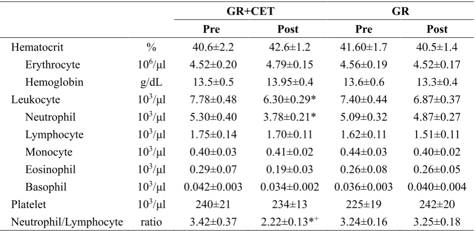
3.2. Blood cell count
There were no significant changes in erythrocyte, hemoglobin, hematocrit, lymphocyte and platelet following 12 weeks of the GR with or without CET (Table 3). However, the GR+CET considerably decreased neutrophil count (counts from 5.30 to 3.78 x10³/μl, P<0.05)
and the ratio of neutrophils to lymphocytes (NLR, ratio from 3.43 to 2.22, P<0.05). Moreover, these blood cell counts were unchanged following the GR alone (Table 3).
3.3. Mitochondrial ETS and OXPHOS in platelets
Either GR+CET or GR for 12 weeks unchanged the values of routine respiration and uncoupling proton leakage of platelets (Figs. 1 and 2). However, the GR with CET significantly increased FADH2 (CII)-related ETS levels in platelet (PMGSE, PMGSOctE, SE, and SGpE) (Fig.
1A, P<0.05). Additionally, the GR+CET enhanced succinate-involved OXPHOS level in platelet, such as OctMPGSP (Fig. 2A, P<0.05) rather than OctMPGP. Furthermore, this exercise
11 101.2 pmol/s /108 cells, P<0.05) and ETS (OctMPGSGp
E, from 106.1 to155.3 pmol/s /108 cells, P<0.05) in platelet, respectively (Fig. 2A). Nevertheless, no significant changes in platelet mitochondrial bioenergetics, that included ETS and OXPHOS capacities, occurred following GR alone (Figs. 1B and 2B).
On the other hand, changes of VO2peak levels were positively associated with changes of
maximal OXPHOS (OctMPGSGpP) (Fig.3A, r= 0.681, P<0.001) and ETS (OctMPGSGpE)
12
4. Discussion
This investigation clearly exhibits that 12-week GR+CET improves systemic aerobic capacity and ventilatory/hemodynamic efficiencies, which is accompanied by improving health-related quality of life in patients with PAD. Notably, this study is the first to demonstrate that the CET regimen effectively enhances capacities of mitochondrial OXPHOS and ETS in platelet through increasing Complex II activity in PAD patients.
4.1. Ventilation/hemodynamic efficiencies and HRQoL
Optimal exercise programs increase the ability of PAD patients to independently perform activities of daily living, thereby further improving their quality of life [2,3]. In this study, 12-week GR+CET increased VO2peak and decreased VE-VCO2 slope, as well as, enhanced
hemodynamic responses (such as CO and SV) to exercise in the PAD patients. The ventilatory parameters obtained from the graded exercise test may convey information regarding prognosis of circulatory disorders [22]. The VO2peak is an indicator of aerobic capacity, respectively [22],
whereasthe VE-VCO2 slope is a powerful predictor of survival in patients with circulatory
disorders [22]. Our previous study using patients with HF reported that these indices of ventilatory efficiency modulated by exercise training were correlated with exercise-induced central and peripheral hemodynamic changes [24]. Accordingly, the GR+CET may effectively improve ventilatory and hemodynamic efficiencies, thereby enhancing systemic aerobic capacity in patients with PAD.
Beside an improvement in systemic aerobic capacity, CET for 12 weeks also increased the distance in 6-minute walk test and consequently heightened the Short Form-36 physical/mental component scores. These findings imply that the exercise regimen effectively enhances the ability of patients to cope with the physical demands of daily activity and subsequently improving psychosocial status in PAD patients. Furthermore, the better HRQoL might exhibit less potential for mortality in PAD patients and simultaneously reduce the financial burden in their health care system [2,3].
4.2. Mitochondrial functionalities in platelets
Progression to PAD is probably associated with a gradual decline in bioenergetic reserve capacity owing to the inability of endogenous homeostatic mechanisms to compensate for the insufficient energy supply [26]. Moreover, mitochondrial dysfunction in PAD patients may result in systemic inflammation that facilitates susceptibility to energy-based pathologies associated with oxidative stress [27]. Additionally, atherosclerosis and thrombosis are reported to be associated with deterioration of platelet mitochondrial function [6,7]. Hence, it is plausible that platelet mitochondrial bioenergetics is a marker for metabolic stress in PAD progression.
13 to inflammatory stress and respond dynamically to changes in their microenvironment [29]. Decreased oxidative capacity due to mitochondrial respiratory chain impairment may be associated with increased release of reactive oxygen species (ROS) and reduction of calcium retention capacity, leading to enhanced apoptosis of skeletal muscles [30]. The present investigation further demonstrated that CET significantly depressed circulatory inflammatory status, indicated by decreased neutrophil count and NLR in patients with PAD. Hence, we posit that the CET-induced physiological adaptations protect against inflammation associated with mitochondrial dysfunction in PAD patients [31].
A previous study reported that ischemia results in accumulation of intracellular succinate, thus leading to elevated mitochondrial ROS production [32]. Elevation of oxidative stress may also induce succinate accumulation by decreasing SDH activity [33]. Recently, our study including sedentary healthy men reported that CET significantly enhanced platelet SDH activity and Complex II-related respiration following hypoxic stress [13]. In clinical investigation, we further demonstrated that CET elevated platelet mitochondrial O2
consumption rate via increasing Complex II activity, which was accompanied by depressed oxidative stress in patients with HF [14]. In the present study, 12-week CET effectively also enhanced platelet mitochondrial OXPHOS and ETS capacities by increasing Complex II activity in the PAD patients. Therefore, CET-induced elevation in Complex II activity in platelets may rapidly eliminate succinate, further reducing ROS production from platelet mitochondria, eventually depressing circulatory oxidative stress and pro-inflammatory status in patients with PAD. Moreover, we further demonstrate that change of VO2peak was positively
correlated with changes of maximal OXPHOS and ETS capacities in platelets. These experimental results suggest that CET-induced platelet metabolic adaptation may be associated with improved systemic aerobic capacity in patients with PAD.
4.3. Limitations of this study
Our small sample size in each group is a major limitation. However, results regarding platelet mitochondrial bioenergetics have high values of statistical power ranging from 0.864 to 1.000. Additionally, this study mainly focused on the effects of CET on platelet mitochondrial bioenergetics rather than platelet reactivity (adhesion and aggregation) or platelet-mediated coagulation or thrombin generation. Our previous studies have investigated the effect of CET on platelet adhesiveness and aggregation and their underling mechanisms in healthy people and patients with cardiovascular disorders [9-12]. Recent study further reported that CET markedly suppressed hypoxia-induced oxidative damage of platelet mitochondria and consequent attenuation of platelet-mediated thrombin generation caused by hypoxia in healthy sedentary men [13]. However, the role of platelet mitochondrial function on CET-mediated platelet reactivity and coagulation in PAD patients warrants further investigation.
15
Author contributions
Jong-Shyan Wang, Tieh-Cheng Fu, and Chih-Chin Hsu was involved in conception and design of research; Ming-Lu Lin, Tieh-Cheng Fu, Shu-Chun Huang, and Yu-Ting Lin performed experiments; Jong-Shyan Wang, Tieh-Cheng Fu and Ming-Lu Lin analyzed data, interpreted results of experiments, prepared the Figures and drafted the paper; Jong-Shyan Wang and Cheng Fu edited and revised the paper; Jong-Shyan Wang, Ming-Lu Lin, Tieh-Cheng Fu, Chih-Chin Hsu, Shu-Chun Huang, and Yu-Ting Lin approved the final version of paper.
Acknowledgments
The authors would like to thank the volunteers for their enthusiastic participation.
Funding
This work was supported by the National Science Council of Taiwan (grant number NSC 180-2314-B-182-039-MY3) and Chang Gung Medical Research Program (grant number CMRPD1J0221).
Conflicts of interest
16
References
1. Kithcart, A.P.; Beckman, J.A. ACC/AHA versus ESC guidelines for diagnosis and management of peripheral artery disease: JACC Guideline Comparison. J. Am. Coll. Cardiol. 2018, 72, 2789-2801.
2. Treat-Jacobson, D.; McDermott, M.M.; Bronas U.G. et al. (2019) Optimal exercise programs for patients with peripheral artery disease: A scientific statement From the American Heart Association. Circulation 2019, 139, e10-e33.
3. Treat-Jacobson, D.; McDermott, M.M.; Beckman, J.A. et al. Implementation of supervised exercise therapy for patients with symptomatic peripheral artery disease: a science advisory from the American Heart Association. Circulation 2019, 140, e700-e710.
4. Klonaris. C.; Patelis. N.; Drebes. A.; Matheiken. S.; Liakakos, T. Antiplatelet treatment in peripheral arterial disease: the role of novel antiplatelet agents. Curr. Pharm. Des. 2016, 22,
4610-4616.
5. Tsigkou. V.; Siasos. G.; Rovos, K.; Tripyla, N.; Tousoulis, D. Peripheral artery disease and antiplatelet treatment. Curr. Opin. Pharmacol. 2018, 39, 43-52.
6. Garcia-Souza, L.F.; Oliveira, M.F. Mitochondria: biological roles in platelet physiology and pathology. Int. J. Biochem. Cell Biol. 2014, 50, 156-160.
7. Zharikov, S.; Shiva, S. Platelet mitochondrial function: from regulation of thrombosis to biomarker of disease. Biochem. Soc. Trans. 2013, 41, 118–123.
8. Kramer, P.A.; Chacko, B.K.; Ravi, S.; Johnson, M.S.; Mitchell, T.; Darley-Usmar, V.M. Bioenergetics and the oxidative burst: Protocols for the isolation and evaluation of human leukocytes and platelets. J. Vis. Exp. 2014, 85, e51301.
9. Wang, J.S. Exercise prescription and thrombogenesis. J. Biomed. Sci. 2006, 13, 753-761. 10. Wang, J.S.; Jen, C.J.; Kung, H.C.; Lin, L.J.; Hsiue, T.R.; Chen, H.I. Different effects of
strenuous exercise and moderate exercise on platelet function in men. Circulation 1994, 90,
2877-2885.
11. Wang, J.S.; Jen, C.J.; Chen, H.I. Effects of exercise training and deconditioning on platelet function in men. Arterioscler. Thromb. Vasc. Biol. 1995, 15, 1668-1674.
12. Wang, J.S.; Li, Y.S.; Chen, J.C. Effects of exercise training and deconditioning on platelet aggregation induced by alternating shear stress in men. Arterioscler. Thromb. Vasc. Biol. 2005, 25, 454-460.
13. Wu, L.H.; Chang, S.C.; Fu, T.C.; Huang, C.H.; Wang, J.S. High-intensity interval training improves mitochondrial function and suppresses thrombin generation in platelets undergoing hypoxic stress. Sci. Rep. 2017, 7, e4191.
17 J. Cardiol. 2019, 274, 214-220.
15. Letts, J.A.; Fiedorczuk, K.; Sazanov, L.A. The architecture of respiratory supercomplexes. Nature 2016, 537, 644-648.
16. Picard, M.; Taivassalo, T.; Ritchie, D.; Wright, K.J.; Thomas, M.M.; Romestaing, C.; Hepple, R.T. Mitochondrial structure and function are disrupted by standard isolation methods. PLoS. One 2011, 6, e18317.
17. Pesta, D.; Gnaiger, E. High-resolution respirometry: OXPHOS protocols for human cells and permeabilized fibers from small biopsies of human muscle. Methods Mol. Biol. 2012,
810, 25-58.
18. Hsu, C.C.; Tsai, H.H.; Fu, T.C.; Wang, J.S. Exercise training enhances platelet mitochondrial bioenergetics in stroke patients: a randomized controlled trial. J. Clin. Med.
2019, 8, 2186.
19. Misra, S.; Shishehbor, M.H.; Takahashi, E.A. et al. Perfusion assessment in critical limb ischemia: principles for understanding and the development of evidence and evaluation of devices: a scientific statement from the American Heart Association. Circulation 2019, 140, e657-e672.
20. Guirguis-Blake, J.M.; Evans, C.V.; Redmond, N.; Lin, J.S. Screening for peripheral artery disease using the ankle-brachial index: updated evidence report and systematic review for the US preventive services task force. JAMA 2018, 320, 184-196.
21. American College of Sports Medicine. General principle of exercise prescription. In ACSM’s Guidelines for Exercise Testing and Prescription (Thompson, W. R., Gordon, N. F. and Pescatello, L. S., eds), Lippincott Williams & Wilkins, Philadelphia 2010, pp. 152– 182.
22. Arena, R.; Guazzi, M.; Myers, J. Ventilatory abnormalities during exercise in heart failure: a mini review. Curr. Respir. Med. Rev. 2007, 3, 179–187.
23. ATS Committee on Proficiency Standards for Clinical Pulmonary Function Laboratories. ATS statement: guidelines for the six-minute walk test. Am. J. Respir. Crit. Care Med. 2002,
166, 111-117.
24. Fu, T.C.; Wang, C.H.; Lin, P.S.; Hsu, C.C.; Cherng, W.J.; Huang, S.C.; Liu, M.H.; Chiang, C.L.; Wang, J.S. Aerobic interval training improves oxygen uptake efficiency by enhancing cerebral and muscular hemodynamics in patients with heart failure. Int. J. Cardiol. 2013, 167, 41-50.
25. Maksimovic, M.; Vlajinac, H.; Marinkovic, J.; Kocev, N.; Voskresenski, T.; Radak, D. Health-related quality of life among patients with peripheral arterial disease. Angiology
2014, 65, 501-506.
18 peripheral artery disease. Front. Physiol. 2017, 8,141.
27. Koutakis, P.; Ismaeel, A.; Farmer, P.; Purcell, S.; Smith, R.S.; Eidson, J.L.; Bohannon, W.T. Oxidative stress and antioxidant treatment in patients with peripheral artery disease. Physiol. Rep. 2018, 6, e13650.
28. Teperman, J.; Carruthers, D.; Guo, Y.; Barnett, M.P.; Harris, A.A.; Sedlis, S.P.; Pillinger, M.; Babaev, A.; Staniloae, C.; Attubato, M.; Shah, B. Relationship between neutrophil-lymphocyte ratio and severity of lower extremity peripheral artery disease. Int. J. Cardiol.
2017, 228, 201-204.
29. Carter, H.N.; Chen, C.C.; Hood, D.A. Mitochondria, muscle health, and exercise with advancing age. Physiology (Bethesda) 2015, 30, 208–223.
30. Anzell, A.R.; Maizy, R.; Przyklenk, K.; Sanderson, T.H. Mitochondrial quality control and disease: insights into ischemia-reperfusion injury. Mol. Neurobiol. 2018, 55, 2547-2564. 31. Pizzimenti, M.; Riou, M.; Charles, A.L.; Talha, S.; Meyer, A.; Andres, E.; Chakfé, N.; Lejay,
A.; Geny, B. The rise of mitochondria in peripheral arterial disease physiopathology: experimental and clinical data. J. Clin. Med. 2019, 8, 2125.
32. Chouchani, E.T.; Pell, V.R.; Gaude, E. et al. Ischaemic accumulation of succinate controls reperfusion injury through mitochondrial ROS. Nature 2014, 515, 431-435.
33. Mills, E.; O'Neill, L.A. Succinate: a metabolic signal in inflammation. Trends Cell. Biol.
19
FIGURE LEGENDS
Figure 1: Effects of general rehabilitation with (A, GR+CET) and without (B, GR) cycling exercise training in platelet mitochondrial O2 consumption rate (OCR) using the
reference protocol 1 (RP1) protocol in patients with peripheral arterial disease. P, pyruvate; M, malate; G, glutamate; S, succinate; Oct, octanoyl-carnitine; Gp, glycerophosphate; Rot, rotenone; Ama, antimycin; ROX, residual O2 consumption; OXPHOS or P, oxidative phosphorylation; ETS or E, electron transport system; LEAK or L, uncoupling proton leakage. Pre-intervention vs. Post-intervention, *P
< 0.05.Values were mean±SEM.
Figure 2: Effects of general rehabilitation with (A, GR+CET) and without (B, GR) cycling exercise training in platelet mitochondrial O2 consumption rate (OCR) using the
reference protocol 2 (RP2) protocol in patients with peripheral arterial disease. P, pyruvate; M, malate; G, glutamate; S, succinate; Oct, octanoyl-carnitine; Gp, glycerophosphate; Rot, rotenone; Ama, antimycin; ROX, residual O2 consumption; OXPHOS or P, oxidative phosphorylation; ETS or E, electron transport system; LEAK or L, uncoupling proton leakage. Pre-intervention vs. Post-intervention, *P
< 0.05. Values were mean±SEM.
Figure 3: Correlations between changes of systemic aerobic capacity (VO2peak) and platelet
maximal (A) oxidative phosphorylation (OXPHOS) and (B) electron transport system (ETS) capacities in patients with peripheral arterial disease. (A) maximal
OXPHOS capacity (OctMPGSGpP) = ADP + Octanoyl-Carnitine + Malate +
Pyruvate + Glutamate + Succinate + Glycerophosphate; (B) maximal ETS capacity (OctMPGSGpE) = ADP + Octanoyl-carnitine + Malate + Pyruvate + Glutamate +
20
SUPPLEMENTARY MATERIALS
Figure S1: Flowchart of enrolled patients with peripheral arterial disease (PAD) during following-up. Inclusion and exclusion criteria listed in the figure were used to recruit eligible candidates. Recruited PAD patients were divided into two groups based on their treatments: general rehabilitation with (GR+CET) and without (GR) cycling exercise training. In addition to general rehabilitation, the GR+CET group performed supervised hospital-based training on a bicycle ergometer (i.e., cycling exercise at ventilation threshold for 30 min/day, 3 days/week for 12 weeks). The GR
group only engaged in general rehabilitation course for 12 weeks, as instructed by their rehabilitation physicians.
Figure S2: Graphs showing a sample of cycling exercise training (CET) effects on platelet mitochondrial oxidative phosphorylation (OXPHOS) (A, B) and electron transport system (ETS) (C, D) in a patient with peripheral arterial disease. The reference protocols [RP1 (A, B) and RP2 (C, D)] are the substrate-uncoupler-inhibitor titration (SUIT) experiments to measure mainly the capacities of mitochondrial oxidative phosphorylation (OXPHOS) (A, B) and electron transport system (ETS) (C, D) in platelets, respectively. Pre-GR+CET, before general rehabilitation with cycling exercise training; Post-GR+CET, after general rehabilitation with cycling exercise training; D, digoxin; P, pyruvate; M, malate; ADP, adenosine diphosphate;
Cyt C, cytochrome C; FCCP, carbonyl cyanide-4-(trifluoromethoxy) phenylhydrazone; G, glutamate; S, succinate; Oct, octanoyl-carnitine; Rot, rotenone;
24
Table 1: Demographic and clinical characteristics in patients with peripheral arterial disease
GR+CET GR
Pre Post Pre Post
Anthropometrics/Clinical Characteristics
Gender n (M/F) 20 (12/8) 20 (12/8) 20 (12/8) 20 (12/8)
Age year 71.1±.1.5 - 70.5±.1.9 -
Height cm 160.2±1.3 - 159.0±2.0 166.8±1.5
Weight kg 69.7±2.4 69.3±2.4 67.6±4.0 67.3±3.9
Heart rate bpm 81±2 81±2 80±2 81±3
Systolic blood pressure mmHg 136±4 133±5 138±4 137±5 Diastolic blood pressure mmHg 81±3 79±4 82±4 81±4 Comorbidity
CVD n (%) 8 (40) - 9 (45) -
Hyperlipidemia n (%) 5 (25) - 6 (30) -
Hypertension n (%) 7 (35) - 8 (40) -
Diabetes mellitus n (%) 14 (70) - 13 (65) -
Medicines
Anti-thrombosis n (%) 8 (40) 8 (40) 9 (45) 9 (45)
Statins n (%) 5 (25) 5 (25) 6 (30) 6 (30)
-blockers n (%) 6 (30) 6 (30) 8 (40) 8 (40)
ACEI/ARB n (%) 4 (20) 4 (20) 4 (20) 4 (20)
Ca2+ CB n (%) 5 (25) 5 (25) 4 (20) 4 (20)
Sulfonylurea n (%) 13 (65) 13 (65) 12 (60) 12 (60) DDP-4 inhibitor n (%) 4 (20) 4 (20) 5 (25) 5 (25) SGLT2 inhibitors n (%) 2 (10) 2 (10) 3 (15) 3 (15) Ankle-brachial index (ABI)
Duplex ultrasonography R (ratio) 0.85±0.02 0.92±0.01*+ 0.87±0.01 0.88±0.01 L (ratio) 0.86±0.01 0.93±0.01*+ 0.85±0.01 0.85±0.01 Sphygmometry R (ratio) 0.86±0.01 0.93±0.02*+ 0.86±0.02 0.89±0.01 L (ratio) 0.87±0.01 0.94±0.02*+ 0.85±0.01 0.86±0.02 Values are mean ± SEM. CET; cycling exercise training; GR, general rehabilitation; Pre, pre-intervention;
Post, post-intervention; BMI, body mass index; CVD, cardiovascular diseases; Ca2+ CB, channel
blockers; DPP-4, Inhibitors of dipeptidyl peptidase 4 inhibitors; ACEI/ARB, angiotensin converting enzyme inhibitor/angiotensin II receptor blocker; R. right; L, left. * P<0. 05, Pre vs. Post; + P<0. 05,
25
Table 2: Effects of general rehabilitation with or without cycling exercise training on cardiopulmonary responses to exercise and health-related quality of life in patients with peripheral arterial disease
GR+CET GR
Pre Post Pre Post
Peak exercise performance
Work-rate watt 52.3±2.5 76.4±2.5*+ 52.4±1.9 57.7±2.1
Hear rate bpm 114±2 128±2*+ 113±2 114±2
Stroke volume mL 64.1±1.8 77.9±3.2*+ 66.2±3.1 72.4±3.8
Cardiac output L/min 7.3±0.2 9.8±0.3*+ 7.4±0.3 7.8±0.3
VE L/min 33.9±1.6 49.4±1.6*+ 33.8±1.2 37.3±1.7
VO2 mL/min 12.3±0.6 18.0±0.6*+ 12.4±0.4 13.6±0.6
VCO2 mL/min 14.5±0.7 20.1±0.9*+ 14.3±0.5 15.6±0.7
VE-VCO2 slope unit 38.8±1.2 34.2±0.8*++ 38.1±1.9 38.3±1.7
MET score 3.52±0.17 5.14±0.17*+ 3.52±0.13 3.88±0.17 6-minute walk test
Initial pain meter 217.6±8.5 323.5±19.8*+ 219.0±11.6 211.2±13.2 Terminal meter 363.0±17.7 451.0±11.0*+ 370.8±18.5 346.8±16.4 Short form-36 health survey questionnaire
Physical score 37.1±2.0 43.1±2.2*+ 38.4±2.2 39.5±2.3 Mental score 35.2±2.2 42.8±2.1*+ 35.2±2.5 36.3±2.5 Values are mean ± SEM. CET; cycling exercise training; GR, general rehabilitation; Pre, pre-intervention; Post, post-intervention; MET, metabolic equivalent of task, VO2, O2
consumption; VE, minute ventilation; VCO2, CO2 production; * P<0.05, Pre vs. Post; + P<0.
26
Table 3: Effects of general rehabilitation with or without cycling exercise training on blood cell counts in patients with peripheral arterial disease
GR+CET GR
Pre Post Pre Post
Hematocrit % 40.6±2.2 42.6±1.2 41.60±1.7 40.5±1.4 Erythrocyte 106/μl 4.52±0.20 4.79±0.15 4.56±0.19
4.52±0.17 Hemoglobin g/dL 13.5±0.5 13.95±0.4 13.6±0.6 13.3±0.4 Leukocyte 103/μl 7.78±0.48 6.30±0.29* 7.40±0.44
6.87±0.37 Neutrophil 103/μl 5.30±0.40 3.78±0.21* 5.09±0.32 4.87±0.27
Lymphocyte 103/μl 1.75±0.14 1.70±0.11 1.62±0.11 1.51±0.11
Monocyte 103/μl 0.40±0.03 0.41±0.02 0.44±0.03 0.40±0.02
Eosinophil 103/μl 0.29±0.07 0.19±0.03 0.26±0.08 0.26±0.05
Basophil 103/μl 0.042±0.003 0.034±0.002 0.036±0.003 0.040±0.004
Platelet 103/μl 240±21 234±13 225±19
242±20 Neutrophil/Lymphocyte ratio 3.42±0.37 2.22±0.13*+ 3.24±0.16 3.25±0.18