Original Article
Ca
2+
-ions and pattern control in Hydra
STEFANIE ZERETZKE
1, FERNANDO PÉREZ
2, KIRSTEN VELDEN
1and STEFAN BERKING*
,11Zoological Institute, University of Cologne, Köln, Germany and 2 Institute of Pathology, Clinical Centre of the University of Cologne,
Köln, Germany
ABSTRACT The fresh water polyp Hydra forms buds which develop a foot at their base and separate from the parent. In the strain H. vulgaris (Zürich), various compounds including phorbolesters, diacylglycerols, cantharidin and Li+-ions were found to prevent foot formation at the bud’s base. Therewith, the bud transforms into a branch which persists at the parent. Other strains were found to be unaffected by such treatments. Here we show that a reduced Ca2+-ion concentration of the culture medium causes branch formation in the H. vulgaris (Zürich) strain but not in the other strains tested. However, all strains tested transformed their buds into branches when the medium was enriched with Ba2+ and Sr2+ ions. We suggest that the various treatments either reduce the internal concentration of Ca2+-ions by stimulating Ca2+-ion export or compete with Ca2+-ions at their target. H. vulgaris (Zürich) is the most sensitive strain tested and appears to have the most efficient Ca2+-pumps. This appears to be necessary for these animals derived from a lake which is extremely rich in Ca2+ ions.
KEY WORDS:
hydrozoa ecology, Ba
2+, Sr
2+, TPA, cantharidin
0214-6282/2002/$25.00
© UBC Press Printed in Spain www.ijdb.ehu.es
*Address correspondence to: Dr. Stefan Berking, Zoological Institute, University of Cologne, Weyertal 119, 50931 Köln, Germany. Fax: +49-221-470-5171. e-mail: [email protected]
Abbreviations used in this paper: DMSO, dimethyl sulfoxide; EDTA, ethylene
diamine tetraacetic acid; TPA, 12-o-tetradecanoylphorbol-13-acetate.
Introduction
In Hydrozoa, one approach to study the control of pattern formation is to treat the animals with compounds known to interfere with signal transduction in bilaterians. The compounds applied include activators of protein kinase C (PKC), like diacylglycerol (DAG) 1,2-dioctanoyl-rac-glycerol (1,2-diC8) and the tumour promoting phorbolester 12-o-tetradecanoylphorbol-13-acetate (TPA), inhibitors of protein kinases like K-252a, the tyrosine kinase inhibitors staurosporine, genistein and H-7, the phosphatase inhibitor cantharidin, xanthate D609 which is sus-pected to inhibit phospholipase C (phosphoinositidase), and Li+
ions. The processes studied include head, foot and bud formation in Hydra (Müller, 1989, Müller, 1990, Hassel and Berking, 1990, De Petrocellis et al., 1993, Hassel et al., 1993, Pérez and Berking, 1994, Pérez, 1996) and the control of metamorphosis in various marine cnidarians (Spindler and Müller, 1972, Müller, 1985, Leitz and Müller, 1987, Henning et al., 1991, Fleck, 1997, Thomas et al., 1997, Kehls et al., 1999, Siefker et al., 2000, for review see Berking, 1998).
The present study concerns budding in Hydra. Hydra has a tube shaped body with a mouth / anus opening surrounded by tentacles at one end, called head, and a foot which ends in a basal disc at the other end. Hydra can reproduce asexually by forming buds. The development visibly starts with a small protrusion of the body wall, and the bud grows by cell multiplication and recruitment of tissue of the parent animal (Tripp, 1928, Burnett, 1961, Sanyal,
1966, Campbell, 1967). The tip of the bud develops into the head. At the bud’s base a foot forms in a ring-shaped manner. This finally causes the separation of the bud from the parent. In the best studied strain, Hydra vulgaris (Zürich), the formation of the basal disc and the separation from the parent can be prevented by most of the agents noted. The point however is that only this strain responds to a short treatment, the others so far tested, do not. With respect to the other developmental processes mentioned, the results obtained are similarly diverse.
In this article we describe experiments which indicate that Ca2+
ions are essential for basal disc formation and bud separation.
Results
In H. vulgaris (Zürich) the formation of a foot at the bud’s base is missing when cultured in medium with a low concentration of Ca2+ ions
Members of the strain H. vulgaris (Zürich) bearing a young bud were cultured in water containing 10 µmol l-1 Ca2+ ions (and 2
mmol l-1 Na+/K+- phosphate buffer, pH 7.4). In about one third of
the treated animals (17 out of 55) the bud was found to develop into a side branch which did not detach from the parent. Controls kept in standard medium (among others 1 mmol l-1 Ca2+ ions, see
to four days. Examples of such malformed animals and an untreated control are shown in Fig. 1. When the Ca2+ ion
concen-tration was adjusted to 100 µmol l-1 all animals (47 out of 47)
developed normal buds. In a further experiment buds of different age were kept in Ca2+ ion reduced culture medium. One to six hour
old buds developed into branches. Older ones were much less sensitive (Table 1).
The effect of a Ca2+ ionreduced culture medium is strain
dependent
The experiments shown above were done with a strain of H. vulgaris which had been collected from the Lake Zurich (Swit-zerland) by P. Tardent in 1966. In the past, experiments with this strain appeared in most publications dealing with Hydra. We also tested H. vulgaris collected from a lake close to Basel (Switzerland) by Thomas Honneger (Technau and Holstein, 1996). In addition, we used the standard wild type strain 105 of H. magnipapillata (which is also one of the best studied strains world-wide) and the multiheaded strain (mh-1), an inbred strain obtained from wild H. magnipapillata (Sugiyama and Fujisawa, 1977). Further, H. viridissima the common green Hydra was treated. We treated animals (H. magnipapillata strain 105: n = 121, H. magnipapillata strain mh-1: n = 188, H. vulgaris (Basel): n = 18, H. viridissima: n = 18) which had just started visibly to form a bud and found none of the polyps of these different strains to produce side branches, neither in Na+/K+ phosphate
buffer depleted of Ca2+ ions nor in this buffer enriched by small
amounts of Ca2+ ions (1 µmol l-1 to 1 nmol l-1).
H. vulgaris (Zürich) was found to suffer most severely from such a treatment: 50 % of these animals disintegrated after 5-9 days of treatment. After 19 days of treatment 95 % (119/125) of the Basel strain were still alive. All members of the Zürich strain had disinte-grated. 50 % of the treated H. magnipapillata disintegrated within about 10 days.
DMSO treatment caused the formation of branched animals in all strains tested
The detergent DMSO is often used to make membranes perme-able to molecules, which would never get into or out of cells on their own. Further, DMSO can be expected to facilitate molecules present within the intercellular space to leak out. The aim of this experiment was to facilitate the leakage of Ca2+ ions. We found that
A
B
Fig. 1. Normal and branched animals of Hydra vulgaris (Zürich). Polyps bearing a 3 to 6 hour old bud were treated for 2 days with 10 µmol l-1 Ca2+
-ions. Three days after treatment the animals were stained according to Hoffmeister and Schaller (1985). (A) Untreated control animal bearing a bud just before separation from the parent (4 days old bud). The foot is indicated by an arrow. (B) Y-shaped animal: the bud has transformed into a branch. (C) H-shaped animal: the branch formed a foot-patch in lateral position (indicated by arrow, 10 day old branch). (D) Y-shaped animal with a branch (10 days old) and a normal bud (5 days old, foot indicated by an arrow). This bud developed following treatment.
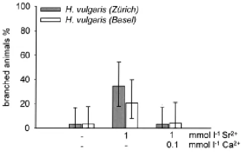
Fig. 2. Treatment with Ba2+ ions causes the formation of branched
animals. Polyps of strain H. vulgaris (Zürich) (grey bars) and polyps of strain H. vulgaris (Basel) (white bars) bearing each a 3 to 6 hour old bud were treated either for 2 days with 1 mmol l-1 BaCl
2. In addition the medium contained 2
mmol l-1 Na+/K+-phosphate buffer and optional 100 µmol l-1 of Ca2+ and Mg2+
ions. Thereafter the animals were transferred to standard culture medium. The result was scored 5 days after onset of treatment. None of the control animals in standard culture medium formed a branch (not shown). Each bar represents three independent experiments with 26 to 43 animals. The vertical bars indicate the respective 95% confidence interval.
a DMSO treatment caused the animals of all strains to branch. However, in the various Hydra strains the efficiency was different: H. vulgaris (Zürich) was the most sensitive one (Table 2).
Ba2+ or Sr2+ treatments caused the formation of branched
animals in all strains tested
Ions of the alkaline earth metals Barium and Strontium are well known to compete with Ca2+ ions at their various targets. If an
internal low level of Ca2+ ions is the cause for the production of
branches, a treatment with these ions should be expected to initiate branching not only in members of H. vulgaris (Zürich) but in members of other strains as well. This indeed was found. Figure 2 shows that a treatment with Ba2+ ions caused members of the
Basel strain to form side branches. Further, the treatment in-creased the frequency of branch formation in the Zürich strain. Ca2+ ions and to a lower extent also Mg2+ ions were found to
antagonise the influence of Ba2+ ions. An identical test with Sr2+
ions showed a similar result, but the efficiency of Sr2+ ions was
lower than that of Ba2+ ions (Fig. 3). A treatment with 300 µmol l-1
Ba2+ ions for one day caused the formation of branched animals in
H. magnipapillata (23%) (not shown).
Mg2+ ions display a dose dependent influence
In the absence of Ca2+ ions buds of H. vulgaris (Zürich) develop
into side branches. When the culture medium is enriched with low amounts of Mg2+ ions (100 µmol l-1) the frequency of side branch
formation is reduced and the animals look much better than those of a control without Mg2+ ions (Fig. 4). However, when the
concen-tration was increased to 1 mmol l-1 the frequency of branch
formation increased (in long treated animals). We conclude that low concentrations of Mg2+ ions stabilise the tissue while high
concentrations are able to antagonise Ca2+ ions at their targets.
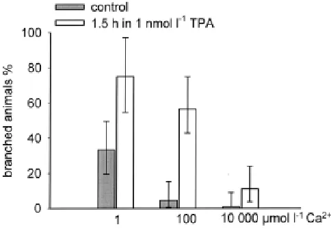
Branching due to TPA and cantharidin treatments is antago-nized by Ca2+ ions
In H. vulgaris (Zürich) a single pulse treatment with 12-o-tetradecanoylphorbol-13-acetate (TPA), a protein kinase C activa-tor, was found to cause animals to branch (Pérez and Berking, 1994). The experiments were done in culture medium which contains 1 mmol l-1 Ca2+ ions. Here we show that in the presence
TABLE 1
LOW AMOUNTS OF CA2+ IONS IN MEDIUM CONTAINING 2 MMOL L-1 NA+/K+
-PHOSPHATE BUFFER CAUSE A YOUNG BUD OF HYDRA VULGARIS (ZÜRICH) TO TRANSFORM INTO A BRANCH (5 DAY CULTIVATION)
bud age (h) Ca2+ (µmol l-1) animals (n) branched animals n (%)
TREATMENT WITH DMSO IN DISTILLED WATER CAUSES BRANCHED ANIMALS IN ALL STRAINS TESTED
Hydra strains animals (n) incubation time (d) DMSO % branched animals, n (%)
H. v. (Zürich) 30 5 0.1 23 (77)
H. v. (Basel) 47 5 0.1 0
H. magnipapillata 10 5 0.1 0
H. v. (Basel) 50 14 1 1 (2)
H. magnipapillata 50 14 1 1 (2)
H. v. (Basel) 100 4 2 2 (2)
H. magnipapillata 120 4 2 1 (1)
H. v. (Basel) 50 2 3 10 (20)
H. magnipapillata 50 2 3 10 (20)
TABLE 3
A HIGH CONCENTRATION OF CA2+ IONS ANTAGONISES BRANCH
FORMATION IN HYDRA VULGARIS (ZÜRICH) TREATED FOR 1H 45 MIN WITH 0.5 µMOL L-1 CANTHARIDIN
Ca2+ (mmol l-1) animals (n) branched animals n (%)
10 68 9 (13)
1 79 52 (66)
of 100 µmol l-1 more than one half of the animals produced a branch
instead of a bud while only some few did so in the control without TPA. When the Ca2+ ion concentration was decreased the effect
increased. When the Ca2+ ion concentration was increased the
effect of TPA decreased down to 10 % branched animals. Thus, the effect of TPA is antagonised by an external high level of Ca2+ ions
(Fig. 5).
In the presence of Ca2+ ions (1 and 10 mmol l-1 CaCl
2,
respec-tively) animals of the strain H. vulgaris (Zürich) bearing a young bud (about 3 hours old) were treated for 1h 45 min with 0.5 µmol l-1
cantharidin. The cantharidin treatment was preceded and followed by a treatment with medium containing the same Ca2+ ion
concentra-tion but no cantharidin (45 and 3 h 15 min, respectively). The group of animals treated with 1 mmol l-1 CaCl
2 represents the control for
standard culture medium which contains among other ions 1 mmol l-1 CaCl
2. It turned out that a high concentration of Ca2+ ions
antagonised the influence of the phosphatase inhibitor cantharidin to transform a bud into a branch (Table 3).
H. vulgaris (Zürich) kept in distilled water reduces it’s Ca2+
and K+ ion content whereas H. vulgaris (Basel) does not
Adult animals of H. vulgaris (Zürich) and H. vulgaris (Basel) have a different body size. Based on the protein content (measured after Lowry et al., 1951) members of the Basel strain are about 2.5 times larger than members of the Zürich strain. The content of K+ ions
reflects this difference: the amount per animal was found to be three time higher in the Basel strain that in the Zürich strain (Table 4).
Fig. 3. Treatment with Sr2+ ions causes the formation of branched
animals. See legend to Fig. 2. The difference is the replacement of Ba2+ ions
However, the content of Ca2+ ions does not reflect this difference: The
amount per animal was almost identical (Table 4). Thus, the concen-tration of free and stored Ca2+ ions appears to be 2 to 3 times higher
in H. vulgaris (Zürich) than in H. vulgaris (Basel). (The measured amount of K+ ions is within the limits determined by others (Lilly 1955,
Steinbach, 1963, Koblick and Yu-tu, 1967)). To our knowledge values for the Ca2+ ion concentration in Hydra are not reported.
When animals were kept in distilled water for 48 hours the amount of K+ ions and Ca2+ ions decreased in members of the
strain H. vulgaris (Zürich) (by 35 and 25%, respectively) but not in members of the strain H. vulgaris (Basel) (Table 4).
Discussion
In the past it was not possible to design a general scheme revealing the role of signal transduction in pattern control in Hydra. Rather, and for unknown reasons, such schemes had to be different for different species of Hydra. The data shown here indicate that one of the central processes in pattern formation is Ca2+ ion dependent. This was found to be true for all species
studied. However, under unfavourable conditions the strains dis-play a considerably different ability to keep their Ca2+ ion content
above a certain threshold.
The by far most sensitive strain is H. vulgaris (Zürich). This could be attributed to the water quality of the Lake Zurich. The lake contains and gets water with an extremely high level of Ca2+ ions
Fig. 5. Calcium ions antagonise branch formation induced by TPA.
Polyps of H. vulgaris (Zürich) bearing a 3 to 6 hour old bud were treated for 2 days with 2 mmol l-1 Na+/K+ phosphate buffer enriched with three different
concentrations of Ca2+ ions (grey bars). Half of the animals were treated in
addition for 1.5 hour with TPA (1 nmol l-1) (open bars). This treatment starts
together with the Ca2+ ion treatment. The result was scored 5 days after
onset of treatment. Each bar represents three independent experiments with 23 to 45 animals. The vertical bars indicate the respective 95% confidence interval.
TABLE 4
H. VULGARIS (ZÜRICH) KEPT IN DISTILLED WATER REDUCE THEIR Ca2+
AND K+ ION CONTENT (AAS- MEASUREMENTS)
Ca 2+ (ng / animal ± SD) K+ (ng / animal ± SD)
Hydra strain std. medium dist. water std. medium dist. water
H. vulgaris (Zürich) 45.3 ± 7.2 33.6 ± 3.2 904 ± 8 590 ± 9 H. vulgaris (Basel) 53.8 ± 6.4 56.8 ± 6.4 2392 ± 30 2344 ± 24
(about 1.2 mmol l-1) which causes a high pH (in the course of a year
from 7.9 to 8.8 in the surface water) and the precipitation of Ca2+
-minerals (Kelts and Hsü, 1978). (The culture medium used in most laboratories also contains similar high concentrations of Ca2+ ions
(CaCl2)). Obviously, the animals in the lake never have any problems to get enough Ca2+ ions. Rather they need -and in
particular such small and thin animals like Hydra- very effective pumps to maintain the low intracellular level necessary for physi-ological processes. Hydra living in this lake may have adapted to these extreme conditions. Other strains may have adapted to the respective low levels of Ca2+ ions in their environment. It would be
interesting to learn whether there is a correlation between the Ca2+
ion content in the water from which a certain Hydra strain was collected and its sensitivity against an extremely low Ca2+ ion
concentration in laboratory experiments.
Short treatments with various compounds affect foot formation at the bud’s base only in members of H. vulgaris (Zürich). The list of effective compounds includes activators of protein kinase C (PKC), such as the diacylglycerol (DAG) 1,2-dioctanoyl-rac-glyc-erol (1,2-diC8), and the tumour promoting phorbolester 12-o-tetradecanoylphorbol-13-acetate (TPA), the tyrosine kinase inhibi-tors staurosporine, genistein and H-7, the phosphatase inhibitor cantharidin (Pérez and Berking, 1994, Pérez, 1996). This exclu-siveness may indicate that all noted treatments funnel into one process which is (quantitatively) different in H. vulgaris (Zürich) compared to the other strains. We suggest this to be the pumping of Ca2+ ions. This suggestion prompted testing the influence of two
members of this list, TPA and cantharidin, in the presence of various Ca2+ ion concentrations.
In H. vulgaris (Zürich) a treatment with TPA or 1,2-dioctanoylglycerol caused animals to branch while two related substances, which however do not stimulate PKC activity in vertebrates, 1,3-dioctanoylglycerol and MPMA
(phorbol-4-o-Fig. 4. The influence of Mg2+ ions on branching in H. vulgaris (Zürich).
Polyps bearing a 3-6 hour old bud were treated for 2 (grey bars) and 5 days (open bars), respectively, with 2 mmol l-1 Na+/K+-phosphate buffer enriched
with different concentration of Mg2+ ions. Thereafter the polyps were
Peroxidase Staining
For demonstrating the formation of foot specific cells we used the peroxidase stain according to Hoffmeister and Schaller (1985).
Ca2+ and K+ Ion Measurements
Hundred animals were cultured for 2 days either in 1 ml standard culture medium or in 1 ml distilled water. The experiment was repeated 6 times. After 48 hours the animals were washed twice with distilled water and then homogenised. Then distilled water and trichloracetic acid (TCA) were added to reach a final concentration of 10% TCA in 800 µl. The homogenate was kept on ice over night. Then the probes were homogenised and centrifuged for 3 minutes at 10,000g. The supernatant was removed and centrifuged through a filter (Micro Spin Filter Tubes, 0,2 µm PVDF; Alltech). Of every probe 3 x 200 µl were analysed by means of Atom Absorption Spectrometry.
Statistics
In the graphs the means of at least three identically performed experi-ments are shown. The bars indicate the respective confidence interval (95%-level).
Acknowledgements
We thank Frank Nicoleni for technical support during the AAS-measure-ments.
References
BERKING, S. (1998). Hydrozoa metamorphosis and pattern formation. Curr. Top. Dev. Biol. 38: 81-131.
BURNETT, A.L. (1961). The growth process in Hydra. J. Exp. Zool. 146: 21-83.
CAMPBELL, R.D. (1967). Tissue dynamics of steady state growth in Hydra littoralis. II Pattern of tissue movement. J. Morph. 121: 19-28.
DE PETROCELLIS, L., DI MARZO, V., GIANFRANI, C. and MINEI, R. (1993). Arachidionic acid, protein kinase C activators and bud formation in Hydra vulgaris. Comp. Biochem. Physiol., 105C: 219-224.
FLECK, J. (1997). Phosphatidylinositol (PI) signaling and subsequent events in metamorphosis induction of cnidarian larvae. Proc. 8th Int. Coral Reef Symp. 2: 1225-1229.
HASSEL, M. (1998). Upregulation of a Hydra vulgaris cPKC gene is tightly coupled to the differentiation of head structures. Dev. Genes Evol. 207: 489-501.
HASSEL, M. and BERKING, S. (1990). Lithium ions interfere with pattern control in Hydra vulgaris. Roux´s Arch. Dev. Biol. 198: 382-388.
HASSEL, M., ALBERT, K. and HOFHEINZ, S. (1993). Pattern formation in Hydra vulgaris is controlled by lithium sensitive processes. Dev. Biol. 156: 362-371.
HASSEL, M., BRIDGE, D.M., STOVER, N.A., KLEINHOLZ, H. and STEELE, R.E. (1998). The level of expression of a protein kinase C gene may be an important component of the patterning process in Hydra. Dev. Genes Evol. 207: 502-14.
HENNING, G., BENAYAHU, Y. and HOFMANN, D.K. (1991). Natural substances, marine bacteria, and a phorbolester induce metamorphosis in the soft coral Heteroxenia fuscescens (Anthozoa, Octocorallia). Verh. Dtsch. Zool. Ges. 84: 486-487.
HOFFMEISTER, S.A.H. and SCHALLER, H.C. (1985). A new biochemical marker for foot-specific cell differentiation in Hydra. Roux´s Arch. Dev. Biol. 194: 453-461.
KEHLS, N.E., HERRMANN, K. and BERKING, S. (1999). The protein phosphatase inhibitor cantharidin induces head and foot formation in buds of Cassiopea andromeda (Rhizostomae, Scyphozoa) Int. J. Dev. Biol. 43: 51-58.
KELTS, K. and HSÜ, K.J. (1978). Freshwater carbonate sedimentation in Lakes -Chemistry, Geology, Physics. Ed. Lerman, A. Springer New York, Heidelberg, Berlin. pp. 295-321.
KOBLICK, D.C. and YU-TU, L. (1967). The osmotic behaviour of digestive cells of Chlorohydra viridissima. J. Exp. Zool., 166: 325-330.
LEITZ, T., and MÜLLER, W.A. (1987). Evidence for the involvement of PI-signalling and diacylglycerol second messengers in the initiation of metamorphosis in the hydroid Hydractinia echinata Fleming. Dev. Biol. 121: 82-89.
LILLY, S.J. (1955). Osmoregulation and ionic regulation in Hydra. J. Exp. Biol., 32: 423-439.
methylether-(12-myristol-13-acetyl)) did not have this effect. It thus appears that a stimulation of a PKC is responsible for the branching (Pérez and Berking, 1994). We found the effect of TPA to be considerably reduced by an increased external concentration of Ca2+ ions. Protein kinases of type C are well studied in Hydra
(Hassel, 1998, Hassel et al., 1998), but a PKC which is inhibited by Ca2+ ions is unknown. A simple interpretation of the results
ob-tained would be that in H. vulgaris (Zürich) PKC stimulation provokes the export of Ca2+ ions out of the cytoplasm into stores
and / or out of the cells. The resultant low internal Ca2+ ion
concentration provokes the observed effect. An increased external level of Ca2+ ions partly compensates the induced loss of Ca2+ ions
from the cytoplasm. The effects observed after a treatment with cantharidin and Ca2+ ions can be explained similarly. Based on the
observation that only in H. vulgaris (Zürich) the other noted compounds cause animals to branch, we suggest that all these substances decrease the internal level of Ca2+ ions by stimulating
a Ca2+-pump. In the non-responding species the compounds may
decrease the Ca2+ ion level as well but not down to the threshold.
Some species were found to respond to certain regimes of treatment with these compounds in a quite different way (see Hassel and Berking, 1990, Müller, 1989, Müller, 1990, De Petrocellis et al., 1993). These responses may be due to different dispositions of the strains tested, so that in these cases other effects of the compounds prevail. For instance, it is well known that the stimula-tion of a PKC can affect a large number of quite different processes. Whether or not in members of a certain species a reduced Ca2+ ion
level contributes to or even is responsible for the effects observed, appears to be accessible by the following experiment: one may treat the animals with the compound in question in the presence of high and low levels of Ca2+ ions, respectively, (with or without
DMSO). In addition the influence of Ba2+ and Sr2+ ions on the
process in question should be tested.
Materials and Methods
Animals
Two strains of Hydra vulgaris were used, the Zürich strain, which was collected by Pierre Tardent (1966) (commonly referred to as “H. vulgaris” in other laboratories) and the Basel strain collected by Thomas Honegger (Technau and Holstein, 1996). Further, we used Hydra magnipapillata strain 105, the standard wild type strain, and the multiheaded mutant strain, mh-1, a sexual inbred of Hydra magnipapillata (Sugiyama and Fujisawa, 1977), and the green Hydra (Chlorohydra viridissima). The animals were maintained at 20°C in standard culture medium (1 mmol l-1 CaCl
2; 0.5 mmol
l-1 MgCl
2; 0.1 mmol l-1 KHCO3; 0.35 mmol l-1 NaHCO3 and 25 µmol l-1 EDTA,
pH = 7.4). They were fed five times a week on freshly hatched nauplii of Artemia salina. All animals used in the experiments were starved for 24 hours prior to the experiment.
Test Assay
If not stated otherwise animals bearing a 3 hour old bud (0 is the visible appearance) were selected and treated with the test-medium (see below) for 48 hours. Then they were washed twice with the culture medium. Three days later the results were scored the first time.
Chemicals
Test substances were applied in distilled water or 2 mmol l-1 Na+/K+
LOWRY, O.H., ROSEBROUGH, N.J., FARR, A.L. and RANDALL, R.J. (1951). Protein measurement with the Folin phenol reagent. J. Biol. Chem. 193: 265-275.
MÜLLER, W.A. (1985). Tumor-promoting phorbol esters induce metamorphosis and multiple head formation in the hydroid Hydactinia. Differentiation 29: 216-222.
MÜLLER, W.A. (1989). Diacylglycerol induced multiple head formation in Hydra. Development 105: 309-316.
MÜLLER, W.A. (1990). Ectopic head and foot formation in Hydra: Diacylglycerol induced increase in positional value and assistance of the head in foot formation. Differentiation 42: 131-143.
PÉREZ, F. (1996). Effects of cantharidin and phorbol ester on bud formation in Hydra vulgaris. Int. J. Dev. Biol. (suppl. 1): 273.
PÉREZ, F. and BERKING, S. (1994). Protein kinase modulators interfere with bud formation in Hydra vulgaris. Roux´s Arch. Dev. Biol. 203: 284-289.
SANYAL, S. (1966). Bud determination in Hydra. Indian J. Exp. Biol. 4: 88-92.
SIEFKER, B., KROIHER, M. and BERKING, S. (2000). Induction of metamorphosis from the larva to the polyp stage is similar in Hydrozoa and a subgroup of Scyphozoa (Cnidaria, Semaeostomeae). Helgol. Mar. Res. 54:230-236.
SPINDLER, K.D. and MÜLLER, W.A. (1972). Induction of Metamorphosis by Bacteria and by a Lithium-pulse in the Larvae of Hydractinia echinata (Hydrozoa). Roux’s Arch. Dev. Biol. 169: 271-280.
STEINBACH, H.B. (1963). Sodium, potassium and chloride in selected hydroids. Biol. Bull. 124: 322-336.
SUGIYAMA, T. and FUJISAWA, T. (1977). Genetic analysis of developmental mechanism in Hydra. Ι Sexual reproduction of Hydra magnipapillata and isolation of mutants. Develop. Growth and Differ. 19: 187-200.
TECHNAU, U. and HOLSTEIN, T. (1996). Phenotypic maturation of neurons and continuous precursor migration in the formation of peduncle nerve net in Hydra. Dev. Biol. 177: 599-615.
THOMAS, M.B., EDWARDS, N.C., BALL, B.E. and MCCAULEY, D.W. (1997). Comparison of metamorphic induction in hydroids. Inv. Biol. 116: 277-285.
TARDENT, P. (1966) Zur Sexualbiologie von Hydra attenuata Pall. Rev. Suisse Zool. 73: 357 - 381.
TRIPP, K. (1928). Die Regenerationsfähigkeit von Hydren in den verschiedenen Körperregionen. Nach Regenerations- und Transplantationsversuchen. Z. Wiss. Zool. 132: 476-525.