A study of Xlim1 function in
the Spemann-Mangold organizer
LAURENT KODJABACHIAN
1,#, ALEXANDER A. KARAVANOV
1,#, HIROKI HIKASA
2,3,
NEIL A. HUKRIEDE
1,
TAZU AOKI
2, MASANORI TAIRA
2,3and IGOR B. DAWID*
,11National Institutes of Health. National Institute of Child Health and Human Development, Laboratory of Molecular Genetics, Bethesda, MD,
USA, 2Laboratory of Molecular Embryology. Department of Biological Sciences, Graduate School of Science, University of Tokyo, Japan and 3Core Research for Evolutional Science and Technology (CREST), Japan Science and Technology Corporation, Japan
ABSTRACT The Spemann-Mangold organizer is required in amphibian embryos to coordinate cell fate specification, differentiation of dorsal cell types and morphogenetic movements at early stages of development. A great number of genes are specifically expressed within the organizer, most of them encoding secreted proteins and transcription factors. The challenge is now to uncover genetic cascades and networks of interactions between these genes, in order to understand how the organizer functions. The task is immense and requires loss-of-function approaches to test the requirement for a given factor in a specific process. For transcription factors, it is possible to generate inhibitory molecules by fusing the DNA binding region to a repressor or activator domain, which should in principle antagonize the activity of the endogenous protein at the level of the DNA targets. We used this strategy to design activated and inhibitory forms of the LIM homeodomain transcription factor Lim1, which is encoded by an organizer gene involved in head development, as revealed by analyses of knockout mice. We found that Lim1 is a transcriptional activator, and can trigger dorso-anterior development upon ventral expression of hyperactive forms, in which Ldb1 is fused to Lim1. Using inhibitory Lim1 fusion proteins, we found that Lim1, or genes closely related to it, is required for head formation as well as for notochord development. Co-expression experiments revealed that Lim1 is required downstream of the early organizer factor Siamois, first, to establish the genetic program of the organizer and second, to mediate the action of organizer agents that are responsible for blocking ventralizing activities in the gastrula.
KEY WORDS:
Spemann-Mangold organizer, Xenopus, Xlim1, anteroposterior axis, axial mesoderm.
0214-6282/2001/$25.00 © UBC Press
Printed in Spain www.ijdb.ehu.es
*Address correspondence to: I.B. Dawid. National Institutes of Health, National Institute of Child Health and Human Development, Laboratory of Molecular Genetics, Bldg 6B, Rm 420, 9200 Rockville Pike, 20892 Bethesda, MD, USA. FAX: +1-301-496-0243. e-mail: [email protected]
# Both authors contributed equally to this work
Introduction
During gastrulation in vertebrates, a complex series of events occurs, which leads to the correct spatial positioning of the three embryonic germ layers along the body axis, and to their patterning by localized and often reciprocal inductions. The Spemann-Mangold organizer (also called the dorsal organizer or gastrula organizer), a relatively small dorsal region of the gastrula in amphibians, coordinates these complex events. The graft of an early gastrula dorsal blastopore lip, where the Spemann-Mangold organizer forms, into the ventral region of a host embryo, leads to the development of a secondary body axis, which includes head features (reviewed in Lemaire and Kodjabachian, 1996; Harland and Gerhart, 1997). In contrast, grafting the same region explanted from a late gastrula only gives rise to tail duplication. These differences served as evidence for the existence of independent
head and trunk organizers. Molecules involved in organizer ontogeny or activity have been isolated on the basis of their early dorsal expression, or their ability to induce aspects of dorsal axis development upon over-expression (reviewed in Lemaire and Kodjabachian, 1996; Niehrs, 1999). So far, the homeoprotein Siamois and its close relative Twin, are unique zygotic factors which can trigger the development of a complete secondary axis, implicating these factors in organizer establishment (Lemaire et al., 1995; Laurent et al., 1997). Various molecules are able to stimulate either head or trunk development, and are therefore believed to act downstream of Siamois in specific compartments of the organizer.
1994a). Analyses of mice mutant for the Lim1 gene have revealed its essential function in the head organizer, as these animals lack most of the anterior brain (Shawlot et al., 1995). This dramatic phenotype indicates that Lim1 is a central regu-lator during the early phases of axial development, and it is therefore important to understand how this gene functions. However, analyses of mutant mouse embryos have only re-vealed partial aspects of the mode of action of this factor, and we reasoned that important information could be gained by studying Lim1 in the more accessible Xenopus system.
Functional domains in the Lim1 protein have been quite exten-sively characterized, allowing three domains to be distinguished: an N-terminal pair of LIM domains which seem to play an adapter role, a central homeodomain with DNA-binding activity, and a C-terminal transactivation domain (Agulnick et al., 1996; Breen et al., 1998). The LIM domain is a double-zinc finger motif, which can interact with various protein domains (reviewed in Dawid et al., 1998). LIM domains can interact with a LIM domain binding protein, Ldb1 or NLI/CLIM-2, which is required to allow Lim1 to exert its dorsalizing potential in frog embryos (Agulnick et al., 1996). A similar situation exists in Drosophila where Chip, a homologue of Ldb1, is required for the normal activity of the LHX factor Apterous (Morcillo et al., 1997; Fernandez-Funez et al., 1998). Ldb1/Chip is known to contain a homodimerization domain (Jurata et al., 1997; Breen et al., 1998), and further studies in Drosophila have revealed that tetramers composed of two Ldb and two LHX molecules are functional in vivo. In particular, it was
shown that a chimera in which the dimerization domain of Chip replaces the LIM domains of Apter-ous, fully rescues the apterous mutant, suggesting that the main role of Chip is to bridge two Apterous molecules via their LIM domains (Milan and Cohen, 1999; Van Meyel et al., 1999).
In the present study we designed activator and inhibitory Lim1 fusion proteins and assayed their function upon ectopic expression in early frog em-bryos. Based on these studies, we present evi-dence that Lim1 is involved at multiple steps of organizer formation and activity in Xenopus.
Results
Xlim1 is a transcriptional activator
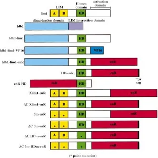
Ectopic expression of mutant forms of Xlim1, where LIM domains have been deleted or mutated, leads to dorsalization of mesoderm and the forma-tion of partial secondary axes (Taira et al., 1994a). Similarly, co-expression of Xlim1 and Ldb1 induces partial ectopic axes, neuralizes ectoderm and in-duces organizer gene expression in animal cells (Agulnick et al., 1996; Breen et al., 1998). In order to test whether vertebrate LHX activity is regulated similarly to Drosophila Apterous, we made a fusion between the dimerization domain of Ldb1, as de-fined by Breen et al. (1998), and the Lim1 protein from which LIM domains were deleted (Ldb1-lim1; Fig. 1). Here, we present data obtained with the zebrafish versions of Ldb1 and Lim1, but similar results were obtained with the Xenopus proteins
Fig. 1. Schematic representation of the constructs used in this study. (See text for details).
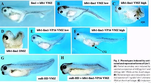
(Hiratani and Taira, submitted). A range of phenotypes was observed upon ventral injection of ldb1-lim1 in frog embryos. Low doses (200 pg to 1 ng) induced partial secondary axes similar to co-injection of ldb1 and lim1 (Fig. 2 A,B), while high doses (4 to 6 ng) led to the formation of ectopic cement gland or eye without clear axis formation (Fig. 2C). In fact, high doses of this construct also impaired axis elongation upon dorsal injection (Fig. 2D), suggesting that Lim1 could repress trunk fates when it is hyper-active. Interestingly, anterior fates were never observed upon injection of Xlim1-3m (Taira et al., 1994a) or co-injection of ldb1 and lim1 (Agulnick et al., 1996), indicating that the Ldb1-lim1 fusion protein is more active in this assay. It is important to note the activity of Lim1 in inducing anterior fates, as it is in agreement with the requirement for this gene in head development in the mouse (Shawlot and Behringer, 1995).
The experiments described above did not reveal whether Lim1 acts as a transcriptional activator or repressor. To address this question, additional constructs were made where various domains of Lim1 are fused to the transactivation domain of the viral protein VP16 (VP16; see Kessler, 1997) or the repressor domain of Drosophila Engrailed (enR; Han and Manley, 1993) (Fig. 1). In a previous study a putative transcriptional activation domain was found in Lim1 between amino acids 266 and 403 (Breen et al., 1998), and this region was therefore deleted in most of our fusion proteins.
result indicates that Lim1 acts as a transcriptional activator, at least in ventral territories of the embryo. Moreover, axis induction by ldb1-lim1-VP16 was found to be antagonized by co-injection of enR-HD encoding one of the repressor versions of Lim1 used in this study (Fig. 2H). Consistently, axis induction by Xlim1-3m or Xlim1 and Ldb1 was also suppressed by the co-expression of this repressor form of Lim1 (not shown). As enR-HD is not believed to contain any domain able to interact with Ldb1-lim1-VP16, the observed antagonism between these molecules is likely to take place at the level of the target DNA. This is illustrated by an experiment where gene expression was measured by northern analysis in animal caps loaded with different combinations of RNAs (Fig. 3). Co-expression of Ldb1 and Xlim1 in animal cells activated the transcription of the organizer genes chordin (chd; Sasai et al., 1994), otx2 (Pannese et al., 1995; Blitz and Cho, 1995) and goosecoid (gsc; Cho et al., 1991), and the presence of the repressive form of XLim1, ∆C Xlim1-enR, blocked this effect (Fig. 3). As Xlim1 in the presence of Ldb1 has been shown to activate directly the transcription of gsc (Mochizuki et al., 2000), we reasoned that the repressive forms of Xlim1 might constitute good reagents to prevent the normal function of this gene in frog embryos.
Xlim1 is required for dorso-anterior development
Similar to findings in Lim1 knockout mice, we observed that dorsal expression of the repressive forms of Xlim1 antagonized
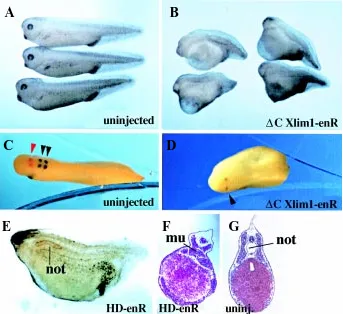
anterior development, as shown in Fig. 4B. Using markers ex-pressed at different positions in the central nervous system, we could determine that the embryonic axis was truncated anterior to rhombomere 5 (Fig. 4 C,D). This position is roughly consistent with the level of truncation observed in mutant mice, as brain was missing rostral to rhombomere 3 (Shawlot et al., 1995). However, we found an additional phenotype, which was not seen in Lim1-/- mouse mutants, as notochord development was strongly im-paired in embryos injected with the HD-enR repressive form of Xlim1 (Fig. 4 E,F). Importantly, injected embryos underwent normal blastopore closure, which ruled out a defect in conver-gence-extension as being the primary cause for defective noto-chord development. In contrast to defective notonoto-chord develop-ment, somite formation could take place in presence of repressive Xlim1, as somitic tissue actually developed at the midline of injected embryos. However, we do not know whether this mis-localized somitic tissue formed de novo at the expense of axial mesoderm, or whether somites simply fused at the midline due to the lack of a physical barrier. As Xlim1 is clearly expressed in the developing notochord in Xenopus (Taira et al., 1994b; Karavanov et al., 1996), it is not surprising to find that this gene is required for normal notochord formation, although this is not apparently the case in mouse.
We next compared the activity of different repressive versions of Xlim1 in antagonizing anterior development, in order to deter-mine which regions are functionally important in the Lim1 molecule.
A
B
D
C
E
F
H
G
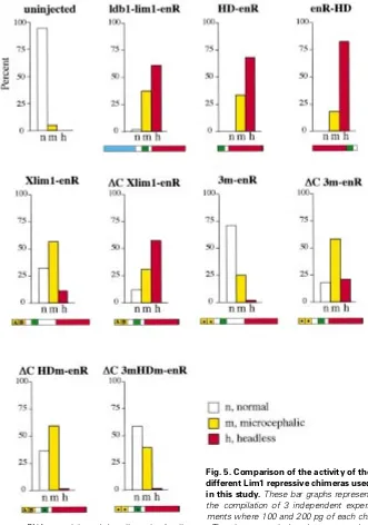
Thus, we injected similar amounts of each of these RNAs (100 and 200 pg/blastomere) repeatedly in three independent experiments, and scored the resulting phenotypes as headless (most severely truncated, with no eyes and no cement gland), microcephalic (no cement gland, small or single eye), or normal (Fig. 5). All repressive forms of Lim1 could trigger a similar range of phenotypes, but with
bryos compared to ∆C Xlim1-enR injected embryos. (3) A mutation in the homeodomain dramatically suppresses the inhibitory activity of ∆C HDm-enR, indicating that the repression requires the DNA binding activity of our chimeras, and does not merely depend on titration of Lim1 cofactors. However, it should be noted that this mutant is not totally inert suggesting that the presence of the LIM domains and possibly other parts of the molecule could sequester cofactors required for normal activity. This is also consistent with the fact that 3m-enR and ∆C 3m-enR are less active than their counterparts without mutation in the LIM domains. Further support for this idea comes from observations made in zebrafish, where over-expression of Islet-3 LIM domains triggered inhibitory effects (Kikuchi et al., 1997), probably by trapping essential LIM binding factors, such as Ldb1. (4) The presence of the amino-terminal region of Lim1 decreases the severity of phenotypes in ∆C Xlim1-enR injected embryos compared to HD-Xlim1-enR injected embryos, suggesting that this region could contain an activator domain. This is supported by the fact that in a yeast one-hybrid system, a fusion of the entire Xlim-1 protein to the GAL4 DNA binding domain is more active than a fusion of the C-terminal activation domain of Xlim-1 to the GAL4 DNA binding domain (Breen et al., 1998). (5) Dimerization is probably not required for repression as HD-enR and enR-HD constructs, which are not believed to dimerize, are as active as the Ldb1-lim1-enR construct. (6) The phenotypes ob-served cannot be attributed to the presence of a myc-tag in some of our constructs, as severe phenotypes are also generated by constructs lacking this epitope.
We next wanted to address the question of the specificity of action of the repressive Lim1 molecules in the embryo. The most
Fig. 3. ∆C Xlim1-enR suppresses the expression of dorsal marker genes activated by Xlim-1 and Ldb1 in animal caps. Animal caps were dissected before gastrulation from embryos injected at the 2-cell stage with mRNAs as indicated. Explants were cultured until control sibling embryos reached stage 11. Total RNA was extracted from 15 animal caps and subjected to Northern blot analysis using chordin (chd), otx2 and goosecoid (gsc) probes. Globin RNA was injected as a control. 18S rRNA stained with ethidium bromide serves as a loading control.
Fig. 4. Phenotypes elicited by expression of repressive forms of Lim1. Dorsal injection at the 4-cell stage of 250 pg ∆C Xlim1-enR mRNA pro-vokes anterior truncation (B,D) compared to uninjected siblings (A,C). Embryos were subjected to two-color whole-mount in situ hybridization analy-sis with two head markers, krox20 (rhombomeres 3 and 5; dark blue) and en2 (isthmus; red) at the tailbud stage. The head in ∆C Xlim1-enR injected embryos is truncated anterior to rhombomere 5 (D). Dorsal injection at the 4-cell stage of 100 pg HD-enR RNA also leads to head truncation and deficiency in notochord, as revealed by staining with the MZ15 antibody (E) or histological section (F). (G) Control section of an uninjected embryo. Note that somitic tissue is present in headless embryos and actually expands in the region nor-mally occupied by the notochord (F). mu, muscle; not, notochord.
variable penetrance. A number of conclu-sions can be drawn from this test, as follows. (1) The respective orientation between the DNA binding domain and the repressor do-main has virtually no influence on the sever-ity of the phenotypes, as HD-enR and enR-HD are about equally active. (2) We con-firmed that the carboxy-terminal region of Lim1 contains an activator domain functional in vivo, as its presence reduces the severity of phenotypes in Xlim1-enR injected
em-A
B
D
C
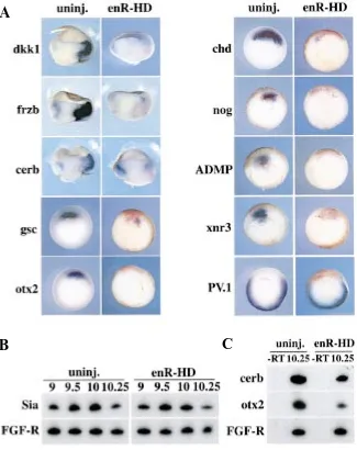
inhibitors Chordin (Piccolo et al., 1996) and Noggin (Zimmerman et al., 1996), as well as the expression of Cerberus which has a triple inhibitory function on BMP, Wnt and Nodal signaling (Pic-colo et al., 1999). In addition the organizer genes goosecoid, otx2 and Xnr3 (Smith et al., 1995) were also repressed, and ADMP, a specific inhibitor of Follistatin recently found to be involved in trunk development (Moos et al., 1995; Dosch and Niehrs, 2000), was repressed as well. In order to study the fate of the cells injected with repressive Lim1, we looked at expression of the ventro-posterior marker PV.1 (Ault et al., 1996) and found that the presumptive dorsal organizer was not ventralized, at least during gastrulation (Fig. 7A). This is consistent with the observation that appropriate test for establishing specificity is
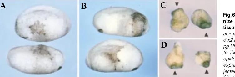
the rescue of mutant phenotypes by co-ex-pression of the normal protein. Despite sub-stantial efforts we could not obtain a signifi-cant level of rescue of anterior structures in whole embryos, or of marker gene expression in explants, by using such a strategy. In short, any of the repressive Lim1 molecules acted dominantly in our assays over any of the activated versions of Lim1 (not shown). Al-though we cannot provide a full explanation for this phenomenon, we do not think that these constructs act in a nonspecific manner, and we carried out a number of tests to sup-port this view. As Xlim1 is a homeodomain factor, we tested whether the repressive forms of Lim1 antagonized the activity of unrelated homeodomain proteins. The homeodomain protein Otx2, when expressed in ventral ectoderm, induces ectopic formation of ce-ment gland (Bradley et al., 1996); this effect was not antagonized by co-expression of re-pressive HD-enR (Fig. 6 A,B). This experi-ment demonstrates that this inhibitory form of Lim1 does not interact with the targets of another homeodomain factor, indicating that repression shows a certain degree of specificity. If repressive versions of Lim1 be-haved specifically they should not trigger any phenotypes in tissues which do not express Xlim1. This is the case when repressive Lim1 is expressed ventrally (Fig. 2G), where very little Xlim1 mRNA is present (Taira et al., 1992). Another such situation can be artifi-cially created when the homeodomain factor Siamois is ectopically expressed in naïve ectoderm, as this protein stimulates the ex-pression of many organizer genes, but not of Xlim1, in animal cap assays (Carnac et al., 1996). Dorsalization of the ectoderm by Siamois results in the formation of large ce-ment glands, and this effect was not sup-pressed upon co-expression of repressive HD-enR (Fig. 6 C,D). Again, this experiment sug-gests that repressive Lim1 constructs act spe-cifically, since cement gland development is impeded when the repressor construct is
ex-Fig. 5. Comparison of the activity of the different Lim1 repressive chimeras used in this study. These bar graphs represent the compilation of 3 independent experi-ments where 100 and 200 pg of each chi-mera RNA were injected dorsally at the 4-cell stage. The phenotypes induced were scored as headless (no cement gland, no eyes) or microcephalic (no cement gland, small or single eye). Refer to the text for an interpretation of these data.
pressed in tissues normally expressing Xlim1 (Fig. 4B).
Xlim1 is required for organizer gene expression
the somites and neural tube still form in HD-enR injected embryos (Fig. 4F). In conclusion, although the exact fate of dorsal cells lacking Xlim1 function is not clear, it appears that organizer formation does not occur in the absence of this gene.
The general requirement for Xlim1 in dorsal gene expression could indicate that this gene is acting very early in the genetic cascade leading to the formation of the Spemann-Mangold organ-izer. The earliest known zygotic actor in this cascade is the gene Siamois, which is directly activated by maternal cues, can trigger complete axis formation, and is required for the establishment of all dorsal fates (Lemaire et al., 1995; Fan and Sokol, 1997; Kessler, 1997; Darras et al., 1997). Although Xlim1 expression is first detected several hours after the onset of Siamois expression (Lemaire et al., 1995), it was possible that repressive forms of Lim1 artificially blocked the activation of Siamois in our injections. However, RT-PCR analysis in embryos injected with high doses of enR-HD demonstrated that Siamois expression was not modi-fied compared to uninjected embryos, over a period of 3 hours. This result indicates that the Lim1 family of genes is required for establishing the organizer program downstream or in parallel to the zygotic factor Siamois.
Xlim1 position in the Spemann-Mangold organizer cascade
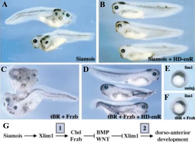
In an effort to determine which steps of organizer function depend on Xlim1 activity, we carried out co-injection experiments with repressive Lim1 and factors known to stimulate sequential steps in axis formation. Ventral expression of Siamois led to the development of a complete secondary axis, and the co-expres-sion of repressive HD-enR completely suppressed this effect (Fig. 8 A,B). This experiment confirms that Lim1 is required down-stream of Siamois during organizer establishment, as suggested by their respective period of expression. It has been shown that activated forms of Lim1 stimulate expression of the BMP inhibitor Chordin in animal cells (Taira et al., 1994a; Fig. 3), and we show here that Lim1 function is required for normal dorsal expression of chordin (Fig. 7A). These data suggest that Lim1 could act downstream of Siamois to establish the program of expression of organizer specific secreted inhibitors. However, it was not known whether these factors can act in absence of Lim1 function to drive dorso-anterior development. Thus, we tested whether axis induc-tion by ventral co-expression of the truncated BMP receptor and the Wnt inhibitor Frzb required Xlim1 function. Unexpectedly, we found that HD-enR could suppress secondary axis development in such conditions, indicating that Lim1 is necessary to relay the
action of organizer agents. Consistent with this result, we could show that expression of the resident Xlim1 gene is activated by this combination of factors (Fig. 8 E,F). In a similar experiment an inhibitory form of Siamois, enR-Sia (Darras et al., 1997), did not antagonize axis formation by these factors (not shown), support-ing the notion that the requirement for Xlim1 in this assay is specific. Hence, it appears that Xlim1 is required sequentially, first to induce organizer gene expression downstream of Siamois, and second to relay the activity of inhibitors of ventralization.
Discussion
Prior to this study it was not known whether Lim1 was a transcriptional activator or repressor. Here, we show that the Ldb1-lim1-VP16 fusion protein could generate phenotypes simi-lar to those induced by the combination of Xlim1 and Ldb1, indicating that Xlim1 acts as a transcriptional activator in this context. However, we cannot rule out that Lim1 could also act as a transcriptional repressor on particular promoters. Further progress into this issue requires the identification of direct tran-scriptional targets of Lim1. Likely candidates for such targets include the organizer genes chordin and otx2, whose expression can be ectopically induced by Xlim1 in animal cells, goosecoid, whose regulatory elements contain Xlim1 binding motifs (Mochizuki et al., 2000), and cerberus, as expression of cerr-l, a murine cerberus homologue, depends on Lim1 activity (Shawlot et al., 1998). Here, we show that putative additional targets may exist as the inhibitory versions of Xlim1 can antagonize expression of most organizer genes examined, with the notable exception of Siamois. Thus, it will be essential to determine which genes Lim1 directly regulates in vivo in order to understand the networks of interactions required for axis formation. This issue can be best addressed in Xenopus with the help of inducible versions of Lim1 fusion proteins (Gammil and Sive, 1997; Tada et al., 1998).
We found that the activated version of Lim1 containing the Ldb1 dimerization domain could induce anterior features upon ventral ectopic expression, which has not been observed in previous studies with wild type or mutant Xlim1 (Taira et al., 1994a; Agulnick et al., 1996). This result suggests that this fusion protein acts more potently in antagonizing ventralizing factors, arguing for the critical importance of dimerization of vertebrate LIM homeodomain proteins as described for their Drosophila counterparts (Milan and Cohen, 1999; Van Meyel et al., 1999). It is interesting to note that activated Lim1 constructs can induce
Fig. 6. Repressive forms of Lim1 do not antago-nize activity of Otx2 and Siamois in animal tissues. (A,B) Embryos were injected in ventral-animal position at the 4-cell stage with (A) 800 pg otx2 RNA or (B) a mixture of 800 pg otx2 and 100 pg HD-enR RNAs. Over-expression of otx2 leads to the formation of cement gland tissue in the epidermis, and this effect is not antagonized by co-expression of HD-enR. (C,D) Embryos were in-jected animally at the 2-cell stage with (C) 20 pg Siamois RNA, or (D) a mixture of 20 pg Siamois and 100 pg HD-enR RNAs. Animal caps were dissected at stage 9 and cultured until siblings reached tadpole stages. Siamois expression in animal cells stimulates cement gland formation, and this effect is not suppressed by co-expression of HD-enR. Arrowheads point at cement glands.
A
B
trunk development at low doses, and anterior features with no signs of normal trunk formation at higher doses (Fig. 2). These data suggest that Lim1 could be involved in initiating both head and trunk development, and that these two developmental programs are mutually ex-clusive, as revealed by studies with normal and inhibitory Otx2 proteins (Andreazzoli et al., 1996; Isaacs et al., 1999). This idea is further sup-ported by our finding that Lim1 is required both for head development and for trunk axial fate determination in Xenopus. Phenotypic analysis in Lim1 mutant mice, however, did not reveal a requirement for this gene in notochord forma-tion. A similar situation has been described for another organizer gene, goosecoid, which is required for notochord development in Xeno-pus, based on the phenotypes induced by in-hibitory versions of this protein (Ferreiro et al., 1998), while no such phenotypes are visible in mutant mice defective for goosecoid (Rivera-Perez et al., 1995). This apparent difference between our study and previous work might come from the respective strategies used to abolish the function of Lim1. In our study, it is likely that the activity of Lim1 related proteins are antagonized by the presence of antimorphic forms of Lim1. Thus, the observed phenotypes could be the result of the blockage of multiple Lim1 related proteins in Xenopus embryos, while inactivation of a single Lim1 gene might not be sufficient to generate the same pheno-types in mouse embryos. Moreover, functional redundancy could also occur between Lim1 and transcription factors belonging to different families if they are collectively required to acti-vate target genes (see Perea-Gomez et al., 1999).
An important flaw to our demonstration of the developmental requirements for Xlim1 is the lack of phenotypic rescue by co-expression of inhibitory and activator forms of the protein. We could show that the two types of proteins acted antagonistically both in animal cells and in ventral marginal cells. However, this mutual antagonism is not apparent upon dorsal co-expression. A possible complication to this as-say is the fact that activated Lim1 molecules generate gastrulation defects on their own, thus combinatorial deleterious effects could prevent
presence of a single repressor form of Lim1 could shut off expression of this gene, making rescue difficult to achieve. Although we cannot provide a totally satisfying explanation for the apparent lack of rescue, we do not think that our chimeras act non-specifically, as they do not antagonize other homeoproteins such as Otx2 and Siamois in contexts where Xlim1 is not normally expressed (Fig. 6). Moreover, the phenotypes generated by expression of inhibitory forms of Xlim1 are consistent with the expression pattern of this gene, in particular from a temporal point phenotypic rescue. However, this is not the only problem, as
co-expression of activated and repressive Lim1 constructs in animal explants treated with Activin did not result in the recovery of dorsal marker gene expression (data not shown). Thus, the inhibitory form of Lim1 is dominant over the activator form in every case tested, suggesting that control elements of target genes are irreversibly resistant to activation once they contact a Lim1-enR chimeric protein. Possibly, in the case when several Lim1 binding sites exist in a target gene (see Mochizuki et al., 2000), the
Fig. 7. Repressive form of Lim1 inhibits expression of organizer genes except Siamois. Embryos were injected at the 4-cell stage with a mixture of 100 pg enR-HD and 400 pg lacZ RNAs. (A) After revealing β-galactosidase activity with a red substrate at stage 10.25/10.5, embryos were processed for in situ hybridization with the indicated probes. In the cases of dkk1, frzb and cerberus, embryos were bisected before hybridization to enhance staining in the deep layers of the embryo. All dorsal genes examined are repressed by enR-HD. However, the ventral-posterior gene PV.1 is not ectopically expressed dorsally. Dorsal is right for dkk1, frzb and cerb panels, and dorsal is up in all other panels. (B,C) RT-PCR experiments on embryos injected at the 4-cell stage in a dorsal vegetal position with 100 pg enR-HD RNA. This site of injection targets cells that express the organizer gene Siamois. Panel B shows that expression of Siamois is unchanged in enR-HD injected embryos between stages 9 and 10.25. Panel C shows that the same embryos exhibit reduced levels of expression of cerberus and otx2, confirming that enR-HD was active in this experiment. FGF-R is used as a loading control.
A
of view. We showed that inhibitory forms of Lim1 do not suppress the expression of Siamois, which appears to be the earliest zygotic gene involved in axis formation, and which is normally expressed prior to Xlim1 (Lemaire et al., 1995). In contrast, inhibitory forms of Lim1, in agreement with the fact that Lim1 is normally required for organizer gene expression, suppress axis induction by Siamois. Less anticipated was the observation that Lim1 is also required downstream of organizer factors whose expression is regulated by this gene. These factors, such as Chordin and Frzb, serve to limit or prevent ventralizing activities of BMP and Wnt acting during gastrulation (Jones et al., 1996). We show here that these dorsal factors can induce the ectopic expression of Xlim1 during gastrulation, thereby suggesting the existence of a positive feedback between these genes, in order to enhance the dorsal expression program. Consistent with this idea, axis development is prevented when inhibitory forms of Lim1 are expressed during gastrulation under the control of the cytomegalovirus promoter (data not shown). Thus, our data indicate that Lim1 is a central regulator, required at two critical steps during early development: First, during the short period of organizer establishment where it participates in the activation of early genes. Second, during the phase where the organizer functions to antagonize ventralizing activities and to allocate dorsal fates. It will be interesting to determine whether Lim1 targets are the same during both phases, which would indicate that this factor is required for initiation and maintenance of organizer gene expression. Alternatively, Lim1 could activate a different set of targets at different times, arguing for a sequential progression towards the acquisition of anterior dorsal fates. Interestingly, the second view is supported by recent observa-tions made in Lim1 mutant mice. Using chimeric mice and explant recombination, Shawlot and colleagues (1999) put forward a double assurance model whereby Lim1 is required in different tissues at different developmental stages in order to impart anterior identity to the embryo. However, targets of Lim1 activity
during this process are not known, and the functional analysis of chimeric Lim1 proteins in Xenopus should help elucidate this question.
Materials and Methods
Methods used in this paper are standard and were previously de-scribed in the following articles. Preparation and microinjection of Xeno-pus embryos: Taira et al., 1994a. Northern blot analysis of animal cap assays: Taira et al., 1992; Taira et al., 1994a. Whole-mount in situ hybridization: Gawantka et al., 1995. Whole-mount immunostaining: Darras et al., 1997. RT-PCR: Darras et al., 1997. The primers used were:
otx2 forward 5’ GCA CCC AGT CGG TGG GAT ATC 3’ otx2 reverse 5 ’CCA CTC TCC GAG CTC ACT TC 3’
siamois forward 5’ AAA CCA CTG ATT CAG GCA GAG G 3’ siamois reverse 5’ GTA GGG CTG TGT ATT TGA AGG G 3’
cerberus forward 5’ GCT TGC AAA ACC TTG CCC TT 3’ cerberus reverse 5’ CTG ATG GAA CAG AGA TCT TG 3’
FGF-R forward 5’ TTG AAG TCT GAT GCG AGT GA 3’ FGF-R reverse 5’ GGG TTG TAG CAG TAC TCC AT 3’
Acknowledgements
We would like to thank Daniel Kessler, Christof Niehrs, Malcolm Moos and Eddy De Robertis for reagents, and Ajay Chitnis for comments on the manuscript.
References
AGULNICK, A.D, TAIRA, M, BREEN, J.J., TANAKA, T., DAWID, I.B. and WESTPHAL, H. (1996). Interactions of the LIM-domain-binding factor Ldb1 with LIM homeodomain proteins. Nature 384: 270-272.
ANDREAZZOLI, M., PANNESE., M. and BONCINELLI, E. (1997). Activating and repressing signals in head development: the role of Xotx1 and Xotx2. Develop-ment 124: 1733-1743.
Fig. 8. Position of Lim-1 in the genetic cascade of the Spemann-Mangold organizer. (A,B) Embryos were injected ventrally at the 4-cell stage with (A) 20 pg Siamois RNA or (B) with a mixture of 20 pg Siamois and 100 pg HD-enR RNAs. Expression of Siamois leads to formation of a complete secondary axis, and co-expression of the repressive form of Lim1 sup-presses this effect. (C,D) Embryos were injected ventrally at the 4-cell stage with (C) 500 pg truncated BMP receptor (tBR) and 400 pg frzb RNAs or (D) with a mixture of 500 pg tBR, 400 pg frzb and 100 pg HD-enR RNAs. Co-expression of the BMP and Wnt an-tagonist’s tBR and frzb promotes formation of a com-plete secondary axis, and this effect is inhibited by co-expression of the repressive form of Lim1. (E,F) in situ hybridization with XLim1 probe on (E) stage 10.5 uninjected embryos or (F) embryos injected as in C, reveals activation of XLim1 by co-expression of BMP and Wnt antagonists. (G) Model for XLim1 position within the organizer genetic cascade based on its requirement downstream of known regulators of or-ganizer function. Lim1 activity is critical downstream of Siamois in establishing the organizer (phase 1), and is needed to relay the activity of organizer’s inhibitors to allow dorso-anterior development (phase 2).
AULT, K.T., DIRKSEN, M.L. and JAMRICH, M. (1996). A novel homeobox gene PV.1 mediates induction of ventral mesoderm in Xenopus embryos. Proc. Natl. Acad. Sci. USA 93: 6415-6420.
BLITZ, I. and CHO, K. (1995). Anterior neurectoderm is progressively induced during gastrulation: the role of the Xenopus homeobox gene orthodenticle. Development 121: 993-1004.
BRADLEY, L., WAINSTOCK, D. and SIVE, H. (1996). Positive and negative signals modulate formation of the Xenopus cement gland. Development 122: 2739-2750.
BREEN, J.J., AGULNICK, A.D., WESTPHAL, H. and DAWID, I.B. (1998). Interac-tions between LIM domains and the LIM domain-binding protein Ldb1. J. Biol. Chem. 273: 4712-4717.
CARNAC, G., KODJABACHIAN, L., GURDON, J.B. and LEMAIRE, P. (1996). The homeobox gene Siamois is a target of the Wnt dorsalisation pathway and triggers organiser activity in the absence of mesoderm. Development 122: 3055-3065.
CHO, K.W., BLUMBERG, B., STEINBEISSER, H. and DE ROBERTIS, E.M. (1991). Molecular nature of Spemann’s organizer: the role of the Xenopus homeobox gene goosecoid. Cell 67: 1111-1120.
DARRAS, S., MARIKAWA, Y., ELINSON, R.P. and LEMAIRE, P. (1997). Animal and vegetal pole cells of early Xenopus embryos respond differently to maternal dorsal determinants: implications for the patterning of the organiser. Develop-ment 124: 4275-4286.
DAWID, I.B., BREEN, J.J. and TOYAMA, R. (1998). LIM domains: multiple roles as adapters and functional modifiers in protein interactions. Trends Genet. 14: 156-162.
DOSCH, R. and NIEHRS, C. (2000). Requirement for anti-dorsalizing morphoge-netic protein in organizer patterning. Mech. Dev. 90: 195-203.
FAN, M., and SOKOL, S. (1997). A role for Siamois in Spemann organizer
formation. Development 124: 2581-2589.
FERNANDEZ-FUNEZ, P., LU, C.H., RINCON-LIMAS, D.E., GARCIA-BELLIDO, A. and BOTAS, J. (1998). The relative expression amounts of apterous and its co-factor dLdb/Chip are critical for dorso-ventral compartmentalization in the Drosophila wing. EMBO J. 17: 6846-6853.
FERREIRO, B., ARTINGER, M., CHO, K. and NIEHRS, C. (1998). Antimorphic goosecoids. Development 125: 1347-1359.
GAMMILL, L.S. and SIVE, H. (1997). Identification of otx2 target genes and restrictions in ectodermal competence during Xenopus cement gland formation. Development 124: 471-481.
GAWANTKA, V., DELIUS, H., HIRSCHFELD, K., BLUMENSTOCK, C. and NIEHRS, C. (1995). Antagonizing the Spemann organizer: role of the homeobox gene Xvent-1. EMBO J. 14: 6268-6279.
GLINKA, A., WU, W., DELIUS, H., MONAGHAN, A. P., BLUMENSTOCK, C. and NIEHRS, C. (1998). Dickkopf-1 is a member of a new family of secreted proteins and functions in head induction. Nature 391: 357-362.
GLINKA, A., WU, W., ONICHTCHOUK, D., BLUMENSTOCK, C. and NIEHRS, C. (1997). Head induction by simultaneous repression of Bmp and Wnt signaling in Xenopus. Nature 389: 517-519.
HAN, K. and MANLEY, J.L. (1993). Functional domains of the Drosophila Engrailed protein. EMBO J. 12: 2723-2733.
HARLAND, R., and GERHART, J. (1997). Formation and function of Spemann’s organizer. Annu. Rev. Cell Dev. Biol. 13: 611-667.
ISAACS, H.V., ANDREAZZOLI, M. and SLACK, J.M.W. (1999). Anteroposterior patterning by mutual repression of orthodenticle and caudal-type transcription factors. Evolution & Development 1: 143-152.
JONES, C.M., DALE, L., HOGAN, B.L., WRIGHT, C.V. and SMITH, J.C. (1996). Bone morphogenetic protein-4 (BMP-4) acts during gastrula stages to cause ventralization of Xenopus embryos. Development 122: 1545-1554.
JURATA, L.W. and GILL, G.N. (1997). Functional analysis of the nuclear LIM domain interactor NLI. Mol. Cell. Biol. 17: 5688-5698.
KARAVANOV, A.A., SAINT-JEANNET, J.P., KARAVANOVA, I., TAIRA, M. and DAWID, I.B. (1996). The LIM homeodomain protein Lim-1 is widely expressed in neural, neural crest and mesoderm derivatives in vertebrate development. Int. J. Dev. Biol. 40: 453-461.
KESSLER, D. S. (1997). Siamois is required for formation of Spemann’s organizer. Proc. Natl. Acad. Sci. USA 94: 13017-13022.
KIKUCHI, Y., SEGAWA, H., TOKUMOTO, M., TSUBOKAWA, T., HOTTA, Y., UYEMURA, K. and OKAMOTO, H. (1997). Ocular and cerebellar defects in zebrafish induced by overexpression of the LIM domains of the islet-3 LIM/ homeodomain protein. Neuron 18: 369-382.
LAURENT, M.N., BLITZ, I.L., HASHIMOTO, C., ROTHBACHER, U. and CHO, K.W. (1997). The Xenopus homeobox gene twin mediates Wnt induction of goosecoid in establishment of Spemann’s organizer. Development 124: 4905-4916.
LEMAIRE, P. and KODJABACHIAN, L. (1996). The vertebrate organizer: structure and molecules. Trends Genet. 12: 525-531.
LEMAIRE, P., GARRETT, N. and GURDON, J. B. (1995). Expression cloning of Siamois, a Xenopus homeobox gene expressed in dorsal-vegetal cells of blastulae and able to induce a complete secondary axis. Cell 81: 85-94.
LEYNS, L., BOUWMEESTER, T., KIM, S. H., PICCOLO, S. and DE ROBERTIS, E. M. (1997). Frzb-1 is a secreted antagonist of Wnt signaling expressed in the Spemann organizer. Cell 88: 747-756.
MILAN, M. and COHEN, S.M. (1999). Regulation of LIM homeodomain activity in vivo: a tetramer of dLDB and apterous confers activity and capacity for regulation by dLMO. Mol. Cell. 4: 267-273.
MOCHIZUKI, T., KARAVANOV, A.A., CURTISS, P.E., AULT, K.T., SUGIMOTO, N., WATABE, T., SHIOKAWA, K., JAMRICH, M., CHO, K.W.Y., DAWID, I.B. and TAIRA, M. (2000). Xlim-1 and LIM domain binding protein 1 cooperate with various transcription factors in the regulation of the goosecoid promoter. Dev. Biol. 224: 470-485.
MOOS, M. Jr., WANG, S. and KRINKS, M. (1995). Anti-dorsalizing morphogenetic protein is a novel TGF-beta homologue expressed in the Spemann organizer. Development 121: 4293-4301.
MORCILLO, P., ROSEN, C., BAYLIES, M.K. and DORSETT, D. (1997). Chip, a widely expressed chromosomal protein required for segmentation and activity of a remote wing margin enhancer in Drosophila. Genes Dev. 11: 2729-2740.
NIEHRS, C. (1999). Head in the WNT: the molecular nature of Spemann’s head organizer. Trends Genet. 15: 314-319.
PANNESE, M., POLO, C., ANDREAZZOLI, M., VIGNALI, R., KABLAR, B., BARSACCHI, G. and BONCINELLI, E. (1995). The Xenopus homologue of Otx2 is a maternal homeobox gene that demarcates and specifies anterior body
regions. Development 121: 707-720.
PEREA-GOMEZ, A., SHAWLOT, W., SASAKI, H., BEHRINGER, R.R. and ANG, S-L. (1999). HNF3beta and Lim1 interact in the visceral endoderm to regulate primitive streak formation and anterior-posterior polarity in the mouse embryo. Development 126: 4499-4511.
PICCOLO, S., AGIUS, E., LEYNS, L., BHATTACHARYYA, S., GRUNZ, H., BOUWMEESTER, T., and DE ROBERTIS, E. M. (1999). The head inducer Cerberus is a multifunctional antagonist of Nodal, BMP and Wnt signals. Nature 397: 707-710.
PICCOLO, S., SASAI, Y., LU, B., and DE ROBERTIS, E. M. (1996). Dorsoventral patterning in Xenopus: inhibition of ventral signals by direct binding of Chordin to BMP-4. Cell 86: 589-598.
RIVERA-PEREZ, J.A., MALLO, M., GENDRON-MAGUIRE, M., GRIDLEY, T. and BEHRINGER, R.R. (1995). Goosecoid is not an essential component of the mouse gastrula organizer but is required for craniofacial and rib development. Development 121: 3005-3012.
SASAI, Y., LU, B., STEINBEISSER, H., GEISSERT, D., GONT, L. K. and DE ROBERTIS, E. M. (1994). Xenopus chordin: a novel dorsalizing factor activated by organizer-specific homeobox genes. Cell 79: 779-790.
SHAWLOT, W. and BEHRINGER, R.R. (1995). Requirement for Lim1 in head-organizer function. Nature 374: 425-430.
SHAWLOT, W., DENG, J. M. and BEHRINGER, R. R. (1998). Expression of the mouse cerberus-related gene, Cerr1, suggests a role in anterior neural induc-tion and somitogenesis. Proc. Natl. Acad. Sci. USA 95: 6198-6203.
SHAWLOT, W., WAKAMIYA, M., KWAN, K.M., KANIA, A., JESSELL, T.M. and BEHRINGER, R.R. (1999). Lim1 is required in both primitive streak-derived tissues and visceral endoderm for head formation in the mouse. Development 126: 4925-4932.
SMITH, W. C., McKENDRY, R., RIBISI, S. Jr. and HARLAND, R. M. (1995). A nodal-related gene defines a physical and functional domain within the Spemann organizer. Cell 82: 37-46.
target of Xenopus T-box genes, causes formation of ventral mesoderm and
endoderm. Development 125: 3997-4006.
TAIRA, M., JAMRICH, M., GOOD, P.J. and DAWID, I.B. (1992). The LIM domain-containing homeobox gene Xlim-1 is expressed specifically in the organizer region of Xenopus gastrula embryos. Genes Dev. 6: 356-366.
TAIRA, M., OTANI, H., SAINT-JEANNET, J.P. and DAWID, I.B. (1994a). Role of the LIM class homeodomain protein Xlim-1 in neural and muscle induction by the Spemann organizer in Xenopus. Nature 372: 677-679.
TAIRA, M., OTANI, H., JAMRICH, M. and DAWID, I.B. (1994b). Expression of the LIM class homeobox gene Xlim-1 in pronephros and CNS cell lineages of Xenopus
embryos is affected by retinoic acid and exogastrulation. Development 120: 1525-36.
VAN MEYEL, D.J., O’KEEFE, D.D., JURATA, L.W., THOR, S., GILL, G.N. and THOMAS, J.B. (1999). Chip and apterous physically interact to form a functional complex during Drosophila development. Mol. Cell 4: 259-265.
WANG, S., KRINKS, M., LIN, K., LUYTEN, F.P. and MOOS, M. Jr. (1997). Frzb, a secreted protein expressed in the Spemann organizer, binds and inhibits Wnt-8. Cell 88: 757-766.