O R I G I N A L R E S E A R C H
Long non-coding RNA NNT-AS1 functions as an
oncogenic gene through modulating miR-485/
BCL9 in cholangiocarcinoma
This article was published in the following Dove Press journal:
Cancer Management and Research
Lining Huang *
Xingming Jiang* Pengcheng Kang Zhidong Wang Kaiming Leng Daolin Ji Yi Xu Hao Wang Yunfu Cui
Department of HPB Surgery, The 2nd Affiliated Hospital of Harbin Medical University, Harbin 150086, People’s Republic of China
*These authors contributed equally to this work
Introduction: Growing evidence suggests that long non-coding RNAs (lncRNAs) could function as important regulators in carcinogenesis and cancer progression. Nicotinamide nucleotide transhydrogenase antisense RNA 1 (lncRNA NNT-AS1) is up-regulated in some human tumors and functions as a tumor promoter. This study aimed to detect the effect of NNT-AS1 on cholangiocarcinoma (CCA) prognosis.
Materials and methods: In this study, we detected NNT-AS1 expression in CCA tissue samples and cell lines, and analyzed the association between NNT-AS1 expression levels and clinical parameters of CCA patients. Moreover, we conducted loss-of-function studies in CCA cancer cells to explore the biological function and molecular mechanism of NNT-AS1. NNT-AS1 was downregulated by using RNAi technology. Cell proliferation was examined by CCK8 and clone formation assays. Cell migration and invasion were determined by wound healing and transwell assays. Western blot assays were used to explore protein expression.
Results:In this study, NNT-AS1 was expressed at high levels in CCA and closely associated with poor prognosis of patients with CCA. NNT-AS1 knockdown impaired cell proliferation, suppressed CCA cell migration and invasion, and restrained tumor growth in vitro. Moreover, NNT-AS1 directly bounded to miR-485 and further regulated BCL9. Finally, rescue assays verified that NNT-AS1 modulated the tumorigenesis of CCA by regulating miR-485.
Conclusion:Taken together, NNT-AS1 played a critical biological role in the development of CCA. Our results elucidated NNT-AS1/miR-485/BCL9 axis might lead to a further understanding of the molecular mechanism of CCA.
Keywords:lncRNA, NNT-AS1, cholangiocarcinoma, BCL9
Introduction
Cholangiocarcinoma (CCA) is the most common biliary tract cancer which repre-sents 3% of all gastrointestinal malignancies, described as a malignancy originating from the ductal epithelial cells lining the intrahepatic and extrahepatic biliary ducts.1–3 Over the past few decades, the overall incidence and mortality rates of CCA have increased worldwide, especially in China and Thailand. Currently, there is no effective chemoprevention or radiotherapy for CCA. Surgical resection and liver transplantation are curative means for treatment, but more than 70% of patients missed surgery opportunities due to early diagnosis failures. Therefore, novel diagnosis and treatment approaches of CCA are urgently demanded.4,5 Correspondence: Yunfu Cui
Department of HPB Surgery, The 2nd Affiliated Hospital of Harbin Medical University, 246 Xuefu-ro, Harbin 150086, People’s Republic of China
Tel +86 4 518 660 5043 Email [email protected]
Cancer Management and Research
Dove
press
open access to scientific and medical research
Open Access Full Text Article
Cancer Management and Research downloaded from https://www.dovepress.com/ by 118.70.13.36 on 20-Aug-2020
Long non-coding RNAs (lncRNAs), which are over 200 nucleotides (nt) in length, have no significant pro-tein-coding potential.6,7Owing to the recent developments in deep-sequencing technologies, lncRNAs have been demonstrated to be capable of serving as pivotal regulators in numerous biological processes, including cellular pro-liferation, invasion, and so on. RNA transcripts contain miRNA response elements that share miRNAs to commu-nicate with and coregulate each other by titrating the availability of miRNAs, which function as competing endogenous RNAs (ceRNAs). Increasing evidences have shown that lncRNAs could act as endogenous miRNA sponges to modulate targets.8,9 It is worth noting that abnormal expression of lncRNAs have been confirmed in many malignant tumors,10such as CCA.
Homo sapiens nicotinamide nucleotide transhydrogen-ase antisense RNA 1 (lncRNA NNT-AS1), locating in 5p12 with 3 exons, is a novel tumor-related lncRNA.11It has been verified that NNT-AS1 is a cancer-promoting gene in gastric cancer,12 cervical cancer.13 However, the functions of NNT-AS1 in CCA are still largely uncertain. Thus, in the present study, we measured the expression of NNT-AS1 in human CCA tissues and adjacent normal bile duct tissues. Furthermore, the correlation between the expression and clinicopathological parameters of CCA patients was examined. Additionally, the effects of NNT-AS1 on CCA malignant biological behaviors were also detected in vitro and vivo, including cell proliferation, migration and invasion.
Materials and methods
Patient samples
A total of 48 CCA tissues and adjacent non-tumorous tissues were collected from CCA patients who was hospi-talized in the Department of HPB Surgery, The 2nd Affiliated Hospital of Harbin Medical University, from August 2014 to July 2015. All the patients did not receive preoperative chemotherapy before enrollment, and their tissue specimens were immediately stored and kept in liquid nitrogen until analysis. Pathological examination was performed immediately after resection of samples. This study was approved by The 2nd Affiliated Hospital of Harbin Medical University. All of the written informed consents were obtained from each patient before recruit-ment, and the local medical ethics committee approved the study protocol.
Cell culture and subcellular fractionation
address
HCCC-9810 and RBE cells used in the study were pur-chased from the Cell Bank of Type Culture of Chinese Academy of Sciences (Shanghai, People’s Republic of China). The other CCA cells including QBC939, Huh-28, HuCCT1, KMBC and CCLP-1 and human intrahepatic biliary epithelial cell (HIBEC) were preserved in our laboratory. Cells were maintained in RPMI-1640 (Gibco, Grand Island, NY, USA) or DMEM (Gibco, Grand Island, NY, USA) containing 10% fetal bovine serum (Invitrogen Life Technologies, Carlsbad, CA, USA) in a humidified atmosphere at 37 °C and 5% CO2. All cell lines were
passaged for less than 6 months. The division of nuclear as well as cytosolic fractions was constructed with the PARIS Kit (Life Technologies, Carlsbad, CA, USA) fol-lowing the producer’s guides.
Transfection
CCA cells were planted in six-well plates till growth to half confluence, then transfected with particular siRNA (100 nM) or scramble negative control siRNA (100 nM) by using Lipofectamine 3000 (Thermo Fisher Scientific, USA) on the basis of the producer’s protocol. Human NNT-AS1-specific sequences were used, sh-NNT-AS1: 5ʹ-AATTCGAGGTGTCTGTGAGGGTGTTTCTGTATTC AAGAGATACAGAAACACCCTCACAGACACCTTTT-TTTG-3ʹ. Small interfering RNA (siRNA) sequences: si-NNT-AS1-1, 5ʹ-CGACAGUGCUIGUGAACUUTT-3ʹ; si-NNT-AS1-2, 5ʹ-ACUGACGCUGACCAUGUGATT-3ʹ; si-NNT-AS1-3, 5ʹ-CCAAGGCUGGAACUGAUATT-3ʹ. And si-NC was obtained from GenePharma (Shanghai, China). MiR-485 mimic and miR-485 inhibitor were pur-chased from Ribobio (Guangzhou, China).
qRT-PCR analysis
Total RNA was extracted from CCA tissue specimens and cells by using Trizol (Thermo Fisher Scientific, Waltham, MA, USA). The Transcriptor First Strand cDNA Synthesis Kit (Roche, Germany) was used to reverse transcribe RNA into cDNA. FastStart Universal SYBR Green Master Kit (Roche, Germany) and a BIO-RAD C1000 Thermal Cycler were used for qRT-PCR experiments. GAPDH was used as the internal control. The primers for PCR were as follows: NNT-AS1, forward: 5ʹ-AGTTCCACC AAGTTTCTTCA-3ʹ, reverse: 5ʹ-AGGTTTTGCCAGCAT AGAC-3ʹ; GAPDH, forward: 5′-GGGAGCCAAAAGG
Cancer Management and Research downloaded from https://www.dovepress.com/ by 118.70.13.36 on 20-Aug-2020
GTCAT-3′, reverse: 5′ -GAGTCCTTCCACGATACCAA-3′. Specifically, the primers for miR-485 were purchased from RiboBio (Guangzhou, China) with the small nuclear RNA (snRNA) U6 as an endogenous control.
CCK-8 assays and colony formation assays
Cell viability was monitored by cell counting kit-8 (CCK8) kit (Houston TX, USA) following the producer’s sugges-tions. The CCLP1 and QBC-939 cells transfected with si-NNT-AS1 or si-NC (3000 cells/well) were cultivated infive 96-well plates with six replicate wells. In terms of the colony formation assay, altogether 200 transfected cells were addressed in a 6-well plate and kept in a medium with 10% FBS for 2 weeks with the replacement of the medium every 4 days. Then, colonies were confirmed with paraformaldehyde and dyed with 0.1% crystal violet (Beyotime, Beijing, China) for 30 min. The quantity of visibly stained colonies was counted in colony formation.Wound-healing assay
4×10⁵ cells were cultured into 12-well plates and grew until 80–90% confluence. The cell monolayer was scratched with a sterile pipette tip. At different times (0, 24 h), the images of cell morphology were captured under an Olympus microscope (10×10). The results were analyzed by software IPP Image-Pro Plus 6.0.
Transwell assay
The migration and invasion of CCA were examined by trans-well assay. After 24 hrs of transfection, cells (5×10⁴cells/ well) were detached with serum free RPMI-1640 and loaded in the upper section of a 24-well transwell unit with 8 μm polycarbonate nucleoporefilters (Corning, NY, USA), while medium with 10% FBS was added to the lower chamber. The transwell unit was incubated for 24 hrs before the cells on the lower surface of the membrane werefixed and stained with crystal violet. The experiment was performed in triplicate. For the invasion assay, 40μL Matrigel (BD Biosciences, San Jose, CA, USA) was coated in the topfilter of the transwell unit and placed in an incubator at 37 °C for 4 h to form a reconstructed basement membrane. The methods used were identical to those applied to the migration assay.
Plasmid construction and luciferase
reporter assay
The sequence of NNT-AS1 contained the predicted binding site with miR-485; then we cloned the putative
sequences of the binding site into a psi-CHECK-2 vec-tor to shape the reporter vecvec-tor psi-CHECK-2-NNT-AS1-wild type (NNT-AS1-WT). NNT-AS1-mutation (NNT-AS1-MUT) psi-ChECK-2 plasmid was con-structed by using the QuikChange® Site-Directed Mutagenesis kit (Stratagene, Wilmington, USA). BCL9-WT and BCL9-MUT reporter plasmid were con-structed by the same method. HEK-293T cells were seeded into 6-well plates and co-transfected with the appropriate plasmids (NNT-AS1-WT, NNT-AS1-MUT; Bcl9-WT, Bcl9-MUT) and miR-485 mimics. After 24 h, cells were collected,firefly as well as Renilla lucifer-ase activities were measured by using the Luciferlucifer-ase Reporter Gene Assay kit from Beyotime Institute of Biotechnology (Nantong, China). Renilla luciferase was regarded as the internal control and the RLU (relative light units) ratio of firefly luciferase relative to Renilla luciferase was calculated.
Western blot assay
Whole-cell lysate preparation and Western blot analysis were performed as previously described.14 Cells’protein lysates were separated by 10% SDS-PAGE, then trans-ferred to a 0.45 μm polyvinylidene fluoride (PVDF) membrane (GE, Piscataway, NJ, USA), and cultivated with specific antibodies. Densitometry (Quantity One software; Bio-Rad) was used to quantify the protein band density. The GAPDH antibody was employed as a control. Anti-BCL9 was obtained from Abcam (Cambridge, UK).
Xenograft mice model
To evaluate the in vivo tumorigenic effects, CCLP1 cells (5×10⁶) transfected with sh-NNT-AS1 and sh-NC were inoculated subcutaneously in the left and right posterior flank of female BALB/c nude mice (6 weeks) separately. Xenograft were measured every 3 days and tumor volume was calculated. 21 days after CCLP1 inoculation, the mice were sacrificed and the samples were collected. All animal experiments were approved and performed in accordance with the guidelines by the Animal Care and Use Committee of The 2nd Affiliated Hospital of Harbin Medical University. All animal experiments were con-ducted according to the Principles of Laboratory Animal Care (National Society for Medical Research). All the experimental procedures were in accordance with the Declaration of Helsinki.
Cancer Management and Research downloaded from https://www.dovepress.com/ by 118.70.13.36 on 20-Aug-2020
Statistical analysis
An independentt-test was performed to analyze the com-parison of continuous data and chi-square test was applied into categorical data. Kaplan-Meier survival analysis and log-rank tests were used to evaluate the correlation betweenNNT-AS1 expression and prognosis of patients with CCA. All statistical analyses were performed by SPSS for Windows v.19.0 (Chicago, IL, USA) and p<0.05 was considered statistically significant for all results.
Results
NNT-AS1 was up-regulated in CCA
tissues and cells
To explore whether NNT-AS1 was related to CCA, we firstly tested the expression in tumor and non-tumor tis-sues. NNT-AS1 expression was relatively high in tumor
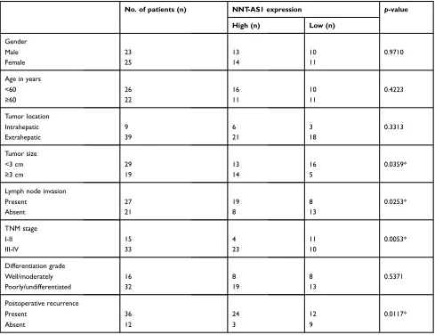
tissues (n=48) (Figure 1A). In addition, the 48 patients were categorized into low (n=21) group and high (n=27) group according to the median expression level of NNT-AS1. Kaplan Meier analysis revealed that higher expression of NNT-AS1 causes lower overall survival rate. (Figure 1B). The clinicopathologic data of CCA patients were presented inTable 1. The high expression level of NNT-AS1 was significantly associated with tumor size (p=0.013), lymph node invasion (p=0.017), and distant metastasis (p=0.009). Meanwhile, NNT-AS1 was present at higher expression in CCA cells (CCLP-1, HCCC-9810, HuCCT1, KMBC, Huh-28, QBC-939 and RBE) compared with HIBEC (Figure 1C). Since cellular location could dictate function of lncRNA, cellular frac-tionation was performed to identify the subcellular locali-zation of NNT-AS1 in CCLP1 and QBC-939 cells. The results showed that NNT-AS1 was mainly expressed in the
5 High NNT-AS1
Low NNT-AS1 Logrank P=0.031
* *
*
*
** **
A
D
E
1.5
1.0
0.5
% (total)
0.0
CCLP1 QBC939
B
C
100
80
60
40
20
0 0
1 2 3 4
0 12 24
Time (months)
Overall survival
36
HIBEC
Emptysi-NC
si-NNT -AS1-1
si-NNT -AS1-2
si-NNT
-AS1-3 Emptysi-NC
si-NNT -AS1-1
si-NNT -AS1-2
si-NNT -AS1-3
Huh28KMBC
HCCC-9810 HuCCT1
GAPDHU6 NNT -AS1
GAPDHU6 NNT -AS1
RBE CCLP1
QBC-939
4
3
2
1
0
0.0 0.4 0.8 1.2 Normal tissues
Nucleus Cytosol CCLP1QBC-939
Relative NNT
-AS1 expression
Relative NNT
-AS1 expression
Relative NNT
-AS1 expression
Tumor tissues ***
Figure 1NNT-AS1 was highly expressed in CCA tissues and cells.
Notes:(A) The qRT-PCR results revealed the expression of NNT-AS1 in 48 CCA samples collected in the research cohort. (B) Kaplan-Meier curves and log-rank test revealed the overall survival of CCA patients with high/low NNT-AS1 expression levels. (C) The differential expression levels of NNT-AS1 in CCA cell lines (Huh28, KMBC, HCCC-9810, HuCCT1, RBE, CCLP1, QBC-939) and HIBEC cell were examined by qRT-PCR. (D) The expression of NNT-AS1 in the nucleus and cytoplasm of CCA cells (CCLP1, QBC-939). U6 (nuclear retained) and GAPDH (exported to cytoplasm) were served as controls. (E) qRT-PCR was used to determine the expression of NNT-AS1 after siRNA transfection in the CCLP1 and QBC-939. The error bars indicate the means ± SD, and each experiment was repeated three times. *p<0.05, **p<0.01, ***p<0.001. Abbreviations:CCA, Cholangiocarcinoma; NNT-AS1, nicotinamide nucleotide transhydrogenase antisense RNA 1; qRT-PCR, quantitative real time polymerase chain reaction; HIBEC, human intrahepatic biliary epithelial cell.
Cancer Management and Research downloaded from https://www.dovepress.com/ by 118.70.13.36 on 20-Aug-2020
cytoplasm (Figure 1D). All these data confirmed that over-expressed NNT-AS1 was closely associated with poor prognosis in CCA.
NNT-AS1 inhibition suppressed CCA cell
proliferation in vitro
To explore the biological functions of NNT-AS1 in CCA progression, three siRNAs targeting the coding region of NNT-AS1 were tested for their knockdown efficiency
(Figure 1E). Being the most efficient one, si-NNT-AS1-1
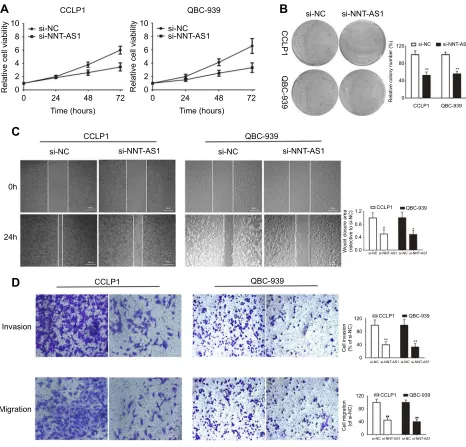
was selected for further experiments. CCK-8 assay showed that NNT-AS1 knockdown suppressed the proliferation ability of CCA cells (CCLP1, QBC-939) compared with control group (Figure 2A). In accordance with prolifera-tion, the data of colony formation were also conspicuous. The clonogenic ability of si-NNT-AS1 transfected CCLP1 and QBC-939 cells was more subdued (Figure 2B).
Knockdown of NNT-AS1 in CCA cell
lines inhibited cell invasion and migration
Further investigated about the potential impacts of NNT-AS1 on the migration and invasion of CCA cells were conducted with wound healing and transwell assays. Knockdown of NNT-AS1 with siRNA significantly reduced wound closure area (Figure 2C). In line with our predictions, as revealed by the transwell assays, knock-down of NNT-AS1 decreased the migration and invasion of CCLP1 and QBC-939 (Figure 2D).NNT-AS1 acted as a ceRNA by sponging
miR-485 and indirectly regulated BCL9
expression
It is well known that lncRNA, as the molecular sponge mechanism of miRNA, is the most common molecular mechanism of lncRNA. NNT-AS1 is mainly located in
Table 1Association between NNT-AS1 expression and clinicopathological characteristics of CCA
No. of patients (n) NNT-AS1 expression p-value
High (n) Low (n)
Gender
Male 23 13 10 0.9710
Female 25 14 11
Age in years
<60 26 16 10 0.4223
≥60 22 11 11
Tumor location
Intrahepatic 9 6 3 0.3313
Extrahepatic 39 21 18
Tumor size
<3 cm 29 13 16 0.0359*
≥3 cm 19 14 5
Lymph node invasion
Present 27 19 8 0.0253*
Absent 21 8 13
TNM stage
I-II 15 4 11 0.0053*
III-IV 33 23 10
Differentiation grade
Well/moderately 16 8 8 0.5371
Poorly/undifferentiated 32 19 13
Postoperative recurrence
Present 36 24 12 0.0117*
Absent 12 3 9
Note:*p<0.05.
Cancer Management and Research downloaded from https://www.dovepress.com/ by 118.70.13.36 on 20-Aug-2020
cytoplasm. Therefore, we verified whether NNt-AS1 played a role as a “sponge” of miRNA in the process of CCA tumorigenesis. Bioinformatics online program (StarBase,
http://starbase.sysu.edu.cn) was used to predict the
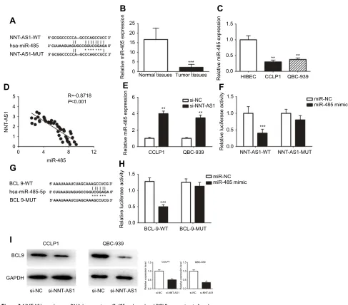
NNT-AS1 and miR-485 potential binding sites (Figure 3A). MiR-485 expression level was much lower than that in CCA tissues than in normal tissues (Figure 3B). MiR-485 was also less expressed in CCLP1 and QBC-939, compared with that in HEBIC (Figure 3C). Spearman’s correlation analysis suggested a significant negative correlation between
NNT-AS1 and miR-485 in CCA patients’ tissues (R=
−0.8717, p<0.0001,Figure 3D). In CCLP1 and QBC-939 transfected with si-NNT-AS1, the expression level of miR-485 was up-regulated (Figure 3E). To investigate the binding ability between miR-485 and NNT-AS1, a luciferase reporter assay was conducted using wild-type and mutant versions of NNT-AS1. The results of luciferase reporter assay showed that ectopic miR-485 expression evidently repressed the luciferase activity (Figure 3F). Moreover, we predicted a target by TargetScan (http://www.targetscan.org) and
A
CCLP1CCLP1
CCLP1
CCLP1 0 40
Relative colony number (%)
80 120
CCLP1
si-NC si-NNT-AS1
0 0 2 4 6
Relative cell viability
8 10
24 48
Time (hours) 72
B
C
D
QBC-939
QBC-939
QBC-939
QBC-939 ** **
QBC-939
si-NC
si-NC
si-NC
si-NC
si-NNT-AS1
si-NNT-AS1
si-NC
0h
24h
Invasion
Migration
si-NNT-AS1 si-NC si-NNT-AS1
si-NNT-AS1
CCLP1
0.0 0.4 0.8 1.2
W
ould closure area
(relective to si-NC)
QBC-939
* *
si-NNT-AS1 si-NC si-NNT-AS1
si-NC CCLP1
0 40 80 120
Cell invasion (% of si-NC)
QBC-939
** **
si-NNT-AS1 si-NC si-NNT-AS1
si-NC CCLP1
0 40 80 120
Cell migration (of si-NC)
QBC-939
** **
si-NNT-AS1 si-NC si-NNT-AS1
0 0 2 4 6
Relative cell viability
8 10
24 48
Time (hours) 72
Figure 2Knockdown of NNT-AS1 inhibited cell proliferation, migration and invasion in vitro.
Notes:(A) CCK-8 assays were used to determine the viability of si-NNT-AS1-transfected cholangiocarcinoma cells. (B) Colony-forming ability was measured in CCLP1 and QBC-939 cells after transfected with si-NNT-AS1 or si-NC by clonogenic assays. (C) The migration capacities were detected in CCLP1 and QBC-939 cells transfected with si-NNT-AS1 or si-NC by wound healing assays. (D) The invasive and migration capacities were detected in CCLP1 and QBC-939 cells transfected with si-NNT-AS1 or si-NC by transwell assays. The error bars indicate the means ± SD. *p<0.05, **p<0.01.
Abbreviations:NC, negative control; NNT-AS1, nicotinamide nucleotide transhydrogenase antisense RNA 1.
Cancer Management and Research downloaded from https://www.dovepress.com/ by 118.70.13.36 on 20-Aug-2020
considered that miR-485 might regulate BCL9 (Figure 3G). By luciferase reporter assay, we confirmed miR-485 could bind to BCL9 mRNA (Figure 3H). Western blot analysis indicated that BCL9 expression was decreased after knock-down NNT-AS1 (Figure 3I).
MiR-485 reversed the carcinogenesis of
NNT-AS1
Rescue assays were carried out to validate the regulatory mechanism of NNT-AS1 in CCA. Wound-healing assay
and transwellassay revealed that silencing of NNT-AS1 repressed the migration and invasion of CCA cells, which could be rescued by suppression of miR-485
(Figure 4A–C). Moreover, CCK8 assays elucidated that
the inhibitory impact of NNT-AS1 knockdown on prolif-eration of CCA cells could be abolished by miR-485 suppression (Figure 4D). In addition, the expression of BCL9 in transfected CCA cells were analyzed. The infl u-ence of si-NNT-AS1 on the expression was reversed by miR-485 inhibitor (Figure 4E). These data suggested that
A
D
E
F
B
C
NNT-AS1-WT
hsa-miR-485
NNT-AS1-MUT
BCL 9-WT
hsa-miR-485-5p
BCL 9-MUT
0
0 1 2 3 4
5 R=-0.8718
P<0.001
miR-485
CCLP1
0 0.0
0.5 1.0
***
Relative luciferase activity
1.5
2 4
Relative miR-485 expression
6
si-NC
**
** si-NNT-AS1
QBC-939 NNT-AS1-WT NNT-AS1-MUT miR-NC miR-485 mimic
H
G
I
CCLP1BCL9
GAPDH
si-NC si-NNT-AS1 si-NC si-NNT-AS1 si-NC si-NNT-AS1 si-NC 0.0 0.5 1.0
Relative expression level
Relative expression level
1.5
0.0 0.5 1.0 1.5
si-NNT-AS1 QBC-939
CCLP1
*
* QBC-939 0.0
0.5 1.0
***
Relative luciferase activity
1.5
BCL-9-WT BCL-9-MUT
miR-NC miR-485 mimic
0 4 8 12
0.0 0.5 1.0 1.5
Normal tissues Tumor tissues HIBEC CCLP1
** **
QBC-939 ***
5 10 15
NNT
-AS1
20 25
Relative miR-485 expression Relative miR-485 expression
Figure 3NNT-AS1 acted as a ceRNA by sponging miR-485 and regulated BCL9 expression indirectly.
Notes:(A) The predicted complementary binding sequences of NNT-AS1 3ʹ-UTR and miR-485. (B) The differential expression of miR-485 in CCA tissues and adjacent normal bile duct tissues was analyzed by qRT-PCR. (C) The miR-485 expression levels were investigated in CCA cells (CCLP1, QBC-939) and HIBEC by qRT-PCR. (D) Pearson’s correlation curve revealed the negative relevance between NNT-AS1 and miR-485 expression. (E) The expression levels of miR-485 in both CCLP1 and QBC-939 transfected with si-AS1 were evaluated by qRT-PCR. (F) Luciferase activity of 293T cells cotransfected with miR-485 mimic and luciferase reporters containing NNT-AS1-WT or NNT-AS1-MUT transcript were analyzed. (G) The predicted binding sites of miR-485 to the BCL9 sequence were shown. (H) Luciferase activity of 293T cells cotransfected with miR-485 mimic and luciferase reporters containing BCL9-WT or BCL9-MUT transcript were performed. (I) The BCL9 expression transfected with si-NNT-AS1 in CCLP1 and QBC-939 were analyzed by Western blot. The error bars in all graphs represented SD. *p<0.05, **p<0.01, ***p<0.001.
Abbreviations:NC, negative control; NNT-AS1, nicotinamide nucleotide transhydrogenase antisense RNA 1; MUT, mutation; WT, wild type.
Cancer Management and Research downloaded from https://www.dovepress.com/ by 118.70.13.36 on 20-Aug-2020
0h
A
B
C
D
E 24h 0h 24h si-NC+ NC inhibitor si-NC+ miR+485 inhibitor si-NC 0.0 0.5 Would closure area
(relective to si-NC)
1.0 1.5 CCLP1 QBC-939 si-NNT-AS1-1 NC inhibitor miR-485 inhibitor + + + + + + + + + + + + + + + + -- -- -- -- -si-NC 0.0 0.5
Cell invasion (% of si-NC)
1.0 1.5 CCLP1 QBC-939 si-NNT-AS1-1 NC inhibitor miR-485 inhibitor + + + + + + + + + + + + + + + + -- -- -- -- -si-NC 0.0 0.5
Cell invasion (% of si-NC)
1.0 1.5 CCLP1 QBC-939 si-NNT-AS1-1 NC inhibitor miR-485 inhibitor + + + + + + + + + + + + + + + + -- -- -- -- -si-NNT-AS1+ NC inhibitor CCLP1 QBC-939 CCLP1 Invasion QBC-939 si-NNT-AS1+ miR-485 inhibitor si-NC+ NC inhibitor si-NC+ miR+485 inhibitor si-NNT-AS1+ NC inhibitor si-NNT-AS1+ miR-485 inhibitor CCLP1 CCLP1 0.0 si-NC+NC inhibitor si-NNT -AS1+NC inhibitor si-NNT
-AS1+miR-485 inhibitorsi-NC+miR-485 inhibitor
si-NC+NC inhibitor
si-NNT
-AS1+NC inhibitor
si-NNT
-AS1+miR-485 inhibitorsi-NC+miR-485 inhibitor 0.5
Relative cell viability (72 h)
1.0 1.5
0.0 0.5
Relative cell viability (72 h)
1.0 1.5 Migration QBC-939 QBC-939 si-NC+ NC inhibitor si-NC+ miR+485 inhibitor si-NNT-AS1+ NC inhibitor si-NNT-AS1+ miR-485 inhibitor BCL9 GAPDH BCL9 GAPDH CCLP1 QBC-939 si-NC+ NC inhibitor si-NC+ miR+485 inhibitor si-NNT-AS1+ NC inhibitor si-NNT-AS1+ miR-485 inhibitor si-NC+ NC inhibitor si-NC+ miR+485 inhibitor si-NNT-AS1+ NC inhibitor si-NNT-AS1+ miR-485 inhibitor
Figure 4MiR-485 reversed the carcinogenesis of NNT-AS1 on CCA cells.
Notes:(A–D) Functional assays identified the phenomenon of NNT-AS1 and BCL9 regulated each other to compete for the binding of miR-485 by wound healing assays, transwell invasion, migration assay and CCK8 in CCLP1 and QBC-939. (E) The expression levels of BCL9 in CCLP1 and QBC-939 cotransfected si-NNT-AS1 with miR-485 inhibitor was analyzed by Western blot. The error bars in all graphs represented SD.
Abbreviations:NC, negative control; NNT-AS1, nicotinamide nucleotide transhydrogenase antisense RNA 1.
Cancer Management and Research downloaded from https://www.dovepress.com/ by 118.70.13.36 on 20-Aug-2020
miR-485 reversed the promoting effect of NNT-AS1 on the proliferation and metastasis of CCA cells.
Ectopic expression of NNT-AS1
promoted CCA growth in vivo
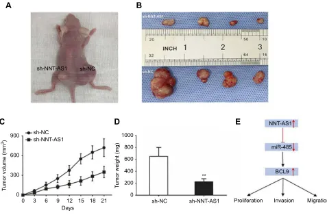
In order to confirm whether NNT-AS1 caused CCA tumori-genesis in vivo, we conducted a subcutaneous tumor formation experiment in nude mice. CCLP1 cells treated with sh-NNT-AS1 or a control vector were injected into nude mice. After 21 days post-injection, the tumors established in the sh-NNT-AS1 group were dramatically smaller than those in sh-NC group
(Figure 5AandB). As shown inFigure 5CandD,
sh-NNT-AS1 injected mice showed a reduction in tumor weight and volume at the end of the in vivo assay compared to the control group (p<0.05). Thesefindings indicated that silencing NNT-AS1 could repress CCA tumor growth in vivo.
Discussion
Cholangiocarcinoma (CCA) is one of the most aggressive malignancies of the digestive system. This disease is difficult
to diagnose at its inchoate stage and has poor prognosis. Therefore, a clear understanding of pathogenesis and major influencing factors is the key to develop effective therapeutic methods for CCA. However, the mechanism of CCA pro-gression, including cell proliferation, invasion, metastasis and anti-apoptosis, has not been elaborated. Increasing evi-dences have indicated that lncRNAs emerge as an important regulator of cancers,15including CCA. NNT-AS1 has been reported to participate in the tumorigenesis of various cancer.16–18 In agreement with most of current studies of tumors on NNT-AS1, our study found that NNT-AS1 was highly expressed in CCA, and its expression level was cor-related with clinical prognosis. We confirmed that NNT-AS1 played an oncogene role in CCA, and the specific molecular mechanism needed to be further explored.
A large number of lncRNAs have been shown to play an important role in the development of cholangiocarci-noma, such as NEAT119and LINC01296.20In the current study, our data showed that the NNT-AS1 expression level was significantly increased of in CCA tissues compared to 0
0 300
T
umor volume (mm
3)
600
900 sh-NC
sh-NC
sh-NNT-AS1 sh-NNT-AS1
A
B
C
D
E
3 6 9 12
Days
sh-NC 0
200 400 600
T
umor weight (mg)
800 1000
sh-NNT-AS1 **
Proliferation Invasion
BCL9 miR-485 NNT-AS1
Migration
15 18 21
Figure 5NNT-AS1 knockdown suppressed tumor growth in vivo.
Notes:(A) Tumors established with the mice in the sh-NC or sh-NNT-AS1 group. (B) Tumors collected from mice were exhibited. (C) Tumor volume curve of mice injected with CCLP1 cells transfected with sh-NC and sh-NNT-AS1 was analyzed. (D) Tumor weight of mice was measured. (E) Summary of the mechanism of NNT-AS1/ miR-485/BCL9 pathway in CCA. The error bars in all graphs represented SD. **p<0.01.
Abbreviations:NC, negative control; NNT-AS1, nicotinamide nucleotide transhydrogenase antisense RNA 1; CCA, cholangiocarcinoma.
Cancer Management and Research downloaded from https://www.dovepress.com/ by 118.70.13.36 on 20-Aug-2020
adjacent non-tumor tissues. High NNT-AS1 expression was associated with advanced clinicopathological features and poor overall survival. Therefore, the evidence proved the oncogenic role of NNT-AS1 in CCA tumorigenesis. Functional experiments results revealed that NNT-AS1 knockdown inhibited the proliferation and invasion ability in vitro, and suppressed the CCA tumor growth in vivo.
Recently, the miRNA‘sponge’theory21is well known. There was a significant negative correlation between NNT-AS1 and miR-485 in CCA. MiR-485 was reported to be abnormally expressed in many cancers. Kang have reported that miR-485 could suppress gastric cancer cell metastasis and sphere formation,22miR-485 was also dysregulated in non-small-cell lung cancer (NSCLC) and markedly inhib-ited NSCLC cell growth and induced cell apoptosis.23
Moreover, miR-485 widely participated in cell proliferation and invasion, as well as autophagy and apoptosis.
Then we identified BCL9 as the potential biological target for miR-485 by using TargetScan Database. Luciferase reporter assay confirmed the molecular binding at 3ʹ-UTR. We hypothesized that there was a‘ceRNA net-work’ among NNT-AS1, miR-485 and BCL9. The results of rescue assays showed that miR-485 reversed the promot-ing effect of NNT-AS1 on the proliferation and metastasis of CCA. NNT-AS1 was up-regulated and competed with BCL9 for binding to miR-485, thereby increasing the pro-tein expression of BCL9 and promoting the proliferation, migration and invasion in CCA (Figure 5E). Published studies have shown that BCL9 was closely related to the development of cancers such as renal cell carcinoma,24 colorectal cancer,25 and gastric cancer.26 Moreover, BCL9 was involved in signal transduction through the Wnt path-way, promotingβ-catenin’s transcriptional activity.27As our knowledge, the Wnt/β-catenin signaling pathway was pathologically activated in CCA, andβ-Catenin was a key mediator of the Wnt/β-catenin pathway. In this regard, NNT-AS1 might promote the CCA tumorigenesis by reg-ulating the Wnt signaling pathway.
Conclusion
We have a conclusion that NNT-AS1 overexpression is associated with tumorigenesis and poor prognosis in CCA, NNT-AS1 promotes proliferation, migration and invasion of CCA via miR-485/BCL9 axis.
Acknowledgments
This work was supported by grants from National Natural Science Foundation of China (No. 81602088), China
Postdoctoral Science Foundation (No. 2017M621305), Heilongjiang Postdoctoral Science Foundation (No. LBH-Z16096), Health and Family Planning Commission Research Project of Heilongjiang Province (No. 2016-049), HMU College Students’ Innovation and Entrepreneurship Training Program (No. 201810226105) and Innovative Science Foundation of Harbin Medical University (No. 2016LCZX09).
Disclosure
The authors report no conflicts of interest in this work.
References
1. Wang WT, Ye H, Wei PP, et al. LncRNAs H19 and HULC, activated by oxidative stress, promote cell migration and invasion in cholan-giocarcinoma through a ceRNA manner. J Hematol Oncol. 2016;9:117. doi:10.1186/s13045-016-0348-0
2. Blechacz B. Cholangiocarcinoma: current knowledge and new developments. Gut and Liver. 2017;11:13–26. doi:10.5009/gnl155 68
3. Afshar M, Khanom K, Ma YT, Punia P. Biliary stenting in advanced malignancy: an analysis of predictive factors for survival. Cancer Manag Res.2014;6:475–479. doi:10.2147/CMAR.S71111
4. Doherty B, Nambudiri VE, Palmer WC. Update on the diagnosis and treatment of cholangiocarcinoma.Current Gastroenterology Reports. 2017;19:2. doi:10.1007/s11894-017-0542-4
5. Gatto M, Bragazzi MC, Semeraro R, et al. Cholangiocarcinoma: update and future perspectives. Dig Liver Dis. 2010;42:253–260. doi:10.1016/j.dld.2009.12.008
6. Zhang Y, Tang L. The application of lncRNAs in cancer treatment and diagnosis. Recent Pat Anticancer Drug Discov.2018;13:292– 301. doi:10.2174/1574892813666180226121819
7. Chen X, Yang Y, Cao Y, et al. lncRNA PVT1 identified as an independent biomarker for prognosis surveillance of solid tumors based on transcriptome data and meta-analysis.Cancer Manag Res. 2018;10:2711–2727. doi:10.2147/CMAR.S166260
8. Huarte M. The emerging role of lncRNAs in cancer. Nat Med. 2015;21:1253–1261. doi:10.1038/nm.3981
9. Li X, Du N, Zhang Q, et al. MicroRNA-30d regulates cyte pyroptosis by directly targeting foxo3a in diabetic cardiomyo-pathy. Cell Death Dis. 2014;5:e1479. doi:10.1038/cddis.2014.4 30
10. Li ZL, Li JL, Ji DL, et al. Overexpressed long noncoding RNA Sox2ot predicts poor prognosis for cholangiocarcinoma and promotes cell proliferation and invasion. Gene. 2018;645:131–136. doi:10.1016/j.gene.2017.12.017
11. Fagerberg L, Hallstrom BM, Oksvold P, et al. Analysis of the human tissue-specific expression by genome-wide integration of transcrip-tomics and antibody-based proteomics. Mol Cell Proteomics. 2014;13:397–406. doi:10.1074/mcp.M113.035600
12. Wang X, Ren M, Li Y, et al. Long noncoding RNA NNT-AS1 promotes gastric cancer proliferation and invasion by regulating microRNA-363 expression. J Cell Biochem. 2018;120(4):5704– 5712. doi:10.1002/jcb.27855
13. Hua F, Liu S, Zhu L, Ma N, Jiang S, Yang J. Highly expressed long non-coding RNA NNT-AS1 promotes cell proliferation and invasion through Wnt/beta-catenin signaling pathway in cervical cancer. Biomed Pharmacother.2017;92:1128–1134. doi:10.1016/j.biopha.2017.03.057 14. Li X, Edwards M, Swaney KF, et al. Mutually inhibitory Ras-PI(3,4)
P2 feedback loops mediate cell migration.Proc Natl Acad Sci U S A. 2018;115:E9125–E9134. doi:10.1073/pnas.1809039115
Cancer Management and Research downloaded from https://www.dovepress.com/ by 118.70.13.36 on 20-Aug-2020
15. Peng WX, Koirala P, Mo YY. LncRNA-mediated regulation of cell signaling in cancer. Oncogene. 2017;36:5661–5667. doi:10.1038/ onc.2017.184
16. Shen Q, Jiang Y. LncRNA NNT-AS1 promotes the proliferation, and invasion of lung cancer cells via regulating miR-129-5p expression. Biomed Pharmacother. 2018;105:176–181. doi:10.1016/j.biopha. 2018.05.123
17. Li Y, Lv M, Song Z, Lou Z, Wang R, Zhuang M. Long non-coding RNA NNT-AS1 affects progression of breast cancer through miR-142-3p/ZEB1 axis. Biomed Pharmacother. 2018;103:939–946. doi:10.1016/j.biopha.2018.04.087
18. Cai Y, Dong ZY, Wang JY. LncRNA NNT-AS1 is a major mediator of cisplatin chemoresistance in non-small cell lung cancer through MAPK/slug pathway.Eur Rev Med Pharmacol Sci.2018;22:4879– 4887. doi:10.26355/eurrev_201808_15624
19. Parasramka M, Yan IK, Wang X, et al. BAP1 dependent expression of long non-coding RNA NEAT-1 contributes to sensitivity to gem-citabine in cholangiocarcinoma. Mol Cancer. 2017;16:22. doi:10.1186/s12943-017-0587-x
20. Zhang D, Li H, Xie J, et al. Long noncoding RNA LINC01296 promotes tumor growth and progression by sponging miR-5095 in human cholangiocarcinoma. Int J Oncol. 2018;52:1777–1786. doi:10.3892/ijo.2018.4362
21. Salmena L, Poliseno L, Tay Y, Kats L, Pandolfi PP. A ceRNA hypothesis: the rosetta stone of a hidden RNA language? Cell. 2011;146:353–358. doi:10.1016/j.cell.2011.07.014
22. Kang M, Ren MP, Zhao L, Li CP, Deng MM. miR-485-5p acts as a negative regulator in gastric cancer progression by targetingfl otillin-1.American Journal of Translational Research.2015;7:2212–2222. 23. Jiang P, Xu C, Chen L, et al. Epigallocatechin-3-gallate inhibited cancer
stem cell-like properties by targeting hsa-mir-485-5p/RXRalpha in lung cancer.J Cell Biochem.2018;119:8623–8635. doi:10.1002/jcb.27117 24. Wang J, Ying Y, Bo S, Li G, Yuan F. Differentially expressed
microRNA-218 modulates the viability of renal cell carcinoma by regulating BCL9.Mol Med Rep. 2016;14:1829–1834. doi:10.3892/ mmr.2016.5403
25. Beaulieu JF. Tuning WNT-beta-catenin signaling via BCL9 proteins for targeting colorectal cancer cells. EBioMedicine. 2015;2:1846– 1847. doi:10.1016/j.ebiom.2015.11.033
26. Han W, Mu Y, Zhang Z, Su X. Expression of miR-30c and BCL-9 in gastric carcinoma tissues and their function in the development of gastric cancer. Oncol Lett. 2018;16:2416–2426. doi:10.3892/ ol.2018.8934
27. Takada K, Zhu D, Bird GH, et al. Targeted disruption of the BCL9/ beta-catenin complex inhibits oncogenic Wnt signaling.Sci Transl Med.2012;4:148ra117. doi:10.1126/scitranslmed.3003808
Cancer Management and Research
Dove
press
Publish your work in this journal
Cancer Management and Research is an international, peer-reviewed open access journal focusing on cancer research and the optimal use of preventative and integrated treatment interventions to achieve improved outcomes, enhanced survival and quality of life for the cancer patient.
The manuscript management system is completely online and includes a very quick and fair peer-review system, which is all easy to use. Visit http://www.dovepress.com/testimonials.php to read real quotes from published authors.
Submit your manuscript here:https://www.dovepress.com/cancer-management-and-research-journal
Cancer Management and Research downloaded from https://www.dovepress.com/ by 118.70.13.36 on 20-Aug-2020