Islet inflammation in type 2 diabetes and
physiology
Kosei Eguchi, Ryozo Nagai
J Clin Invest.
2017;
127(1)
:14-23.
https://doi.org/10.1172/JCI88877
.
The finding of islet inflammation in type 2 diabetes (T2D) and its involvement in
b
cell
dysfunction has further highlighted the significance of inflammation in metabolic diseases.
The number of intra-islet macrophages is increased in T2D, and these cells are the main
source of proinflammatory cytokines within islets. Multiple human studies of T2D have
shown that targeting islet inflammation has the potential to be an effective therapeutic
strategy. In this Review we provide an overview of the cellular and molecular mechanisms
by which islet inflammation develops and causes
b
cell dysfunction. We also emphasize the
regulation and roles of macrophage polarity shift within islets in the context of T2D
pathology and
b
cell health, which may have broad translational implications for
therapeutics aimed at improving islet function.
Review Series
Find the latest version:
Series Editors: Alan R. Saltiel and Jerrold M. Olefsky
Introduction
Metabolic syndrome comprises a cluster of diseases associated with excess nutrition and insufficient physical activity. Studies over the last two decades have shown that chronic inflammation is a common and potentially unifying mechanistic cause of these diseases (1–4). Inflammation can be viewed as an evolutionari-ly selected protective response enabling the host organism to cope with stresses from external factors, and can be classified as acute or chronic (5). Acute inflammation is characterized by prominent local and systemic signs, as well as infiltration of the affected area by immune cells, mainly neutrophils. By contrast, chronic inflammation is characterized by less prominent local and systemic signs, enhanced tissue injury and fibrosis, and infiltration of the affected area mainly with monocytes/macro-phages and lymphocytes (5).
Inflammation in the context of metabolic syndrome largely exhibits the characteristics of chronic inflammation and is thus often accompanied by tissue infiltration by monocytes/mac-rophages (6) and lymphocytes (7, 8). Type 2 diabetes (T2D) is a common and serious complication of metabolic syndrome, and although the disease can exist in isolation, many T2D patients meet the diagnostic criteria for metabolic syndrome (9). Given that chronic metabolic stress induced by excess nutrition was not a driv-ing force durdriv-ing evolution, it is perhaps not surprisdriv-ing that inflam-mation in response to this stress eventually results in deleterious effects on tissue function and contributes to T2D pathology.
Insulin resistance and β cell dysfunction are the two major
components of T2D pathology, and β cell function starts to
decline even before the onset of impaired glucose tolerance (10, 11). Histologic changes characteristic of inflammation occur within the islets of T2D subjects, including immune cell infiltra-tion (12–16), amyloid deposiinfiltra-tion (14, 17, 18), cell death, and
fibro-sis (18, 19). These reports suggest inflammation is involved in β
cell dysfunction, though inflammatory pathologic changes have
been observed in only a portion of T2D patients, suggesting that islet inflammation and its contribution to T2D pathology may vary among patients (12–14, 18).
Several rodent experimental models (20–23) as well as observa-tions in humans (12–14) have made it clear that macrophages play a key role in the islet inflammation seen in T2D. The most
well-stud-ied mechanism by which islet macrophages cause β cell dysfunction
is through secretion of IL-1β, and it has been demonstrated that
interference with the IL-1 pathway relieves T2D and restores β cell
function in both rodents (24, 25) and humans (26–28). Factors that
stimulate islet macrophages to secrete IL-1β in vivo include human
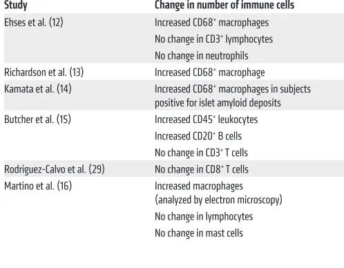
islet amyloid polypeptide (hIAPP) (20, 23), palmitate (21), and endocannabinoid (22). The possibility that other immune cell types are involved in islet inflammation in T2D remains to be confirmed; while one study reported an increase in the B cell number (15), other groups have not observed changes in the number of immune cell types including neutrophils, lymphocytes, and mast cells (refs. 12– 16, 29, 30, and Table 1). It has also been suggested that some T2D patients develop islet autoimmunity during the course of disease and that contributes to β cell functional decline (31).
There is significant heterogeneity among macrophages in terms of both their function and origin in vivo, and the transition between functional states occurs along a continuum regulated by the micro-environment, especially in the context of sterile inflammation (32– 34). Using the concept of M1-like and M2-like polarization of mac-rophages to describe an essentially heterogeneous population of tissue macrophages is a simplified operational framework (35). Dur-ing islet inflammation, overall macrophage polarity shifts toward the proinflammatory classically activated M1-like phenotype, and
this shift has been shown to contribute to β cell dysfunction in T2D
mouse models (21–23). Additionally, alternatively activated M2-like macrophages, which include macrophages with antiinflammatory (36), pro-fibrosis (37), and pro-angiogenic (38) phenotypes, were recently found to be the key mediators of β cell proliferation during both development (39, 40) and adulthood (41–44) in mice.
In this Review we present an overview of the links between
islet inflammation and β cell dysfunction in T2D, with a focus
on the regulation of islet macrophage polarity through
commu-The finding of islet inflammation in type 2 diabetes (T2D) and its involvement in β cell dysfunction has further highlighted the significance of inflammation in metabolic diseases. The number of intra-islet macrophages is increased in T2D, and these cells are the main source of proinflammatory cytokines within islets. Multiple human studies of T2D have shown that targeting islet inflammation has the potential to be an effective therapeutic strategy. In this Review we provide an overview of the cellular and molecular mechanisms by which islet inflammation develops and causes β cell dysfunction. We also emphasize the regulation and roles of macrophage polarity shift within islets in the context of T2D pathology and β cell health, which may have broad translational implications for therapeutics aimed at improving islet function.
Islet inflammation in type 2 diabetes and physiology
Kosei Eguchi1 and Ryozo Nagai2
1Department of Genetics and Complex Diseases and Sabri Ülker Center, Harvard T.H. Chan School of Public Health, Boston, Massachusetts, USA. 2Jichi Medical University, Tochigi, Japan.
mice or KKAy mice (mouse model described in ref. 51), contain
more CD11b+Ly-6C+ macrophages without a significant change in
islet-resident CD11b+Ly-6C– macrophages, compared with their
respective controls (21). We observed that CD11b+Ly-6C+ cells
from islets of obese mice do not express the cell surface proteins CD206 and CD301, which are characteristic of M2-like alter-natively activated macrophages, and exhibit greater expression of Il1b and Tnf and lower expression of Il10 than islet-resident
CD11b+Ly-6C– macrophages. These findings demonstrate that
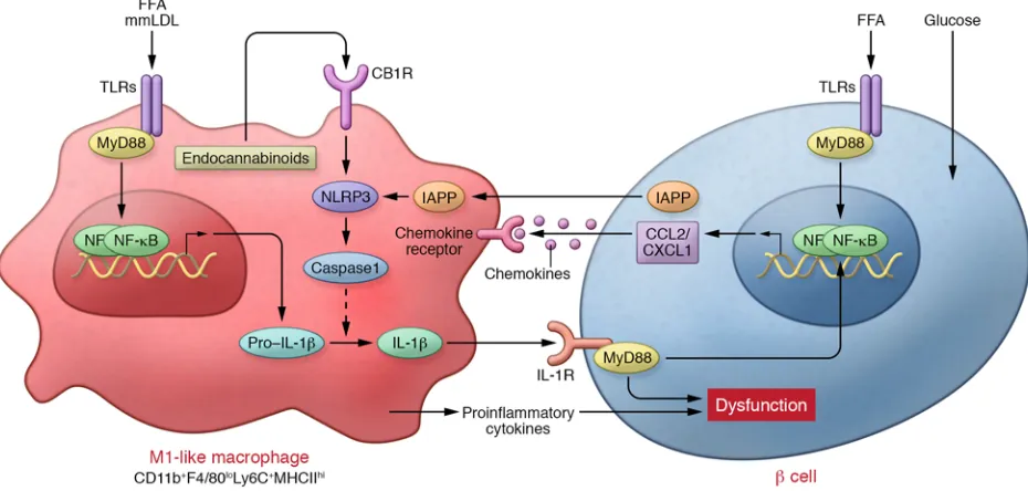
macrophages shift to an inflammatory M1-like phenotype within the islets of at least two different T2D mouse models (Figure 1).
Another report found that M1-like CD68+F4/80– macrophages
are present only in the islets of db/db mice and islet-resident
M2-like CD206-expressing CD68+F4/80+ macrophages in the
islets of both db/+ and db/db mice, confirming that islet macro-phages in db/db mice undergo a shift in polarization to an M1-like phenotype (52). In contrast, one recent study described Il1b and Tnf expression in non-diabetic islet-resident macrophages, although this study did not compare these macrophages with the macrophages of inflamed islets (53).
Islet macrophages link
β
cell dysfunction
and islet inflammation
The first in vivo evidence that islet inflammation plays a causative role in T2D was derived from a human clinical study on the effect of anakinra, a recombinant human IL-1 receptor antagonist (IL-1Ra) that blocks both IL-1α and IL-1β signaling, in 70 patients with T2D (26). Thirteen weeks of treatment with anakinra significantly reduced serum IL-6 and C-reactive protein levels, improved glyce-mia and C-peptide secretion, and reduced the serum proinsulin/ insulin ratio without significantly affecting homeostatic model assessment of insulin resistance (HOMA-IR). These effects of anakinra on insulin secretion were also seen in prediabetic patients with impaired glucose tolerance (54). Further, studies utilizing
mul-tiple mAbs against IL-1β confirmed the contribution of IL-1β
sig-naling to systemic inflammation, glycemic control (55), and β cell
dysfunction (27, 28) in T2D (56, 57). A dose-escalation study of
gov-ekizumab, a recombinant humanized mAb against IL-1β,
demon-strated that an intermediate dose (0.03–0.1 mg/kg) but not a high dose (>0.3 mg/kg) of gevokizumab significantly improved glycated
hemoglobin A1C (HbA1C), suggesting the effects of IL-1β in T2D are
multifaceted (see below) (27). For greater mechanistic insight into
the role of IL-1 signaling in β cell dysfunction in T2D, IL-1Ra was
administered to mice fed a high-fat diet (HFD) (25) and to GK rats (24). Ehses and colleagues clearly showed that treating GK rats with IL-1Ra resulted in decreased hyperglycemia that was accompanied by increased insulin mRNA levels within islets, reduced serum pro-insulin/insulin ratios, reduced macrophage infiltration into islets and mRNA levels of chemokine/proinflammatory cytokine within islets, and ameliorated insulin resistance (24). There was no change in islet mass with IL-1Ra treatment (24, 25), suggesting that a func-tional decline of β cells rather than an apoptotic effect is the primary mechanism by which islet inflammation decreases insulin secretion in T2D (Figure 1). This finding is consistent with the earlier observa-tion that in T2D individuals, islets containing increased numbers of macrophages did not contain higher numbers of TUNEL-positive cells and macrophages were not observed in proximity to apoptotic nications among multiple cell types within the islet and its roles
in T2D pathology and physiology. From this viewpoint we argue that immune cell function within islets is seamlessly regulated and thus plays an indispensable role in maintaining β cell health as well as serving as a primary source of proinflammatory cytokines that
cause β cell dysfunction in T2D. Expanding our understanding of
the cells that mediate islet inflammation may have broad transla-tional implications for therapeutics to improve islet function.
Evidence for the involvement of islet inflammation
and islet macrophage polarity shift in T2D
Initial evidence of immune cell infiltration of the pancreatic islets in T2D came from immunostaining analyses showing increased numbers of islet macrophages in rodent T2D models (the Goto-Kakizaki [GK] rat and the db/db [leptin-deficient] mouse; refs. 12, 45), as well as in human T2D patients (12, 13). Additionally,
micro-array profiling of human β cells obtained through laser-capture
microdissection revealed a three-fold increase in the expression of chemokines CCL2 and CCL13 in T2D subjects compared with controls, providing molecular evidence of immune cell infiltra-tion and islet inflammainfiltra-tion in human T2D patients (46). These results suggest that islet inflammation in T2D is mainly driven by innate immunity, whereas inflammation in type 1 diabetes (T1D) is known to be mainly driven by adaptive immunity (47, 48). Several studies also showed that prolonged exposure to high glucose levels causes β cells to secrete IL-1β, which contributes to islet inflamma-tion in human islets in vitro (49) and in mice (50).
Given the heterogeneity of macrophages (32) and the impor-tance of macrophage polarization in the pathology of obese adi-pose tissue (AT) (6), we asked whether there are multiple subtypes of macrophages within the islets and analyzed the extent to which their characteristics are altered in T2D islets. Compared with islets from control db/+ mice, islets from db/db mice expressed several-fold higher levels of such chemokines and cytokines as
Ccl2, Cxcl1, Il1b, and Tnf. Flow cytometric (FCM) analysis
[image:3.585.39.288.86.280.2]dem-onstrated that islets from two murine models of T2D, obese db/db
Table 1. Analysis of immune cells in islets of T2D subjects
Study Change in number of immune cells
Ehses et al. (12) Increased CD68+ macrophages No change in CD3+ lymphocytes No change in neutrophils Richardson et al. (13) Increased CD68+ macrophage
Kamata et al. (14) Increased CD68+ macrophages in subjects positive for islet amyloid deposits Butcher et al. (15) Increased CD45+ leukocytes
Increased CD20+ B cells No change in CD3+ T cells Rodriguez-Calvo et al. (29) No change in CD8+ T cells Martino et al. (16) Increased macrophages
Islet inflammation induced by TLR activation in T2D. In
deter-mining the mechanisms by which inflammation is induced in obese AT, the roles of pattern-recognition receptors, including TLRs (58, 59) and NLRs (60) have been extensively analyzed. Several fac-tors, including saturated FFAs, minimally modified LDL (mmLDL) cholesterol, advanced glycation end products of LDL cholesterol, and damage-associated molecular patterns (61) such as HSP60, S100 calcium-binding protein A8 (62), and high-mobility group B (HMGB) proteins (63) all reportedly activate TLRs in obese AT (64). Indeed, saturated FFAs have been reported to induce chemo-kine (Cxcl1 and Ccl3) expression within islets in vitro (12, 46).
Addi-tionally, HMGB1 has been shown to activate the NF-κB pathway
in isolated murine islets via TLR2 and TLR4 pathways (65).
Fur-thermore, Tlr2–/– mice fed a HFD for 20 weeks were protected from
HFD-induced β cell dysfunction and insulin resistance (66),
indi-cating a critical role for the TLR2 pathway in islet inflammation. Treatment of islets isolated from healthy human control sub-jects with palmitate, but not oleate or high glucose, increased secretion of cytokines and chemokines, including IL-6 and CXCL1, mimicking the inflammation seen in islets from T2D patients. Pal-mitate-induced expression of inflammatory genes (IL6 and IL8) in human islets was IL-1 dependent, as IL-1Ra abolished the effects of palmitate (46). Similarly, in both human and mouse islets,
exposure to FFAs induced Il1b and Cxcl1 expression and pro–IL-1β
expression. This inflammatory response was partially inhibited
by IL-1Ra (67). Consistently, exposure of MIN6 β cells to culture
medium conditioned by palmitate-treated macrophages, with or
without addition of IL-1β– and TNF-α–neutralizing antibodies,
confirmed that those proinflammatory cytokines are produced
by macrophages, not by β cells, and that they promote β cell
dys-function and enhance chemokine expression in β cells (21). These
reports demonstrate the ability of saturated FFAs to initiate an
β cells (12). This may be an important distinction from the effect of
islet inflammation in T1D, where the primary mechanism of disease is the autoimmune destruction of β cells (47).
The actions of macrophages leading to β cell dysfunction in
obese mice were further interrogated using clodronate liposome– mediated suppression of macrophage recruitment to islets in
db/db and KKAy mice. In both models, mice treated with
clodro-nate liposomes showed improved glucose tolerance with improved insulin secretion in an oral glucose tolerance test (OGTT), and islets isolated from these mice exhibited increased glucose-stim-ulated insulin secretion (GSIS) as compared to the blunted GSIS seen in islets from mice treated with empty liposomes. Levels of pancreatic and duodenal homeobox-1 (Pdx1) and insulin mRNA were also increased in islets from clodronate liposome–treated mice (21). These data clearly demonstrate the causative
involve-ment of islet inflammation and macrophage infiltration in β cell
failure in T2D rodent models (Figure 1).
Mechanisms of islet inflammation and
inflammation-induced
β
cell dysfunction
[image:4.585.57.522.59.281.2]The aforementioned studies support the therapeutic strategy of tar-geting islet inflammation to ameliorate T2D. To gain mechanistic insight into the pathophysiology of islet inflammation, experiments using a specific factor that initiates inflammation have several advan-tages. These include the ability to set up in vitro preparations to dis-sect the complex communication between multiple cell types within islets, easy utilization of mutant mice, and the ability to set up short-term in vivo models to eliminate confounding effects from insulin sensitization due to systemic antiinflammatory manipulations. With these advantages in mind, in the next section we review reports analyzing islet inflammation induced by specific stimuli, including hIAPP, saturated free fatty acids (FFAs), and endocannabinoids.
Islet inflammation induced by IAPP in T2D. Amyloid
depos-its are commonly observed within islets from T2D patients (68). Islet amyloid deposits are composed of IAPP (69, 70), which
is produced in β cells and secreted in a monomeric form along
with insulin (71). In pathologic conditions, secreted hIAPP is
converted to a β-sheet structure and oligomerized (72, 73). The
monomeric form of IAPP does not itself affect β cell function,
and only the aggregated form — particularly with non-fibrillar hIAPP oligomers, less with mature amyloid fibrils — exhibits tox-icity toward β cell function in vitro (74, 75). Given this fact, it is noteworthy that the oligomerization of IAPP differs among spe-cies. For example, human and feline IAPP can be oligomerized but rat and mouse IAPP cannot (74, 76). Thus hIAPP transgenic (hIAPP-Tg) mice (77) exhibit islet amyloid deposition, while transgenic mice expressing rat IAPP (rIAPP-Tg) do not (78). Fur-ther analysis of hIAPP-Tg mice revealed that dietary fat promotes
islet amyloid formation and that one year on a HFD caused β cell
loss and impaired insulin secretion (79). Islets in hIAPP-Tg mice exhibited larger F4/80-positive areas and greater expression of chemokines (Ccl2 and Cxcl1) and macrophage markers (Emr1 and
Itgax) than non-transgenic mice after one year on either a low-fat
diet or HFD (80). Additionally, immunostaining studies revealed the presence of IAPP within the lysosomes of islet macrophages in hIAPP-Tg mice but not rIAPP-Tg mice, suggesting that macro-inflammatory response in the islets, and also demonstrate a role
for the autocrine or paracrine effects of IL-1β. In vivo, short-term
intravenous ethyl-palmitate infusion induced Ccl2, Cxcl1, Il1b, and
Tnf expression and reduced insulin and Pdx1 expression within
islets, mimicking the observations from db/db and KKAy mice (21) and individuals with T2D (46). FCM analysis of islets from these
mice revealed that ethyl-palmitate infusion recruited CD11b+
Ly-6C+ classically activated M1-like monocytes/macrophages to
the islets. Tlr4–/– and Myd88–/– mice were completely protected from
both M1-like macrophage recruitment and β cell dysfunction. Bone
marrow transplantation studies revealed that TLR4 in islet cells, but not in bone marrow cells, was required for M1-like macrophage recruitment. The short-term nature of the ethyl-palmitate experi-ment supports the causative contribution of M1-like
macrophage-induced islet inflammation to palmitate-macrophage-induced β cell dysfunction
without the confounding effect of insulin sensitization (21). These reports demonstrate that TLR-mediated sensing of
pal-mitate in β cells is responsible for the initial chemokine secretion
that induces macrophage recruitment. Thereafter, communication via proinflammatory cytokines and chemokines between M1-like
macrophages and β cells form a vicious cycle that amplifies islet
inflammation (Figure 1). These findings are consistent with the previous finding that antagonizing IL-1 signaling inhibits both
[image:5.585.88.500.62.364.2]macrophage recruitment and β cell dysfunction in GK rats (24).
deposits and with macrophages, but not with β cells. These studies
show that macrophages are the source of IL-1β within islets both in
vivo and in vitro (ref. 20 and Figure 1).
Several studies have sought to determine the identity of the
priming factors that induce production of pro–IL-1β and the
inflammasome component NLRP3 (88), which leads to inflamma-some activation within T2D islets. Among several factors reported to stimulate TLR4 in metabolic syndrome (58, 89), Masters and colleagues confirmed that mmLDL cholesterol (90) can prime macrophages via TLR4 for inflammasome activation by hIAPP (20). More recent reports indicate that soluble hIAPP species pro-duced during early hIAPP aggregation are themselves able to acti-vate the TLR2/MyD88 pathway to prime macrophages (82, 91), and palmitate priming of inflammasomes has also been reported (92). Additionally, an independent group confirmed the presence of inflammasome markers (Nlrp3, Pycard, and Casp1) and inflam-matory cytokines (Il1b, Tnf, and Il6) within islets of hIAPP-Tg mice fed a HFD (80).
Depletion of islet macrophages from hIAPP-Tg mice fed a HFD improved systemic glucose tolerance and GSIS of isolated islets, and almost completely blocked islet inflammation, despite greater deposition of amyloid within islets, demonstrating the causal role of islet macrophages in hIAPP-induced islet
inflam-mation and β cell dysfunction in vivo (23). hIAPP-induced IL-1β
secretion and NLRP3/caspase-1 induction within isolated islets was completely abolished by clodronate liposome–mediated macrophage depletion (23), which demonstrates the contribution made by resident macrophages to inflammasome activation and
IL-1β secretion induced by hIAPP in vitro. In this experiment, in
contrast to complete inhibition of Il1b expression, macrophage depletion led to an 80% reduction in hIAPP-induced Ccl2 expres-sion, suggesting that nonphagocytic cells (likely β cells) contribute to islet chemokine production (81). Although there was no obvi-ous infiltration of islets by different macrophages subsets, FCM analysis showed that macrophages within islets from hIAPP-Tg phages are involved in the toxic effects of islet amyloid deposition
in T2D (78). Finally, in a human study using
immunohistochemi-cal analysis of islets, there were three-fold more CD68+ cells per
unit area in islets from diabetic patients with islet amyloid deposi-tion than in islets from non-diabetic patients or diabetic patients without amyloid deposition (14).
Masters and colleagues have presented a clear perspective on how hIAPP activates macrophages and induces islet inflammation (20). This group focused on the finding that inflammasome acti-vation is the critical event in the initiation of islet inflammation by
hIAPP, as hIAPP induced IL-1β but had no effect on IL-6 or TNF
secretion from mmLDL-primed dendritic cells in vitro. The sig-nificance of the inflammasome/IL-1 pathway to hIAPP-induced islet inflammation in vitro was further demonstrated by the find-ing that TNF secretion from macrophages and chemokine secre-tion from isolated islets induced by long-term stimulasecre-tion with hIAPP was strongly suppressed by IL-Ra (81, 82). In vivo, IL-1Ra treatment improved glucose tolerance in recipients of hIAPP-Tg islets and conferred nearly complete protection from macrophage recruitment to the transplanted hIAPP-Tg islets (81). IL-1Ra also improved islet function without affecting insulin sensitivity in hIAPP-Tg KKAy mice (83).
In accordance with the earlier electron microscopic observa-tion of amyloid within macrophage lysosomes (84), the involve-ment of phagolysosomes in hIAPP-mediated inflammasome activation was demonstrated in an experiment in which admin-istration of the phagocytosis inhibitors cytochalasin D and bafilo-mycin A blocked inflammasome activation in macrophages in vitro (20). These mechanisms are consistent with the mechanisms of inflammasome activation in other amyloid-induced patholo-gies including Alzheimer’s disease (85, 86). It was also shown that fibril aggregates accumulate within macrophage lysosomes, fur-ther supporting the involvement of hIAPP phagocytosis by mac-rophages (87). Immunostaining of islets from hIAPP-Tg mice fed
[image:6.585.46.539.66.209.2]a HFD for one year revealed that IL-1β colocalized with amyloid
mice fed a HFD express more CD11b, CD11c, and Ly-6C as well as more Il1b mRNA and less Il10 mRNA than macrophages within islets from wild-type mice fed a HFD. From these findings it was speculated that hIAPP aggregation may affect the differentiation state of monocytes entering islets and that hIAPP polarizes resi-dent macrophages toward a proinflammatory M1-like phenotype (ref. 23 and Figure 1).
Islet inflammation induced by endocannabinoid activation of inflammasomes in islet macrophages. Cannabinoid receptors are
expressed in both nervous systems and the peripheral tissues. The most potent endocannabinoids (i.e., endogenous agonist for the cannabinoid receptors) are the lipid ligands anandamide and 2-arachidonoylglycerol. Endocannabinoids play multiple roles, including the regulation of appetite and mood in the nervous sys-tems and the regulation of energy homeostasis in peripheral tis-sues (93, 94). The cannabinoid 1 receptor (CB1R) antagonist ibip-inabant reportedly attenuates β cell loss in diabetic Zucker diabetic fatty (ZDF) rats (95). Investigation into this observation revealed
a new mechanism for β cell loss involving CB1R signaling in islet
macrophages. Jourdan and colleagues first confirmed that treat-ing ZDF rats with the non–brain-penetrant CB1R inverse agonist
JD5037 (96) preserved pancreatic β cell function and protected
against β cell loss (22). Immunohistochemical analysis of the
iso-lated islets revealed that JD5037 reduced macrophage infiltration into islets. mRNA analysis demonstrated that JD5037 reversed the M1-like polarity shift of islet macrophages, characterized by decreased expression of Tnf, Nos2, Nlrp3, and Cnr1 and increased expression of Tgfb1, Il10, and Arg1 within ZDF islets. Moreover,
CB1R and NLRP3 were expressed in CD68+ macrophages but not
in insulin-producing cells. Macrophage depletion using clodro-nate liposomes demonstrated that macrophages are responsible for the increases in Cnr1, Nlrp3, Txnip, and Tnf expression and for the reduced insulin secretion capacity in ZDF rats. Additionally, the effect of macrophage CB1R on islet inflammasome activation and diabetes development was confirmed through macrophage-selective knockdown of Cnr1 in ZDF rats. Finally, macrophage production of anandamide was stimulated by high glucose and palmitate in vitro. With these experiments, the authors demon-strated that in ZDF rats, autocrine activation of CB1R on
macro-phages by anandamide causes inflammasome activation and β cell
dysfunction and apoptosis (Figure 1).
Beneficial and physiologic functions of islet
macrophages
The studies summarized above suggest there is therapeutic poten-tial in targeting cytokines from islet macrophages as well as the mechanisms responsible for M1-like polarization of islet macro-phages. However, the fact that lean Il1b-deficient mice exhibit glucose intolerance with reduced islet insulin and Pdx1 transcrip-tion (97) clearly indicates that these cytokines and the cells that secrete them also exert beneficial effects. Several analyses on the
beneficial effects of low-concentration IL-1β on β cells have been
performed, including investigation into the mechanisms involving the Fas-FLIP pathway (97) and insulin granule docking to the plas-ma membrane (98). Further studies of the physiologic and benefi-cial functions of islet macrophages and cytokines will enhance the safety and efficacy of therapeutics that target M1-like polarization
and cytokine secretion. Beyond that, these studies would aid in the development of new therapeutic strategies aimed at enhanc-ing the beneficial aspects of macrophage functions.
It has long been known that macrophages are present within healthy islets (99) and are important for the expansion of β cell mass (39, 40, 100). These studies demonstrated the indispensable role of CSF1/CSF1R signaling from duct epithelial cells for islet-resident macrophage development and the essential contribution made by
islet-resident macrophages to efficient β cell proliferation during
embryonic development and adulthood. Therefore, characterization of islet-resident macrophages has translational possibilities. Recent studies have shown that there are at least two distinct lineages of macrophages, and that lineage may affect their functional poten-tial. The first macrophage lineage, which is derived from the yolk sac (YS), is established before the appearance of hematopoietic stem
cells (HSCs) and persists as a population of F4/80bright macrophages
that self-renew throughout adulthood in many tissues (101, 102). In some tissues, like intestine, YS-derived macrophages are replaced by HSC-derived cells; consequently, adult mice lacking CCR2, which is required for macrophage trafficking from the bone marrow, have markedly reduced tissue macrophages (103). The second macro-phage lineage, which is derived from HSCs, gives rise to circulating
monocytes that differentiate into CD11bhiF4/80lo macrophages,
which are dependent on the transcription factor Myb. Analysis of
mouse pancreas showed that pancreatic F4/80bright macrophages
found in proximity to β cells at E16.5 are Myb independent, indicating that islet macrophages originate from the YS (101, 104, 105). Recent studies also showed that islet macrophages are absent in Csf1 mutant
op/op mice in adulthood, are self-maintained independently from
bone marrow–derived circulating monocytes, have a low prolifera-tion rate (53), and are not affected by Ccl2 and Ccr2 deficiency (21), characteristics that are consistent with a YS origin. In contrast, lin-eage-tracing studies have shown that islet macrophages exhibit high positivity for HSC progeny markers (53). Therefore, the origin of islet resident macrophages remains to be confirmed by further analyses.
Recent studies have indicated that macrophages also play an
important role in the maintenance of β cell mass and proliferation
of β cells in response to increased workload or damage to islets.
In a pancreatitis model it was shown that CSF1R signaling in
M2-like macrophages is important for β cell proliferation and islet
angiogenesis (41). In a study analyzing the streptozotocin model of diabetes, stromal cell–derived factor 1 recruited M2-like mac-rophages via CXCR4, after which these macmac-rophages activated
Wnt signaling in β cells, making M2-like macrophages
indispens-able for β cell replication (44). In the partial pancreatic ductal liga-tion (PDL) model, a well-established model of damage-induced islet proliferation, clodronate liposome–mediated macrophage
ablation revealed the indispensability of F4/80+ macrophages
recruited to islets for β cell proliferation (42). The majority of the
recruited macrophages were CD206+F4/80+ macrophages that
exhibited increased Arg1 expression and decreased Nos2 expres-sion, which is indicative of the alternatively activated M2-like phenotype. These macrophages contributed to the
microenviron-ment necessary for β cell proliferation by releasing TGF-β1 (106),
which induced SMAD7 in β cells. SMAD7 promotes replication of
pre-existing β cells by increasing cyclin D1 and cyclin D2 and by
Recently, studies of VEGF in β cells clarified the mechanisms underlying M2-like macrophage activation and the requirement
for M2-like macrophages in β cell proliferation in mice. It was
previously shown that Vegfa deficiency in β cells causes impaired
islet vascularization and impaired insulin secretion (107), while
Vegfa overexpression in β cells results in islet
hypervasculariza-tion, inflammahypervasculariza-tion, and β cell dysfunction (108). In a transient β
cell–specific Vegfa-overexpressing mouse model, which demon-strates intra-islet endothelial cell proliferation accompanied by
macrophage recruitment and decreased β cell proliferation and
mass, withdrawal of the Vegfa overexpression stimulated β cell
proliferation. Inhibiting macrophage recruitment through par-tial bone marrow ablation revealed the importance of M2-like
CD45+CD11+Gr1– macrophage recruitment to islets for β cell
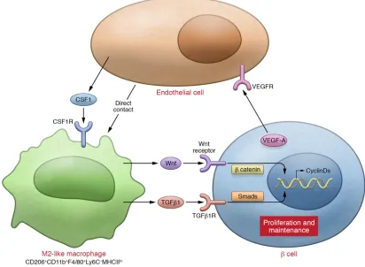
pro-liferation (109). Interestingly, it was reported that endothelial cells could provide a niche for M2-like macrophage polarization through their direct contact with macrophages and also by pro-viding paracrine factors, including CSF1 (110). It therefore seems that quiescent endothelial cells provide the local environment that enables M2-like activation of macrophages, and the environment established by these endothelial cells and M2-like macrophages promotes β cell proliferation (Figure 2).
To further analyze the origin and mechanisms of the M2-like
macrophage activation required for β cell proliferation, Van Gassen
and colleagues utilized adoptive transfer of GFP+CD11b+Ly6Chi
monocytes in a PDL model (111). This study demonstrated that
circulating CD11b+Ly6Chi monocytes are recruited to the
pancre-as, where they differentiate from CD11b+Ly6ChiMHCIIhi cells into
CD11b+Ly6CloMHCIIlo M2-like or tissue-resident
macrophage-like cells. When Ccr2–/– mice were used, though the
CCL2/CCR2-mediated Ly-6Chi monocyte recruitment was blocked, there was
compensatory proliferation of MHC-IIlo tissue-resident
macro-phages and PDL did not significantly alter β cell proliferation
(42). Because CSF1R signaling reportedly mediates local
macro-phage replication and promotes differentiation of MHC-IIhi into
M2-like MHC-IIlo macrophages in general settings (112), the effect
of CSF1R neutralization was analyzed in the PDL model. CSF1R neutralization not only inhibited tissue-resident macrophage
pro-liferation but also reduced differentiation of recruited MHC-IIhi
macrophages into MHC-IIlo macrophages (ref. 111 and Figure 2).
These reports demonstrate that both macrophages recruit-ed from bone marrow and islet-resident macrophages exhibit a
capacity to support β cell proliferation through acquisition of an
M2-like macrophage phenotype supported by CSF1R signaling. Furthermore, these studies suggest that multiple cytokines,
includ-ing TGF-β1, VEGFA, and CSF1, secreted from multiple cell types,
including macrophages, β cells, and quiescent endothelial cells,
contribute to establishing a microenvironment that maintains the
macrophage M2-like phenotype and β cell proliferation (Figure 2).
Conclusions
In addition to the contribution of inflammation to insulin resistance, the existence of islet inflammation and its causative involvement in
β cell dysfunction in T2D is now well appreciated. Islet cells, includ-ing β cells, play an important role in the initiation of islet inflam-mation, as they have the ability to sense stimuli and secrete che-mokines as well as hIAPP to activate macrophages. Most studies
indicate that macrophages are the main source of proinflammatory cytokines within islets. Among these proinflammatory cytokines,
IL-1β secreted from M1-like macrophages plays a crucial role in the
initiation and amplification of islet inflammation. In comparison, M2-like macrophages are indispensible for both islet development
and β cell proliferation in adults. Sterile factors stimulating
mac-rophage activation, the origin of the macmac-rophages, and the local milieu established by multiple cytokines from several cell types within islets are all key determinants of macrophage polarization and function in both normal physiology and T2D pathology.
The new understanding of β cell failure in T2D gained from
studies of islet inflammation provides clues to translational
pos-sibilities. For example, as inflammasome/IL-1β signaling is the
most common and impactful pathway activated in islets of mul-tiple T2D models, strategies targeting IL-1 signaling have pro-duced encouraging results in clinical studies (27, 28, 54, 55). In
addition, because the correct dosage of IL-1β–neutralizing
anti-body is critical for recovery of β cell function in humans, a greater
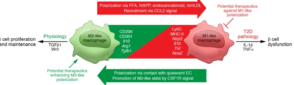
understanding of the macrophages’ role in islet physiology should help to optimize IL-1–targeted strategies. Given that inflamma-tory pathologic changes have been observed in only a portion of T2D patients (12–14, 18), the identification of biomarkers that cor-relate with islet inflammation will help to enhance the effective-ness of therapeutics that target islet inflammation by allowing for appropriate patient selection. Also, in order to avoid systemically suppressing the function of M1-like macrophages, targeting the mechanisms of macrophage M1-like activation that are unique to islets could potentially lead to safe and effective strategies for sup-pressing islet inflammation (Figure 3).
It would also be desirable to support processes related to the normal physiology and beneficial aspects of islet macrophage
behavior, such as their facilitation of β cell proliferation. The key
components necessary to establish a physiological islet microen-vironment remain unclear. The fact that a microenmicroen-vironment
sup-porting β cell proliferation is established through complex
com-munication among several cell types via multiple cytokines makes development of translational approaches difficult. However, CSF1 signaling appears to be a core component, as its importance has been demonstrated in multiple settings, including development,
a pancreatitis model, and a PDL model. Furthermore, targeting β
cell proliferation may be effective in both T1D and T2D settings. Therefore, future studies of the beneficial aspects of islet macro-phage biology could be highly productive (Figure 3).
An improved understanding of islet macrophage biology should enable development of strategies for blocking pathologic
T2D islet inflammation to ameliorate β cell dysfunction while
pro-moting physiologic immune cell function to enhance β cell
prolifer-ation, which may have broad translational implications (Figure 3).
Acknowledgments
We thank Alexander Bartelt, Kathryn C. Claiborn, Kacey Prentice, Lauren T. Robertson, and Scott B. Widenmaier for helpful discussions.
1. Hotamisligil GS, Shargill NS, Spiegelman BM. Adipose expression of tumor necrosis factor-alpha: direct role in obesity-linked insulin resis-tance. Science. 1993;259(5091):87–91. 2. Hotamisligil GS. Inflammation and metabolic
disorders. Nature. 2006;444(7121):860–867. 3. Olefsky JM, Glass CK. Macrophages,
inflamma-tion, and insulin resistance. Annu Rev Physiol. 2010;72:219–246.
4. Lumeng CN, Saltiel AR. Inflammatory links between obesity and metabolic disease. J Clin Invest. 2011;121(6):2111–2117.
5. Kumar V, Abbas AK, Aster JC. Robbins Basic Pathology. 9th ed. Philadelphia, Pennsylvania, USA: Saunders; 2013.
6. Lumeng CN, Bodzin JL, Saltiel AR. Obesity induces a phenotypic switch in adipose tis-sue macrophage polarization. J Clin Invest. 2007;117(1):175–184.
7. Winer S, et al. Normalization of obesity-associ-ated insulin resistance through immunotherapy. Nat Med. 2009;15(8):921–929.
8. Nishimura S, et al. CD8+ effector T cells
con-tribute to macrophage recruitment and adi-pose tissue inflammation in obesity. Nat Med. 2009;15(8):914–920.
9. Grundy SM, et al. Diagnosis and management of the metabolic syndrome: an American Heart Association/National Heart, Lung, and Blood Institute Scientific Statement. Circulation. 2005;112(17):2735–2752.
10. Ferrannini E, Gastaldelli A, Miyazaki Y, Matsuda M, Mari A, DeFronzo RA. beta-Cell function in subjects spanning the range from normal glucose tolerance to overt diabetes: a new analysis. J Clin Endocrinol Metab. 2005;90(1):493–500.
11. Mari A, et al. Impaired β cell glucose sensitivity
rather than inadequate compensation for insulin resistance is the dominant defect in glucose intolerance. Diabetologia. 2010;53(4):749–756. 12. Ehses JA, et al. Increased number of islet-asso-ciated macrophages in type 2 diabetes. Diabetes. 2007;56(9):2356–2370.
13. Richardson SJ, Willcox A, Bone AJ, Foulis AK, Mor-gan NG. Islet-associated macrophages in type 2 diabetes. Diabetologia. 2009;52(8):1686–1688. 14. Kamata K, et al. Islet amyloid with macrophage
migration correlates with augmented β-cell
deficits in type 2 diabetic patients. Amyloid. 2014;21(3):191–201.
15. Butcher MJ, et al. Association of proinflamma-tory cytokines and islet resident leucocytes with islet dysfunction in type 2 diabetes. Diabetologia. 2014;57(3):491–501.
16. Martino L, et al. Mast cells infiltrate pancreatic islets in human type 1 diabetes. Diabetologia. 2015;58(11):2554–2562.
17. Westermark P. Quantitative studies on amy-loid in the islets of Langerhans. Ups J Med Sci. 1972;77(2):91–94.
18. Zhao HL, et al. Prevalence and clinicopatho-logical characteristics of islet amyloid in chi-nese patients with type 2 diabetes. Diabetes. 2003;52(11):2759–2766.
19. Hayden MR. Islet amyloid and fibrosis in the cardiometabolic syndrome and type 2 diabetes mellitus. J Cardiometab Syndr. 2007;2(1):70–75. 20. Masters SL, et al. Activation of the NLRP3
inflam-masome by islet amyloid polypeptide provides a
mechanism for enhanced IL-1β in type 2
diabe-tes. Nat Immunol. 2010;11(10):897–904. 21. Eguchi K, et al. Saturated fatty acid and TLR
signaling link β cell dysfunction and islet
inflam-mation. Cell Metab. 2012;15(4):518–533. 22. Jourdan T, et al. Activation of the Nlrp3
inflam-masome in infiltrating macrophages by endo-cannabinoids mediates beta cell loss in type 2 diabetes. Nat Med. 2013;19(9):1132–1140. 23. Westwell-Roper CY, Ehses JA, Verchere CB. Resident macrophages mediate islet
amyloid polypeptide-induced islet IL-1β
production and β-cell dysfunction. Diabetes.
2014;63(5):1698–1711.
24. Ehses JA, et al. IL-1 antagonism reduces hyper-glycemia and tissue inflammation in the type 2 diabetic GK rat. Proc Natl Acad Sci U S A. 2009;106(33):13998–14003.
25. Sauter NS, Schulthess FT, Galasso R, Castellani LW, Maedler K. The antiinflammatory cytokine interleukin-1 receptor antagonist protects from high-fat diet-induced hyperglycemia. Endocrinol-ogy. 2008;149(5):2208–2218.
26. Larsen CM, et al. Interleukin-1-receptor antago-nist in type 2 diabetes mellitus. N Engl J Med. 2007;356(15):1517–1526.
27. Cavelti-Weder C, et al. Effects of gevokizumab on glycemia and inflammatory markers in type 2 diabetes. Diabetes Care. 2012;35(8):1654–1662. 28. Rissanen A, Howard CP, Botha J, Thuren T,
Glob-al I. Effect of anti-IL-1β antibody (canakinumab)
on insulin secretion rates in impaired glucose tolerance or type 2 diabetes: results of a random-ized, placebo-controlled trial. Diabetes Obes Metab. 2012;14(12):1088–1096.
29. Rodriguez-Calvo T, Ekwall O, Amirian N, Zapardiel- Gonzalo J, von Herrath MG. Increased immune cell infiltration of the exocrine pancreas: a possible contribution to the pathogenesis of type 1 diabetes. Diabetes. 2014;63(11):3880–3890. 30. Marchetti P. Islet inflammation in type 2 diabetes.
Diabetologia. 2016;59(4):668–672.
31. Brooks-Worrell BM, Boyko EJ, Palmer JP. Impact
of islet autoimmunity on the progressive β-cell
functional decline in type 2 diabetes. Diabetes Care. 2014;37(12):3286–3293.
32. Davies LC, Jenkins SJ, Allen JE, Taylor PR. Tissue-resident macrophages. Nat Immunol. 2013;14(10):986–995.
33. Gosselin D, et al. Environment drives selec-tion and funcselec-tion of enhancers controlling tissue-specific macrophage identities. Cell. 2014;159(6):1327–1340.
34. Lavin Y, et al. Tissue-resident macrophage enhancer landscapes are shaped by the local microenvironment. Cell. 2014;159(6):1312–1326. 35. Mantovani A, Sica A, Locati M.
Macro-phage polarization comes of age. Immunity. 2005;23(4):344–346.
36. Murray PJ, Wynn TA. Protective and pathogenic functions of macrophage subsets. Nat Rev Immu-nol. 2011;11(11):723–737.
37. Lech M, Anders HJ. Macrophages and fibrosis: How resident and infiltrating mononuclear phagocytes orchestrate all phases of tis-sue injury and repair. Biochim Biophys Acta. 2013;1832(7):989–997.
38. Kelly J, Ali Khan A, Yin J, Ferguson TA, Apte RS. Senescence regulates macrophage activation and angiogenic fate at sites of tissue injury in mice. J Clin Invest. 2007;117(11):3421–3426. 39. Geutskens SB, Otonkoski T, Pulkkinen MA,
Drexhage HA, Leenen PJ. Macrophages in the murine pancreas and their involvement in fetal endocrine development in vitro. J Leukoc Biol. 2005;78(4):845–852.
40. Banaei-Bouchareb L, et al. Insulin cell mass is altered in Csf1op/Csf1op macrophage-deficient mice. J Leukoc Biol. 2004;76(2):359–367. 41. Tessem JS, et al. Critical roles for
macro-phages in islet angiogenesis and maintenance during pancreatic degeneration. Diabetes. 2008;57(6):1605–1617.
42. Xiao X, et al. M2 macrophages promote beta-cell proliferation by up-regulation of SMAD7. Proc Natl Acad Sci U S A. 2014;111(13):E1211–E1220. 43. Criscimanna A, Coudriet GM, Gittes GK,
Pigan-elli JD, Esni F. Activated macrophages create lineage-specific microenvironments for
pan-creatic acinar- and β-cell regeneration in mice.
Gastroenterology. 2014;147(5):1106–18.e11. 44. Cao X, Han ZB, Zhao H, Liu Q. Transplantation
of mesenchymal stem cells recruits trophic mac-rophages to induce pancreatic beta cell regen-eration in diabetic mice. Int J Biochem Cell Biol. 2014;53:372–379.
45. Homo-Delarche F, et al. Islet inflammation and fibrosis in a spontaneous model of type 2 diabe-tes, the GK rat. Diabetes. 2006;55(6):1625–1633. 46. Igoillo-Esteve M, et al. Palmitate induces a
pro-inflammatory response in human pan-creatic islets that mimics CCL2 expression by beta cells in type 2 diabetes. Diabetologia. 2010;53(7):1395–1405.
47. Atkinson MA, Maclaren NK. The pathogenesis of insulin-dependent diabetes mellitus. N Engl J Med. 1994;331(21):1428–1436.
48. Wallberg M, Cooke A. Immune mecha-nisms in type 1 diabetes. Trends Immunol. 2013;34(12):583–591.
49. Maedler K, et al. Glucose-induced beta cell production of IL-1beta contributes to glucotox-icity in human pancreatic islets. J Clin Invest. 2002;110(6):851–860.
50. Zhou R, Tardivel A, Thorens B, Choi I, Tschopp J. Thioredoxin-interacting protein links oxidative stress to inflammasome activation. Nat Immunol. 2010;11(2):136–140.
51. Ikeda H. KK mouse. Diabetes Res Clin Pract. 1994;24(suppl):S313–S316.
52. Cucak H, Grunnet LG, Rosendahl A. Accumulation of M1-like macrophages in type 2 diabetic islets is followed by a systemic shift in macrophage polar-ization. J Leukoc Biol. 2014;95(1):149–160. 53. Calderon B, et al. The pancreas anatomy
condi-tions the origin and properties of resident macro-phages. J Exp Med. 2015;212(10):1497–1512. 54. van Asseldonk EJ, Stienstra R, Koenen TB,
Joosten LA, Netea MG, Tack CJ. Treatment with Anakinra improves disposition index but not insulin sensitivity in nondiabetic subjects with the metabolic syndrome: a randomized, double-blind, placebo-controlled study. J Clin Endocrinol Metab. 2011;96(7):2119–2126.
ran-domized study evaluating the glycemic and anti-inflammatory effects of subcutaneous
LY2189102, a neutralizing IL-1β antibody, in
patients with type 2 diabetes. Diabetes Care. 2013;36(8):2239–2246.
56. Donath MY. Targeting inflammation in the treat-ment of type 2 diabetes: time to start. Nat Rev Drug Discov. 2014;13(6):465–476.
57. Herder C, Dalmas E, Böni-Schnetzler M, Donath MY. The IL-1 pathway in type 2 diabetes and cardiovascular complications. Trends Endocrinol Metab. 2015;26(10):551–563.
58. Shi H, Kokoeva MV, Inouye K, Tzameli I, Yin H, Flier JS. TLR4 links innate immunity and fatty acid-induced insulin resistance. J Clin Invest. 2006;116(11):3015–3025.
59. Jia L, et al. Hepatocyte Toll-like receptor 4 regu-lates obesity-induced inflammation and insulin resistance. Nat Commun. 2014;5:3878. 60. Vandanmagsar B, et al. The NLRP3 inflammasome
instigates obesity-induced inflammation and insu-lin resistance. Nat Med. 2011;17(2):179–188. 61. Kono H, Rock KL. How dying cells alert the
immune system to danger. Nat Rev Immunol. 2008;8(4):279–289.
62. Vogl T, et al. Mrp8 and Mrp14 are endogenous activators of Toll-like receptor 4, promoting lethal, endotoxin-induced shock. Nat Med. 2007;13(9):1042–1049.
63. Klune JR, Dhupar R, Cardinal J, Billiar TR, Tsung A. HMGB1: endogenous danger signaling. Mol Med. 2008;14(7–8):476–484.
64. Eguchi K, Manabe I. Toll-like receptor, lipotoxic-ity and chronic inflammation: the pathological link between obesity and cardiometabolic dis-ease. J Atheroscler Thromb. 2014;21(7):629–639. 65. Krüger B, et al. Islet-expressed TLR2 and
TLR4 sense injury and mediate early graft failure after transplantation. Eur J Immunol. 2010;40(10):2914–2924.
66. Ehses JA, et al. Toll-like receptor 2-deficient mice are protected from insulin resistance and beta cell dysfunction induced by a high-fat diet. Dia-betologia. 2010;53(8):1795–1806.
67. Böni-Schnetzler M, et al. Free fatty acids induce a proinflammatory response in islets via the abundantly expressed interleukin-1 receptor I. Endocrinology. 2009;150(12):5218–5229. 68. Maloy AL, Longnecker DS, Greenberg ER. The
relation of islet amyloid to the clinical type of diabetes. Hum Pathol. 1981;12(10):917–922. 69. Cooper GJ, Willis AC, Clark A, Turner RC, Sim
RB, Reid KB. Purification and characterization of a peptide from amyloid-rich pancreases of type 2 diabetic patients. Proc Natl Acad Sci U S A. 1987;84(23):8628–8632.
70. Westermark P, Wernstedt C, Wilander E, Hayden DW, O’Brien TD, Johnson KH. Amy-loid fibrils in human insulinoma and islets of Langerhans of the diabetic cat are derived from a neuropeptide-like protein also present in normal islet cells. Proc Natl Acad Sci U S A. 1987;84(11):3881–3885.
71. Ogawa A, Harris V, McCorkle SK, Unger RH, Lus-key KL. Amylin secretion from the rat pancreas and its selective loss after streptozotocin treat-ment. J Clin Invest. 1990;85(3):973–976. 72. Wei L, et al. The molecular basis of distinct
aggre-gation pathways of islet amyloid polypeptide. J Biol Chem. 2011;286(8):6291–6300. 73. Buchanan LE, et al. Mechanism of IAPP amyloid
fibril formation involves an intermediate with
a transient β-sheet. Proc Natl Acad Sci U S A.
2013;110(48):19285–19290.
74. Lorenzo A, Razzaboni B, Weir GC, Yankner BA. Pancreatic islet cell toxicity of amylin associ-ated with type-2 diabetes mellitus. Nature. 1994;368(6473):756–760.
75. Haataja L, Gurlo T, Huang CJ, Butler PC. Islet amyloid in type 2 diabetes, and the toxic oligomer hypothesis. Endocr Rev. 2008;29(3):303–316. 76. Green J, et al. Full-length rat amylin forms
fibrils following substitution of single residues from human amylin. J Mol Biol. 2003;326(4):1147–1156.
77. Fox N, et al. Human islet amyloid polypeptide transgenic mice as a model of non-insulin-dependent diabetes mellitus (NIDDM). FEBS Lett. 1993;323(1–2):40–44.
78. de Koning EJ, et al. Human islet amyloid poly-peptide accumulates at similar sites in islets of transgenic mice and humans. Diabetes. 1994;43(5):640–644.
79. Hull RL, et al. Increased dietary fat promotes islet amyloid formation and beta-cell secretory dysfunction in a transgenic mouse model of islet amyloid. Diabetes. 2003;52(2):372–379. 80. Meier DT, Morcos M, Samarasekera T,
Zraika S, Hull RL, Kahn SE. Islet amyloid formation is an important determinant for inducing islet inflammation in high-fat-fed human IAPP transgenic mice. Diabetologia. 2014;57(9):1884–1888.
81. Westwell-Roper C, et al. IL-1 blockade attenuates islet amyloid polypeptide-induced proinflammatory cytokine release and pan-creatic islet graft dysfunction. J Immunol. 2011;187(5):2755–2765.
82. Westwell-Roper C, Denroche HC, Ehses JA, Verchere CB. Differential activation of innate immune pathways by distinct islet amyloid polypeptide (IAPP) aggregates. J Biol Chem. 2016;291(17):8908–8917.
83. Westwell-Roper CY, Chehroudi CA, Denroche HC, Courtade JA, Ehses JA, Verchere CB. IL-1 mediates amyloid-associated islet dysfunc-tion and inflammadysfunc-tion in human islet amyloid polypeptide transgenic mice. Diabetologia. 2015;58(3):575–585.
84. de Koning EJ, et al. Macrophages and pancreatic islet amyloidosis. Amyloid. 1998;5(4):247–254. 85. Halle A, et al. The NALP3 inflammasome is
involved in the innate immune response to amyloid-beta. Nat Immunol. 2008;9(8):857–865. 86. Heneka MT, et al. NLRP3 is activated in Alzheim-er’s disease and contributes to pathology in APP/ PS1 mice. Nature. 2013;493(7434):674–678. 87. Badman MK, Pryce RA, Chargé SB, Morris JF,
Clark A. Fibrillar islet amyloid polypeptide (amylin) is internalised by macrophages but resists proteolytic degradation. Cell Tissue Res. 1998;291(2):285–294.
88. Bauernfeind FG, et al. Cutting edge: NF-kappaB activating pattern recognition and cytokine receptors license NLRP3 inflammasome activa-tion by regulating NLRP3 expression. J Immunol.
2009;183(2):787–791.
89. Miller YI, Viriyakosol S, Binder CJ, Feramisco JR, Kirkland TN, Witztum JL. Minimally modi-fied LDL binds to CD14, induces macrophage spreading via TLR4/MD-2, and inhibits phagocytosis of apoptotic cells. J Biol Chem. 2003;278(3):1561–1568.
90. Abderrahmani A, et al. Human high-density lipo-protein particles prevent activation of the JNK pathway induced by human oxidised low-density lipoprotein particles in pancreatic beta cells. Dia-betologia. 2007;50(6):1304–1314.
91. Hutton MJ, Westwell-Roper C, Soukhatcheva G, Plesner A, Dutz JP, Verchere CB. Islet allograft rejection is independent of toll-like receptor signaling in mice. Transplantation. 2009;88(9):1075–1080.
92. Wen H, et al. Fatty acid-induced NLRP3-ASC inflammasome activation interferes with insulin signaling. Nat Immunol. 2011;12(5):408–415. 93. Pacher P, Bátkai S, Kunos G. The
endocannabi-noid system as an emerging target of pharmaco-therapy. Pharmacol Rev. 2006;58(3):389–462. 94. Maccarrone M, et al. Endocannabinoid signal-ing at the periphery: 50 years after THC. Trends Pharmacol Sci. 2015;36(5):277–296.
95. Rohrbach K, et al. Ibipinabant attenuates β-cell
loss in male Zucker diabetic fatty rats indepen-dently of its effects on body weight. Diabetes Obes Metab. 2012;14(6):555–564.
96. Tam J, et al. Peripheral cannabinoid-1 receptor inverse agonism reduces obesity by reversing leptin resistance. Cell Metab. 2012;16(2):167–179. 97. Maedler K, et al. Low concentration of
interleu-kin-1beta induces FLICE-inhibitory
protein-mediated β-cell proliferation in human
pancre-atic islets. Diabetes. 2006;55(10):2713–2722. 98. Hajmrle C, et al. Interleukin-1 signaling
con-tributes to acute islet compensation. JCI Insight. 2016;1(4):e86055.
99. Hume DA, Robinson AP, MacPherson GG, Gordon S. The mononuclear phagocyte system of the mouse defined by immunohistochemi-cal loimmunohistochemi-calization of antigen F4/80. J Exp Med. 1983;158(5):1522–1536.
100. Banaei-Bouchareb L, Peuchmaur M, Czernichow P, Polak M. A transient microenvironment loaded mainly with macrophages in the early developing human pancreas. J Endocrinol. 2006;
188(3):467–480.
101. Schulz C, et al. A lineage of myeloid cells inde-pendent of Myb and hematopoietic stem cells. Science. 2012;336(6077):86–90.
102. Gomez Perdiguero E, et al. Tissue-resident macrophages originate from yolk-sac-derived erythro-myeloid progenitors. Nature. 2015;518(7540):547–551.
103. Bain CC, et al. Constant replenishment from cir-culating monocytes maintains the macrophage pool in the intestine of adult mice. Nat Immunol. 2014;15(10):929–937.
104. Eguchi K, Manabe I. Macrophages and islet inflammation in type 2 diabetes. Diabetes Obes Metab. 2013;15 Suppl 3:152–158.
105. Morris DL. Minireview: Emerging concepts in islet macrophage biology in type 2 diabetes. Mol Endocrinol. 2015;29(7):946–962.
essen-tial for inflammation-induced but not β-cell
workload-induced β-cell proliferation. Diabetes.
2013;62(4):1217–1226.
107. Brissova M, et al. Pancreatic islet produc-tion of vascular endothelial growth fac-tor — a is essential for islet vascularization, revascularization, and function. Diabetes. 2006;55(11):2974–2985.
108. Agudo J, et al. Vascular endothelial growth
factor-mediated islet hypervascularization and inflammation contribute to progressive reduction
of β-cell mass. Diabetes. 2012;61(11):2851–2861.
109. Brissova M, et al. Islet microenvironment, modu-lated by vascular endothelial growth factor-A
sig-naling, promotes β cell regeneration. Cell Metab.
2014;19(3):498–511.
110. He H, et al. Endothelial cells provide an instruc-tive niche for the differentiation and functional
polarization of M2-like macrophages. Blood. 2012;120(15):3152–3162.
111. Van Gassen N, et al. Macrophage dynamics are regulated by local macrophage proliferation and monocyte recruitment in injured pancreas. Eur J Immunol. 2015;45(5):1482–1493.