http://dx.doi.org/10.4236/crcm.2014.312140
How to cite this paper: Cosme, J., Branco, J., Carneiro, H., Diviza, G., Chagas, C., Velhinho, E. and Silva, G. (2014) Spinal Epi- dural Abscess: When a Fast Diagnosis Is Necessary. Case Reports in Clinical Medicine, 3, 664-668.
http://dx.doi.org/10.4236/crcm.2014.312140
Spinal Epidural Abscess: When a Fast
Diagnosis Is Necessary
Joana Cosme
*, Joana Branco, Hermana Carneiro, Gheorghe Diviza, Carlos Chagas,
Emília Velhinho, Glória Silva
Serviço Medicina III, Hospital Pulido Valente, Centro Hospitalar Lisboa Norte, Lisboa, Portugal Email: *[email protected]
Received 15 November 2014; revised 10 December 2014; accepted 24 December 2014
Academic Editor: Jung Ho Park, Hallym University Sacred Heart Hospital, Republic of Korea
Copyright © 2014 by authors and Scientific Research Publishing Inc.
This work is licensed under the Creative Commons Attribution International License (CC BY). http://creativecommons.org/licenses/by/4.0/
Abstract
A 61 years old man with fatty liver disease and history of recent urinary infection was admitted to the Department of Internal Medicine for severe back pain and fever. The blood culture exams were positive for methicillin-resistant Staphylococcus aureus and later the cervical spine MRI showed an anterior epidural abscess associated with medullary compression. The patient was kept under antibiotic therapy and a decompressive surgery was required. Spinal epidural abscess presentation can be subtle with its presenting complaints ranging in severity from nonspecific back pain to marked weakness. This case report highlights the importance of an early diagnosis in order to avoid a devastating outcome.
Keywords
Spinal Epidural Abscess, Magnetic Resonance Imaging, Early Diagnosis
1. Introduction
Spinal epidural abscess (SEA) is a rare pyogenic infectious disease with a potentially devastating outcome, lo-cated between the spinal dura mater and the vertebral periosteum [1] [2].
It was first described by Morgagni in 1761 but it was clearly defined only in 1820 by Bergamaschi [2]. The prevalence of this condition has increased in the last decades [1] [2] from 0.2 - 1.2 cases per 10,000 hos-pital admissions in the mid-1970s to a current estimated incidence of 2 - 12.5 per 10,000 admissions [2]. Popu-lation aging, increased prevalence of injection drug use, of spinal instrumentation and of vascular access, as well
as, greater availability of magnetic resonance imaging (MRI) and greater awareness of SEA are probably the main responsible for this increase [2]. It occurs in patients from 10 days to 87 years, with most cases occurring in those 30 - 60 years old [1]. The increasing patient age has been shown to be a poorer prognostic factor [1].
Staphylococcus aureus is the most common pathogen involved in this infection, responsible for approximately two-thirds of spinal abscesses [1]-[3]. Other pathogens including gram-positive and gram-negative bacteria, an-aerobic bacteria, mycobacteria, fungi, and parasites have also been implicated [3]. The most common abscess location is the lumbar spine (48%), followed by the thoracic spine (31%) and then the cervical spine (24%) [2].
The treatment of SEA is two-pronged: medical with antibiotics and surgical with laminectomy [3]. Despite being a rare condition SEA is associated with high mortality rate, ranging from 15% to 23% [1]. As SEA has a wide variation of causes, clinical presentation and locations. A high level of alert and suspicion are essential for an accurate diagnosis. In this article, we empathize the importance of a fast diagnosis in avoid-ing a devastatavoid-ing outcome.
2. Case Report
A 61 years old man, caucasian, farmer with arterial hypertension and dyslipidemia and with history of acute myocardial infarction three years before, was admitted to the Department of Internal Medicine in Hospital Pulido Valente in North Lisbon Hospital Center. His daily medications were hydrochlorothiazide/olmesartan, carvedilol, pitavastatin, acetilsalicilic acid, clopidogrel, omeprazol, bromazepam and naproxen. He referred al-cohol consumption (approximately 35 grams of alal-cohol per day) and fatty liver history.
His symptoms at admission were back severe pain with 5 days of evolution and fever (maximum axillary temperature measured of 38.5˚C). Five days prior to admission it was prescribed amoxicillin/clavulanic acid for an urinary infection. There was no history of previous spinal trauma or surgery, drug usage or other invasive exams besides coronarography with angioplasty 3 years before.
The physical examination revealed redness on his back. Body temperature and others vital parameters were stable at admission. Neurological examination revealed sensory motor quadriparesis. Power was grade 2 in the lower limbs and grades 1in the upper limbs.
Laboratory analyses revealed the following: White blood cell count: 10.59 × 10⁹ cells/L with differential count: neutrophils, 64%; lymphocytes 25%; monocytes 9%; eosinophils 1%; basophils 1%; red blood cell count 5.34 × 10¹² cells/L; haemoglobin 15.0 g/L; platelets, 163 × 109 platelets/L; alanine aminotransferase 90 U/L; C-reactive protein 3.4 mg/dL, creatinine 0.9 mg/dL. The patient was HIV negative. All viral serologies were negative.
The cervical radiography showed a loss/straightening of cervical lordosis (Figure 1) and the cervical and lumber computerized tomography scan did not showed other aspects besides this.
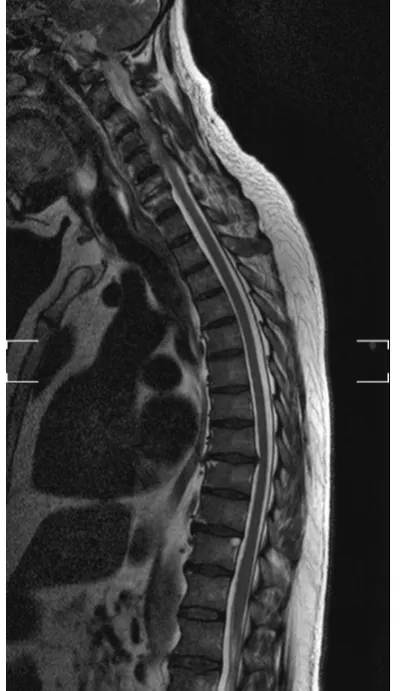
The urine culture was negative and all the blood culture exams (two in aerobiosis and one in anaerobiosis) were positive for methicillin resistant Staphylococcus aureus. The patient was immediately treated with vanco-mycin and further imaging was performed with cervical spine MRI that showed anterior epidural abscess ex-tending from C2 to C6 vertebral bodies low signal intensity on T1-weighted images, and high signal intensity on T2-weighted images and STIR and with gadolinium enhancement, associated with medullary compression (Figure 2 and Figure 3). The serial levels of vancomycin where monitorized.
These findings as well as the persistent and worsening back pain associated with a sudden onset of flaccid tetraparesis led to the decision to urgently transfer the patient for specialised surgical intervention.
The patient underwent spinal decompression. After the surgery the patient continued the antibiotic therapy and physical rehabilitation for more than a month. After surgery, repeated spinal imaging exams and neurologi-cal examinations were made. There was a progressive resolution of the pain and a partial recovery of the limbs strength (power improved for 3 - 4) and of the ability for walking. After clinical and analytic stability he was transferred to a physiatrist unit to continue his neurologic recovery. The patient will continue his follow up evaluations in neurosurgery and infectious diseases units.
3. Discussion
Despite the advances in its medical and surgical treatment, SEA remains a morbid disease process.
The learning point from this case is the need for early diagnosis and urgent intervention. A complete history and clinical examination and a high level of suspicion are essential in the diagnosis of this condition.
Figure 1.Patient cervical radiography.
Figure 3.Patient cervical spine magnetic resonance imaging.
although the pain associated with a spinal abscess typically becomes severe and unrelenting, the presentation of this condition can be subtle and insidious. It is import to be aware that patients’ presenting complaints rang in se-verity from nonspecific back pain to marked weakness [1].
Secondly, there are several risk factors and medical comorbidities associated with the development of SEA. L. Arko et al. (2014) showed that intravenous drug use was the most frequently reported risk factor (22% of pa-tients with SEA) and diabetes (27%) and hepatic disease (14%) the most common medical comorbidities [2]. O. Adogwa et al. (2014) pointed out other risk factors: diabetes mellitus (38%), end-stage renal disease (24%), his-tory of endocarditis (9%), recent hishis-tory of urosepsis (6%), rheumatoid arthritis (5%) and hishis-tory of spinal trauma (2.4%) [1]. According to S. Zimmerer et al. (2011) the risk of SEA is higher in immunocompromised person (e.g. persons who have the acquired immune deficiency syndrome or are under immunosuppressive treatment after organ transplantation) or in patients with history of drug addiction, alcoholism, cancer, and sys-temic inflammation or infection [4]. Despite this, SEA can also occur in the absence of predisposing factors. Verma R et al. (2014) described a case of a 35-year-old-man with an extensive SEA that, like the patient de-scribed above, had no risk factors commonly associated with the development of SEA. These authors labelled this condition as “spontaneous SEA” [5].
Thirdly, although the changes on the initial cervical spine radiograph and spine computerized tomography are subtle and did not point to a specific diagnosis, MRI is essential for the establishment of the diagnosis.
The patients’ prognosis in this type of condition is dependent not only on the clinical and neurological condi-tion of the patient at presentacondi-tion but also on the timing for instigacondi-tion of an appropriate treatment [6]. For this reason, in a neurosurgical emergency like this, a prompt diagnosis and urgent treatment are essential to a suc-cessful management and to avoid high rates of morbidity and mortality [5] [6].
4. Conclusions
SEA is a rare event with significant variation in its causes, anatomical locations and rate of progression. A highest degree of suspicion and appropriate investigations and treatment without delay are essential to re-duce the morbidity and mortality associated with this condition. MRI is essential to an early diagnosis.
References
(2014) Spontaneous Spinal Epidural Abscess in Patients 50 Years of Age and Older: A 15-Year Institutional Perspec-tive and Review of the Literature. Journal of Neurosurgery: Spine, 20, 344-349.
http://dx.doi.org/10.3171/2013.11.SPINE13527
[2] Arko, L., Quach, E., Nguyen, V., Chang, D., Sukul, V. and Kim, B.S. (2014) Medical and Surgical Management of Spinal Epidural Abscess: A Systematic Review. Neurosurgical Focus, 37, E4.
http://thejns.org/doi/pdf/10.3171/2014.6.FOCUS14127
[3] Tuchman, A., Pham, M. and Hsieh, P.C. (2014) The Indications and Timing for Operative Management of Spinal Epi-dural Abscess: Literature Review and Treatment Algorithm. Neurosurgical Focus, 37, E8.
http://dx.doi.org/10.3171/2014.6.FOCUS14261
[4] Zimmerer, S., Conen, A., Muller, A., Sailer, M., Taub, E., Fluckiger, U. and Schwenzer-Zimmerer, K. (2011) Spinal Epidural Abscess: Aetiology, Predisponent Factors and Clinical Outcomes in a 4-Year Prospective Study. European Spine Journal, 20, 2228-2234.
http://www.ncbi.nlm.nih.gov/pmc/articles/PMC3229722/pdf/586_2011_Article_1838.pdf http://dx.doi.org/10.1007/s00586-011-1838-y
[5] Verma, R., Chaudhari, T.S. and Lachuriya, G. (2014) Spontaneous Extensive Spinal Epidural Abscess Presenting as Acute Quadriparesis. BMJ Case Reports. http://casereports.bmj.com/content/2014/bcr-2014-204892.full.pdf+html
[6] Mackenzie, A., Laing, R.B.S., Smith, C.C., Kaar, G.F. and Smith, F.W. (1998) Spinal Epidural Abscess: The Impor-tance of Early Diagnosis and Treatment. Journal of Neurology, Neurosurgery & Psychiatry, 65, 209-212.