Open Access
Vol 11 No 5Research article
Hypertrophy is induced during the
in vitro
chondrogenic
differentiation of human mesenchymal stem cells by bone
morphogenetic protein-2 and bone morphogenetic protein-4 gene
transfer
Andre F Steinert
1,2, Benedikt Proffen
1, Manuela Kunz
1, Christian Hendrich
1,
Steven C Ghivizzani
2,3, Ulrich Nöth
1, Axel Rethwilm
4, Jochen Eulert
1and Christopher H Evans
21Orthopaedic Center for Musculoskeletal Research, Orthopaedic Clinic, König-Ludwig-Haus, Julius-Maximilians-University, Brettreichstrasse 11,
97074 Würzburg, Germany
2Center for Molecular Orthopaedics, Harvard Medical School, 221 Longwood Avenue, BLI 152, Boston, MA 02115, USA 3Department of Orthopaedics and Rehabilitation, University of Florida, 3450 Hull Road, Gainesville, FL 32607, USA 4Institut für Virologie und Immunbiologie, Julius-Maximilians-University, Versbacherstrasse 7, 97078 Würzburg, Germany
Corresponding author: Christopher H Evans, [email protected]
Received: 1 May 2009 Revisions requested: 10 Jun 2009 Revisions received: 15 Sep 2009 Accepted: 2 Oct 2009 Published: 2 Oct 2009
Arthritis Research & Therapy 2009, 11:R148 (doi:10.1186/ar2822) This article is online at: http://arthritis-research.com/content/11/5/R148 © 2009 Steinert et al.; licensee BioMed Central Ltd.
This is an open access article distributed under the terms of the Creative Commons Attribution License (http://creativecommons.org/licenses/by/2.0), which permits unrestricted use, distribution, and reproduction in any medium, provided the original work is properly cited.
Abstract
Introduction The present study compares bone morphogenetic protein (BMP)-4 and BMP-2 gene transfer as agents of chondrogenesis and hypertrophy in human primary mesenchymal stem cells (MSCs) maintained as pellet cultures.
Methods Adenoviral vectors carrying cDNA encoding human BMP-4 (Ad.BMP-4) were constructed by cre-lox combination and compared to previously generated adenoviral vectors for BMP-2 (Ad.BMP-2), green fluorescent protein (Ad.GFP), or firefly luciferase (Ad.Luc). Cultures of human bone-marrow derived MSCs were infected with 5 × 10² viral particles/cell of
Ad.BMP-2, or Ad.BMP-4, seeded into aggregates and cultured for three weeks in a defined, serum-free medium. Untransduced cells or cultures transduced with marker genes served as controls. Expression of BMP-2 and BMP-4 was determined by ELISA, and aggregates were analyzed histologically, immunohistochemically, biochemically and by RT-PCR for chondrogenesis and hypertrophy.
Results Levels of BMP-2 and BMP-4 in the media were initially 30 to 60 ng/mL and declined thereafter. BMP-4 and BMP-2 genes were equipotent inducers of chondrogenesis in primary MSCs as judged by lacuna formation, strong staining for proteoglycans and collagen type II, increased levels of GAG synthesis, and expression of mRNAs associated with the chondrocyte phenotype. However, BMP-4 modified aggregates showed a lower tendency to progress towards hypertrophy, as judged by expression of alkaline phosphatase, annexin 5, immunohistochemical staining for type X collagen protein, and lacunar size.
Conclusions BMP-2 and BMP-4 were equally effective in provoking chondrogenesis by primary human MSCs in pellet culture. However, chondrogenesis triggered by BMP-2 and BMP-4 gene transfer showed considerable evidence of hypertrophic differentiation, with, the cells resembling growth plate chondrocytes both morphologically and functionally. This suggests caution when using these candidate genes in cartilage repair.
Introduction
Mesenchymal progenitor cells, also referred to as mesenchy-mal stem cells (MSCs), provide an attractive alternative to chondrocytes with regard to cell-based approaches to carti-lage repair [1]. With the use of the proper three-dimensional serum-free culture conditions, expanded MSCs can be stimu-lated to differentiate along the chondrogenic pathway when the appropriate factors, such as certain members of the trans-forming growth factor (TGF)-β superfamily, are present [2-4]. This research has led to the first clinical application of autolo-gous bone marrow stromal cells for the repair of full-thickness articular cartilage defects in humans [5,6]. However, to date, the delivery of MSCs into cartilaginous lesions has neither clin-ically nor experimentally resulted in sustained regeneration of hyaline cartilage in vivo [7]. Inadequate delivery of the soluble factors necessary to drive the chondrogenic differentiation of the transplanted cells in vivo is a major impediment to effective chondrogenesis in situ [7]. To overcome this limitation, gene transfer approaches are being explored clinically [8] and experimentally [9-12] to enable the sustained delivery of chon-drogenic and anti-inflammatory factors to cartilage defects.
Another obstacle was identified from studies of in vitro chon-drogenesis using MSCs or chondrocytes treated with bone morphogenetic proteins (BMPs), members of the TGF-β superfamily. BMPs are a group of secreted polypeptides with pleiotropic roles in many different cell types and were originally identified by their ability to induce endochondral bone forma-tion in ectopic extraskeletal sites in vivo [1,7-10]. Among other BMPs, BMP-2 and BMP-7 are known to induce differentiation of mesenchymal progenitor cells and preosteoblasts into mature osteoblasts, and to enhance the differentiated function of osteoblasts, which have led to the clinical application of these proteins for bone regeneration [1,7-10]. We and others have tested several BMPs for their potential use in cartilage regeneration including BMP-2, BMP-4, BMP-6 and BMP-7, which were shown to induce chondrogenic differentiation of mesenchymal progenitor cells and to up regulate the levels of type II collagen and aggrecan in chondrocytes and chondro-progenitor cells [1,7-11]. During development of the limbs, however, BMPs along with other regulators also mediate the replacement of chondrogenesis by endochondral ossification comprising chondrocyte maturation, hypertrophy, transition from type II to type X collagen with subsequent chondrocyte apoptosis, while osteoprogenitor cells differentiate into oste-oblasts and replace the cartilage with mineralized bone tissue. Equivalently, chondrogenic cultures induced by BMPs showed high expression of genes associated with chondro-cyte hypertrophy, including collagen type (COL) X and indian hedgehog (IHH), among others [1,7-11,13]. This suggests that the chondrogenic differentiation of the MSCs advanced to the end stage, hypertrophic state that is typical of endochon-dral ossification during skeletal development. This conclusion correlates well with existing in vivo data. For example, delivery of BMP-2 expressing MSCs resulted in tissue hypertrophy and
the formation of osteophytes, when transplanted orthotopically to osteochondral defects [14] or ectopically [15,16] in small animal models. Moreover, such hypertrophy-associated changes are not exclusively found in terminal differentiated growth plate chondrocytes, but are also present in pathologi-cal conditions such as osteoarthritis [17,18].
Inspired by these observations, we aim to further explore the effects of chondrogenic-induction by BMPs on hypertrophy, maturation and apoptosis. We have previously shown that adenoviral delivery of individual cDNAs encoding BMP-2 or TGF-β1 into primary MSCs is capable of driving chondrogen-esis in culture [19,20]. In the present study, using adenoviral-mediated gene transfer our aim was to compare the effects of BMP-4 and BMP-2 expression on chondrogenesis of primary MSCs and to investigate whether levels and extent of hyper-trophy in vitro is influenced by the choice of transgene.
Materials and methods
Construction and preparation of recombinant adenoviral vectors
The complete coding sequence of the human BMP-4 gene [GenBank:M22490] cloned into λ gt10 bacteriophage vec-tors (ATCC No. 40342; Manassas, VA, USA) was isolated and purified according to standard protocols [21]. The iso-lated λ gt10 DNA was then digested with EcoRI to release the 1.7 kB sized BMP-4 cDNA insert, which was then cloned into the EcoRI site of the pAdlox shuttle vector, and first-genera-tion, E1, E3-deleted, serotype 5 adenoviral vectors carrying the cDNAs for human BMP-4 were constructed by cre-lox
recombination as previously described [22]. The vectors encoding BMP-2, firefly luciferase (Luc) or green fluorescent protein (GFP) from jellyfish were generated previously [22]. The resulting vectors were designated Ad.BMP-2, Ad.BMP-4, Ad.Luc and Ad.GFP, and suspensions of recombinant adeno-virus were prepared by amplification in 293 cells followed by purification using three consecutive CsCl gradients [22]. Viral titers were estimated to be between 1012 and 1013 particles/
mL by optical density at 260 nm and standard plaque assay.
Culture of human bone marrow-derived MSCs and adenoviral transduction
Bone marrow was harvested from the surgical waste of femurs of six patients, aged 48 to 63 years (mean age 55 years), undergoing total hip arthroplasty, after informed consent was given and as approved by the institutional review board of the University of Wuerzburg as described earlier [23]. The col-lected cells were spun at 1 × 10³ rpm for five minutes,
resus-pended in complete DMEM (containing 10% fetal bovine serum (FBS) and 1% penicillin/streptomycin), and plated at 4 to 6 × 10⁷ nucleated cells per 75 cm2 flask (Falcon, Beckton
Dickinson Labware, Franklin Lakes, NJ, USA). Unattached cells were removed after three days, and adherent colonies were cultured at 37°C, 5% CO2 in DMEM with 10% FBS
expansion of chondroprogenitor cells. Medium changes were performed every three to four days, and after 14 days adherent colonies were trypsinized and replated in several 75 cm2
tis-sue culture flasks. At confluence (approximately 1.2 × 10⁶
cells/T-75 flask), the cultures were infected in 750 μL serum-free DMEM for two hours at a dose of 5 × 10³ vp/cell of
Ad.BMP-2, or Ad.BMP-4. Control cultures were similarly infected with Ad.GFP or Ad.Luc at 5 × 10³ vp/cell, or
remained uninfected. For comparison, an additional set of untransduced recombinant human protein controls were main-tained, which were cultured in the presence of 10 ng/mL
TGF-β1 protein, or 25 ng/mL BMP-2, or 25 ng/mL BMP-4 (all R&D Systems, Minneapolis, MN, USA). Following viral infection, the supernatant was aspirated and replaced with 10 mL complete DMEM.
Aggregate culture and transgene expression
Twenty-four hours post-infection, the MSC cultures were trypsinized, washed and placed in aggregate culture as described previously [24], and as modified by Penick and col-leagues [25]. Briefly, MSCs were suspended to a concentra-tion of 1 × 10⁶ cell/mL in serum-free DMEM containing 1 mM
pyruvate, 1% ITS + Premix (insulin, transferrin and selenous acid containing culture supplement), 37.5 mg/mL ascorbate-2-phosphate and 10-7 M dexamethasone (all Sigma, St. Louis,
MO, USA), and 200 μL aliquots (2 × 10⁵ cells) were
distrib-uted to a polypropylene, v-bottom 96-well plate (Corning, Corning, NY, USA) to promote aggregate formation. As men-tioned above, to particular control aggregates 25 ng/mL BMP-2, 25 ng/mL BMP-4, or 10 ng/mL TGF-β1 recombinant pro-tein (all R&D Systems, Minneapolis, MN, USA) was added to induce chondrogenesis. The cell pellets were cultured at 37°C, 5% CO2 and formed spherical aggregates within 24
hours. Changes of media were performed every two to three days, with the recombinant protein being also freshly added to the respective controls. The aggregates were harvested at var-ious time points for further analyses.
Media conditioned by the aggregates over a 24-hour period were collected at day 3, 7, 14 and 21 of culture and assayed for BMP-2 and BMP-4 expression using the appropriate com-mercially available ELISA kits (R&D Systems, Minneapolis, MN, USA).
Cell proliferation, glycosaminoglycan and alkaline phosphatase assays
For analysis of cell proliferation in aggregates, the WST1 test was performed at day 3, 7, 14 and 21 of culture according to the directions of the supplier (Boehringer, Ingelheim, Ger-many). Briefly, at time points indicated, pellets were washed twice with PBS and incubated with the WST1 reagent for two hours at 37°C. After this incubation, the formazan dye pro-duced by metabolically active cells was quantified by measur-ing the absorbance at 450/690 nm in 96-well plates (Falcon).
Cell proliferation in aggregates was further assessed by quan-titative detection of adenosine 5'-triphosphate (ATP), which correlates with the number of viable cells present using the CellTiter-Glo® Luminescent Cell Viability Assay (Promega,
Mannheim, Baden-Würtemberg, Germany) according to the manufacturer's instructions. Briefly, pellets were homogenized mechanically using a pellet pestle and mixed with 100 μL of CellTiter-Glo® reagent, which was generated by reconstitution
of CellTiter-Glo® substrate with CellTiter-Glo® buffer. After
incubation for 10 minutes at room temperature luminescence was measured using a plate-reading luminometer.
For analysis of glycosaminoglycan (GAG) content, aggregates were washed with PBS, digested with 200 μL of papain digest solution (1 μg/mL, Sigma, St. Louis, MO, USA), and incubated for 16 hours at 65°C. Total GAG content was measured by reaction with 1,9-dimethylmethylene blue using the Blyscan™ Sulfated Glycosaminoglycan Assay (Biocolor Ltd., New-townabbey, Northern Ireland) as directed by the supplier. For normalization, DNA content of aggregates was also deter-mined fluorometrically using the Quant-iT™ PicoGreen® kit as
directed by the supplier (Invitrogen GmbH, Karlsruhe, Ger-many).
Alkaline phosphatase (ALP) activity was measured densito-metrically using change in absorbance at 405 nm by the con-version of p-nitrophenyl phosphate to p-nitrophenol and inorganic phosphate, as described previously [26]. Briefly, aggregates were homogenized mechanically and incubated with 0.1 mL of alkaline lysis buffer (0.1 M glycin, 1% triton X-100, 1 mM MgCl2, 1 mM ZnCl2) at room temperature for one hour. Thereafter 100 μL of lysis buffer was added which was supplemented with p-nitrophenylphosphate (2 mg/mL; Sigma, St. Louis, MO, USA), and stopped after 15 minutes with 50 μL 50 mM NaOH before optical densities were determined at 405 nm in an ELISA reader. ALP activity was referred to a standard curve made from p-nitrophenol (Sigma, St. Louis, MO, USA), and normalized to the DNA content and given as relative ALP activity in U/μg.
Histological and immunohistochemical analyses
For histological analyses, aggregates were fixed in 4% para-formaldehyde for one hour before tissue processing. After dehydration in graded alcohols, the aggregates were paraffin embedded, and sectioned to 5 μm. Representative sections were stained using H&E for evaluation of cellularity and alcian blue (Sigma, St. Louis, MO, USA) for the detection of matrix proteoglycan. ALP activity was also detected by a histochem-ical assay performed according to the manufacturer's protocol (Sigma, St. Louis, MO, USA) and alternate sections were used for immunohistochemistry.
sections were pre-digested with pepsin at 1 mg/mL in Tris-HCl (pH 2.0) for 15 minutes at room temperature for COL II detection, or with chondroitinase ABC (Sigma, St. Louis, MO, USA) for 10 minutes for chondroitin-4-sulfate (CS4) detection (5 U/mL in distilled water), or with 0.25% trypsin containing 1 mM EDTA for 15 minutes at 37°C for COL X detection, before sections were incubated overnight at 4°C primary antibodies diluted in 0.5% BSA. As primary antibodies monoclonal anti-COL II (Acris Antibodies GmbH, Hiddenhausen, Germany), CS4 (Millipore GmbH, Schwalbach, Germany) or anti-COL X antibodies (Calbiochem, Bad Soden, Germany) were used. Immunostaining was visualized by treatment with perox-idase-conjugated antibodies (Dako, Hamburg, Germany) fol-lowed by diaminobenzidine staining (DAB kit; Sigma, St. Louis, MO, USA). The slides were finally counterstained with hemalaun (Merck, Darmstadt, Germany). For all immunohisto-chemical analyses, controls with non-immune immunoglobulin (Ig) G (Sigma, St. Louis, MO, USA) instead of the primary anti-bodies were performed.
Although more sophisticated and accurate methods of lacu-nae size determination have been described [27], we used a simple random field histomorphometric cell surface area measurement procedure to approximate cell sizes in aggre-gates. For each aggregate analyzed, three individual mid-sec-tions stained with H&E or alcian blue were taken, and the surface areas of 10 randomly chosen lacunae by two inde-pendent investigators (AFS and BP) in a blinded fashion were measured from each of three representative microscope views taken from the center or the periphery (outer 200 μm area) section using the KS 400® computerized image analysis
sys-tem (Carl Zeiss GmbH, Jena, Germany). At least three differ-ent aggregates per group and bone marrow preparations from five different preparations were analyzed.
For comparison, we also analyzed the sizes of the lacunae within different zones of growth plate cartilage obtained from a four-year-old child, from whom a sixth toe was removed. Spe-cifically, from the toe we obtained four physes (two joints) and at least three sections per physis were analyzed by measuring the surface areas of 10 randomly chosen lacunae from each of three representative microscope views by two independent investigators (AFS and BP). The lacunae were taken from the reserve, proliferative, hypertrophic or calcifying zone.
Cell viability and apoptosis assay
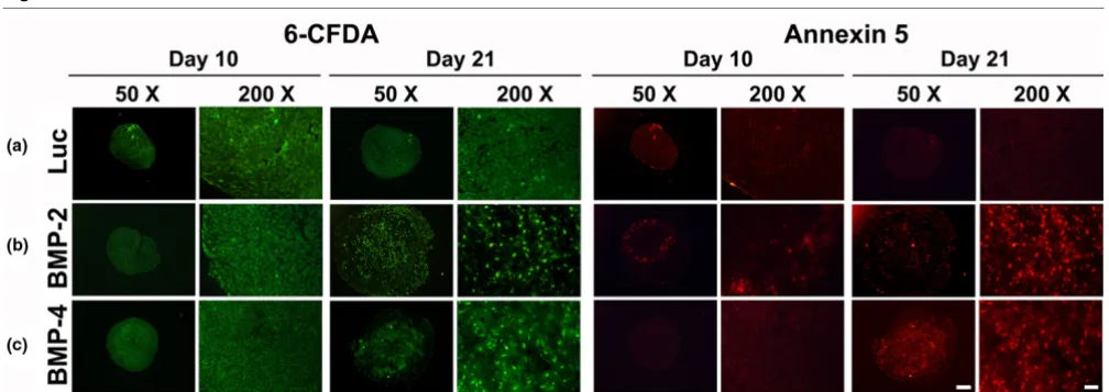
As annexin 5 (Ann5) is expressed by hypertrophic chondro-cytes and in osteoarthritic cartilage [17], we were next inter-ested in the appearance of live and apoptotic cells within our aggregate system after 10 and 21 days, which was visualized using the Ann5-Cy3 apoptosis detection kit (APOAC; Sigma, St. Louis, MO, USA) as directed by the supplier. The assay uses the Cy3.18 dye as red fluorochome conjugated with Ann5-Cy3 for apoptosis detection through binding to phos-phatidylserine epitopes on the plasma membrane of early
apoptotic cells, and the hydrolysis of the non-fluorescent 6-carboxyfluorescein diacetate (6-CFDA) to the green fluores-cent compound 6-carboxyfluorescein by the esterases of living cells to label viable cells. This combination allows the differen-tiation among early apoptotic cells (Ann5 positive, 6-CFDA positive), necrotic cells (Ann5 positive, 6-CFDA negative), and viable cells (Ann5 negative, 6-CFDA positive). Aggregates were incubated with 50 μL of the double labelling staining solution for 10 minutes at room temperature. After staining, aggregates were washed five times with 100 μL of binding buffer, fixed overnight in PBS-buffered 4% paraformaldehyde, dehydrated, infiltrated with isoamylacetate (Merck, Hohenb-runn, Germany), embedded in paraffin, and sectioned to 4 μm. Viable and non-viable cells were observed on the respective mid-sections using a fluorescence microscope and the appro-priate green and red filters.
Total RNA extraction, semi-quantitative and real-time RT-PCR
RNA was extracted from MSC aggregates at the indicated time-points. For this, 6 to 10 pellets per group and time point for each donor were pooled and homogenized using a pellet pestle and repeated titration in 1 mL of Trizol reagent (Invitro-gen, Karlsruhe, Germany). Total RNA was subsequently extracted using Trizol reagent with an additional purification step using separation columns (NucleoSpin RNA II kit; Mach-erey-Nagel GmbH, Düren, Germany) including a DNase treat-ment step according to the manufacturer's instructions. RNA from aggregates of each condition (2 μg each group) was used for random hexamer primed cDNA synthesis using Bio-Script reverse transcriptase (Bioline GmbH, Luckenwalde, Germany).
For semi-quantitative PCR analyses equal amounts (100 ng) of each cDNA were used as templates for amplification in a 30
expres-sion of the EF1α housekeeping gene, was calculated from three bands (one per patient).
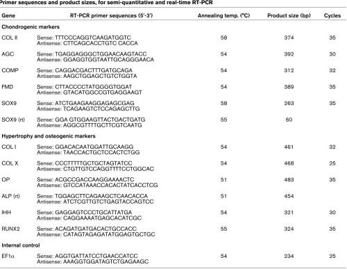
For a more detailed mRNA expression profile of chondrogenic and hypertrophy associated genes, genetically-modified MSC aggregates were subjected to real-time quantitative PCR anal-yses. One microliter of each cDNA was used as template for amplification in a 50 μL reaction volume using BioTaq DNA Polymerase Taq (Bioline GmbH, Luckenwalde, Germany) and 50 pmol of gene-specific primers was used for COL II, SOX9, ALP and COL X as listed in Table 1. Real-time PCR conditions were as follows: 30 seconds at 94°C, 30 seconds at anneal-ing temperature, 60 seconds at 72°C (see Table 1 for PCR conditions). Real-time PCR was performed with the DNA Engine Opticon system (MJ Research, Waltham, MA, USA) using SYBR Green (Biozym Scientific GmbH, Hessisch Old-endorf, Germany) as fluorescent dye allowing determination of
the threshold cycle at which exponential amplification of PCR products begins. Specificities of amplicons were confirmed by melting curve analyses by gel electrophoresis of test PCR reactions. For quantification mRNA expression was normal-ized to the expression levels of the housekeeping gene EF1α and relative expression levels compared with values from undifferentiated monolayer MSCs are shown using the relative expression software tool (REST) [28]. Each PCR was per-formed in triplicate on three separate bone marrow prepara-tions for each independent experiment.
Statistical analysis
The data from the ELISA, WST1, ATP, GAG, DNA, and ALP content, cell surface area and RT-PCR analyses were expressed as mean values ± standard deviation (SD). Each experiment was performed in quadruplicate (n = 4) and repeated on at least three and up to six individual marrow prep-Table 1
Primer sequences and product sizes, for semi-quantitative and real-time RT-PCR
Gene RT-PCR primer sequences (5'-3') Annealing temp. (°C) Product size (bp) Cycles
Chondrogenic markers
COL II Sense: TTTCCCAGGTCAAGATGGTC Antisense: CTTCAGCACCTGTC CACCA
58 374 35
AGC Sense: TGAGGAGGGCTGGAACAAGTACC
Antisense: GGAGGTGGTAATTGCAGGGAACA
54 392 30
COMP Sense: CAGGACGACTTTGATGCAGA
Antisense: AAGCTGGAGCTGTCTGGTA
54 312 32
FMD Sense: CTTACCCCTATGGGGTGGAT
Antisense: GTACATGGCCGTGAGGAAGT
54 389 35
SOX9 Sense: ATCTGAAGAAGGAGAGCGAG
Antisense: TCAGAAGTCTCCAGAGCTTG
58 263 35
SOX9 (rt) Sense: GGA GTGGAAGTTACTGACTGATG Antisense: AGGCGTTTTGCTTCGTCAATG
55 60
Hypertrophy and osteogenic markers
COL I Sense: GGACACAATGGATTGCAAGG
Antisense: TAACCACTGCTCCACTCTGG
54 461 32
COL X Sense: CCCTTTTTGCTGCTAGTATCC
Antisense: CTGTTGTCCAGGTTTTCCTGGCAC
54 468 25
OP Sense: ACGCCGACCAAGGAAAACTC
Antisense: GTCCATAAACCACACTATCACCTCG
51 483 35
ALP (rt) Sense: TGGAGCTTCAGAAGCTCAACACCA Antisense: ATCTCGTTGTCTGAGTACCAGTCC
51 454
IHH Sense: GAGGAGTCCCTGCATTATGA
Antisense: CAGGAAAATGAGCACATCGC
54 321 30
RUNX2 Sense: ACAGATGATGACACTGCCACC
Antisense: CATAGTAGAGATATGGAGTGCTGC
55 324 35
Internal control
EF1α Sense: AGGTGATTATCCTGAACCATCC
Antisense: AAAGGTGGATAGTCTGAGAAGC
54 234 25
rt: primer pairs, that have been used for real-time PCR only.
[image:5.612.58.558.104.491.2]arations from different patients (m = 3 to 6), as indicated in the respective experiments. All numerical data were subjected to variance analysis (one or two factor analysis of variance) and statistical significance was determined by student's t-test, and level of P < 0.05 was considered significant.
Results
Transgene expression by aggregates of genetically modified MSCs
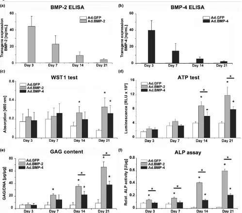
[image:6.612.58.557.186.625.2]Consistent with previous findings [21], cultures infected with these doses of Ad.BMP-2 and Ad.BMP-4 generated approxi-mately 30 to 60 ng/mL of gene product per 24 hours at day 3 post-infection (Figures 1a, b). The amount of each transgene
Figure 1
Transgene expression and biochemical composition of MSCs during 21 days of aggregate culture following BMP-2 and BMP-4 gene transfer
Transgene expression and biochemical composition of MSCs during 21 days of aggregate culture following BMP-2 and BMP-4 gene transfer. Pri-mary MSCs were infected with Ad.BMP-2, Ad.BMP-4 or Ad.GFP at 5 × 10² vp/cell, seeded into aggregates and analyzed biochemically during a
product steadily decreased thereafter, and reached levels of about 3 to 6 ng/mL at day 21 (Figures 1a, b). Levels of BMP-2 and BMP-4 in media conditioned by Ad.GFP or Ad.Luc infected cultures were below 200 pg/mL (Figures 1a, b), equivalent to the levels observed in the naïve controls (data not shown).
Cell proliferation, GAG content and ALP activity
As primary MSCs were shown to be capable of expressing the BMP-2 or the BMP-4 transgene in aggregate culture, we examined the effects of BMP-2 and BMP-4 gene delivery on cell proliferation using the WST1 cell proliferation assay. At day 3 and 7 of culture the cell proliferation rate in MSC aggre-gates was approximately equal in all groups tested (Figure 1c). BMP-2 and BMP-4 transduced MSC aggregates maintained their proliferation rate over 21 days while Ad.GFP cells (Figure 1c) and unmodified control cultures (not shown) decreased rate of proliferation (Figure 1c). The same pattern was observed using the ATP test, where sustained high cell prolif-eration rates were observed at day 14 and 21 in BMP-2- and BMP-4-modified aggregates compared with the controls, while at the same time points, levels in the BMP-2-modified aggregates were significantly elevated compared with the BMP-4 cultures (Figure 1d). To quantitatively compare extra-cellular matrix synthesis among treatment groups, GAG levels
in the aggregates after 21 days in culture were determined (Figure 1e). All aggregates infected with Ad.BMP-2 or Ad.BMP-4 showed significantly increased GAG production relative to those receiving Ad.GFP (Figure 1e), Ad.Luc or untransduced aggregates (not shown), which showed no evi-dence of chondrogenesis. At days 14 and 21, significantly ele-vated levels of GAG synthesis in the BMP-2 compared with the BMP-4 transduced cultures became apparent (Figure 1e). Indicative of hypertrophic chondrocytes we analyzed ALP activity, which was found to be significantly elevated at all time points in the BMP-2-modified aggregates compared with the GFP controls and BMP-4 transduced cultures, whereas signif-icantly higher values in the BMP-4 modified cultures compared with the GFP controls could only be resolved at day 14 and 21 (Figure 1f).
Histological and immunohistochemical analyses of chondrogenesis
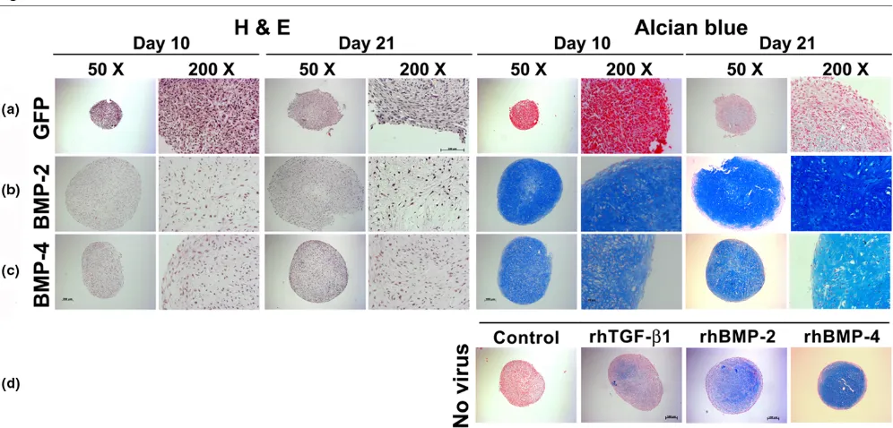
Transduction of MSCs with adenoviral vectors encoding BMP-2 (Figure 2b) or BMP-4 (Figure 2c) using viral doses suf-ficient to generate 30 to 60 ngs transgene product at day 3 induced a significant chondrogenic response in the respective aggregate cultures compared with the controls (Figure 2a), which were not chondrogenic. This was demonstrated by increased aggregate size and strong production of
proteogly-Figure 2
Histological appearance of MSC pellets after chondrogenic induction with BMP-2 or BMP-4 gene transfer
Histological appearance of MSC pellets after chondrogenic induction with BMP-2 or BMP-4 gene transfer. Monolayer cultures of MSCs were infected with (a) Ad.GFP, (b) Ad.BMP-2 or (c) Ad.BMP-4 at 5 × 10² vp/cell as indicated, seeded into aggregates 24 hours after infection and
[image:7.612.58.556.398.637.2]cans as indicated by metachromatic staining with alcian blue in the Ad.BMP-2 or Ad.BMP-4 transduced cultures (Figures 2b, c) compared with the Ad.GFP controls (Figure 2a). Inter-estingly, the phenotype of the Ad.BMP-4 (Figure 2c) infected aggregates appeared chondrogenic but less hypertrophic at day 10 and 21 compared with the Ad.BMP-2 cultures in that the BMP-2-modified cells were more rounded with greater cytoplasmic volume (Figure 2b).
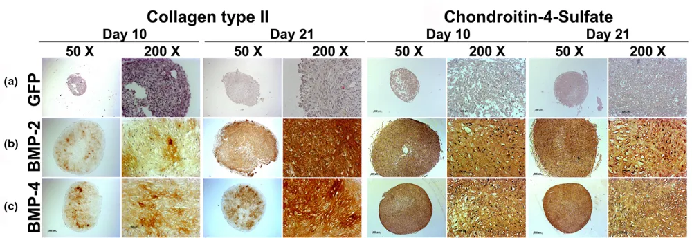
Correspondingly, immunohistochemical analyses for COL II, the predominant collagen type in cartilage, and CS4, one of the monomers of the polysaccharide portion of proteoglycan, showed significantly enhanced production of these cartilage matrix proteins at days 10 and 21 of culture in the aggregates receiving Ad.BMP-2 (Figure 3b) or Ad.BMP-4 (Figure 3c) rel-ative to the Ad.GFP (Figure 3a) controls.
Uninfected aggregates maintained in the presence of recom-binant BMP-2, BMP-4, or TGF-β1 protein were also chondro-genic as evidenced by lacunae formation, positive staining for alcian blue (Figure 2d), COL II and CS4 (not shown), although the stage of chondrogenesis seemed less progressed com-pared with that in the aggregates genetically modified with BMP-2 or BMP-4 (Figures 2b, c) after 21 days, while control cultures where growth factor supplementation was absent were non-chondrogenic.
Hypertrophic differentiation and apoptosis
We used staining for ALP and immunohistochemistry for COL X as markers for chondrocyte hypertrophy (Figure 4). No detectable ALP and only weak COL X immunostaining was seen in the control aggregates transduced with Ad.GFP
(Fig-ure 4a). ALP staining was primarily pericellular in the aggre-gates infected with Ad.BMP-4 (Figure 4c). In contrast, aggregates transduced with Ad.BMP-2 showed more abun-dant staining for ALP throughout the extracellular matrix at day 10 and was most extensive at day 21 of culture (Figure 4b). Correspondingly, immunohistochemical analyses of the Ad.BMP-2 infected aggregates revealed strong abundant staining for COL X in the aggregate matrix at day 10 and 21 of culture (Figure 4b). In the Ad.BMP-4-modified cultures COL X immunostaining of the matrix was strongly observed at day 21 in the aggregate matrix, while staining tended to be pericellular at day 10 of culture (Figure 4c); no significant differences were noted among the aggregates. Notably, the distribution pattern of the hypertrophy markers was somewhat heteroge-neous in the aggregates, which we attribute to the rather inho-mogeneous aggregate morphologies obtained during culture in v-bottom plates as opposed to more homogeneous aggre-gate morphologies seen after centrifugation and culture in 15 mL conical tubes [20].
Double fluorescence staining with Ann5-Cy3/6-CFDA allowed visualisation of Ann5 expressions. The high levels of green fluorescence found in BMP-modified (Figures 5b, c) and control groups (Figure 5a) revealed high viability of adenoviral infected MSCs in aggregate cultures after 10 and 21 days. At day 10, only very few cells in the Luc (Figure 5a) and BMP-2 (Figure 5b) and BMP-4 (Figure 5c) modified aggregates appeared to be annexin 5 positive. At day 21, the BMP-2 (Fig. 5B) and the BMP-4 (Fig. 5C) modified groups showed many Ann5-positive cells, as evidenced by red fluorescence, com-pared with the Ad.Luc transduced (Figure 5a) and
untrans-Figure 3
Immunohistochemical analyses for cartilage matrix proteins of MSC pellets after chondrogenic induction with BMP-2 or BMP-4 gene transfer
Immunohistochemical analyses for cartilage matrix proteins of MSC pellets after chondrogenic induction with BMP-2 or BMP-4 gene transfer. Mon-olayer cultures of MSCs were infected with (a) Ad.GFP, (b) Ad.BMP-2 or (c) Ad.BMP-4 at 5 × 10² vp/cell as indicated and placed into aggregate
[image:8.612.56.556.483.655.2]duced (not shown) cultures where only very few such cells were seen.
A similar pattern of hypertrophy and apoptosis was observed in the untransduced control aggregates that were maintained in the presence or absence of recombinant BMP-2, BMP-4 or TGF-β1 protein (not shown).
Comparison of BMP-2 and BMP-4 modified MSC aggregates with immature growth plate chondrocytes
[image:9.612.56.560.94.270.2]In the different types of aggregates examined in Figures 2 to 5, different cell morphologies were apparent, especially with respect to incidence and extent of lacunae formation. Thus we were next interested to know if it was possible to distinguish the types of aggregates produced by measuring the sizes of the respective lacunae, approximated by simple histomorpho-metric cell surface area measurement on aggregate sections. For comparison, we first analyzed the sizes of the lacunae Figure 4
Histological and immunohistochemical analyses for hypertrophy of MSC pellets after chondrogenic induction with BMP-2 or BMP-4 gene transfer
Histological and immunohistochemical analyses for hypertrophy of MSC pellets after chondrogenic induction with BMP-2 or BMP-4 gene transfer. Following genetic modification with (a) Ad.GFP, (b) Ad.BMP-2 or (c) Ad.BMP-4 aggregates after 10 and 21 days of culture stained for ALP (left panels) and collagen type X (right panels) are shown. Regions of positive immunostaining appear brown. Panels are reproduced at low (50×; bar = 200 μm) or high (200×; bar = 50 or 100 μm) magnification as indicated. Ad = adenoviral vector; ALP = alkaline phosphatase; BMP = bone morpho-genetic protein; GFP = green fluorescent protein; MSC = mesenchymal stem cell.
Figure 5
Analyses for cell viability and apoptosis within MSC pellets after chondrogenic induction with BMP-2 or BMP-4 gene transfer
[image:9.612.55.559.479.657.2]within different zones of growth plate cartilage obtained from a four-year-old child, from whom a sixth toe was removed. These measurements were compared with those of the lacu-nae found in the center and periphery of the different treatment groups of genetically modified aggregates.
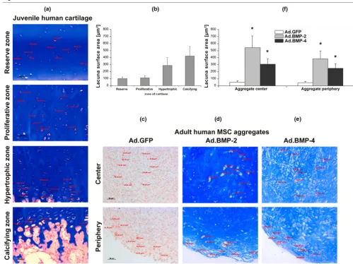
As shown in Figure 6a, the reserve, proliferative, hypertrophic and calcifying zone of cartilage could be clearly separated by the proximity of the cells to the joint space and the bone
respectively, alignment of the chondrocytes along the arcades of Benninghoff [29] and by the appearance of hypertrophic cells. Analyses of lacunae surface areas in the different growth plate zones revealed mean lacunae surface areas ± SD of 100.8 ± 25.8 μm2 in the reserve zone, 113.3 ± 25.5 μm2 in the
proliferative zone, 288.5 ± 111.0 μm2 in the hypertrophic zone
and 421.8 ± 131.9 μm2 in the calcifying zone of growth plate
[image:10.612.53.552.92.466.2]cartilage (Figure 6b). The mean values ± SD represent meas-urements of 10 lacunae per zone, which were performed on Figure 6
Analysis of hypertrophic cell morphology in MSC pellets after chondrogenic induction with BMP-2 or BMP-4 gene transfer in comparison to growth plate chondrocytes
three representative mid-sections per growth plate. A total of four physes (1 digit, 2 joints) was examined.
In contrast the GFP-modified aggregates showed no lacuna formation, either in the center or in the periphery of the pellets (Figure 6c). However the BMP-2- and BMP-4-modified aggre-gates displayed a chondrogenic phenotype with lacunae for-mation throughout the aggregates (Figures 6d, e). Analyses of cell surface areas in the different aggregate types revealed a mean value of 60.6 ± 14.5 μm2 in the center and 57.3 ± 12.4
μm2 the periphery of the Ad.GFP transduced aggregates,
which showed no lacunae formation, of 541.3 ± 166.3 μm2 in
the center and 386.1 ± 108.7 μm2 the periphery of the
Ad.BMP-2 transduced aggregates, and of 307.8 ± 75.6 μm2
in the center and 248.7 ± 65.4 μm2 the periphery of the
Ad.BMP-4 transduced aggregates (Figure 6f). Thus lacunae formed in both the BMP-2 and BMP-4 transduced pellets and led to significantly larger cell surface areas compared with the non-chondrogenic controls. Nevertheless, the lacunae formed in the presence of BMP-2 were larger than those formed by BMP-4 and approximated the size of lacunae noted in the cal-cifying zone of the human growth plate. In contrast, the lacu-nae that formed in the presence of BMP-4 were closer in size to those of the hypertrophic zone (Figures 6e, f).
Time course of chondrocytic and hypertrophic marker gene expression
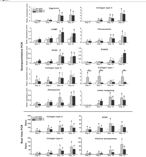
To examine further the effects of BMP-2 and BMP-4 gene delivery on hypertrophic differentiation, we analyzed the tem-poral expression profiles of genes associated with chondro-cyte maturation and osteogenic differentiation using semi-quantitative and real-time RT-PCR (Figure 7). These genes included AGC, COL II, COMP, FMD, SOX9, RUNX2, COL X, COLI, ALP, OP and IHH. Consistent with preceding analyses [30], the aggregate cultures transduced with BMP-2 showed evidence of chondrogenic differentiation at the RNA level with upregulation of the chondrogenic markers AGC, COL II, FMD, COMP and SOX-9 over time, compared with the non-chon-drogenic Ad.GFP controls where these markers were expressed only at low levels (Figure 7). Expression of these genes was upregulated to a similar degree in the BMP-4- and BMP-2-modified aggregates and marked differences between the BMP-2 and BMP-4 groups were not observed (Figure 7).
Evidence of chondrocyte hypertrophy at the mRNA level in the BMP-2- and BMP-4-modified aggregates was reflected by a subsequent upregulation of COL X and OP at day 3, IHH and ALP at day 7 and RUNX2 at day 14 compared with Ad.GFP controls (Figure 7). These results suggest that BMP-2 and BMP-4 gene transfer induced a significant chondrogenic and hypertrophic response in MSC aggregates on mRNA level over time.
Discussion
We and others have shown previously that primary MSCs undergo chondrogenesis following genetic modification with Ad.BMP-2 or Ad.TGF-β1 in aggregate culture in vitro [30-32] or when transplanted into chondral defects in vivo [14]. In the present study we adapted the MSC aggregate culture system to determine whether adenoviral delivery of BMP-4 can lead to chondrogenesis of primary MSCs in vitro, and to evaluate the extent of hypertrophy compared with BMP-2-modified cul-tures.
Adenoviral delivery of BMP-4 led to reliable chondrogenesis in human MSC aggregate cultures in a fashion comparable with that noted when the same dose of the BMP-2 transgene was administered as shown by staining with alcian blue, COL II and CS4 and the quantitative GAG assay, indicating increased GAG levels at days 14 and 21 in the BMP-2-modified aggre-gates. Notably, chondrogenic differentiation induced by either transgene increase levels of metabolic activity and cell prolif-eration compared with controls as evidenced by the WST1 and ATP assays. Moreover, high levels of chondrocyte hyper-trophy occurred in MSC pellet cultures modified with either BMP transgene, as assessed by lacunar size, and expression of ALP, COL X and Ann5, and was overall slightly more advanced in the BMP-2-modified cultures compared with the BMP-4 modified cultures reaching significance levels in the ALP assay at all time points. Notably, exact the lacunar size comparisons between growth plate tissues and in vitro cell pellets might be inaccurate (Figure 6) due to artifacts that may appear during fixation and processing of these different types of tissues.
The RT-PCR data are in general agreement with the biochem-ical and histologbiochem-ical observations, showing high levels of chondrogenic mRNAs in aggregates after BMP-stimulation, such as AGC, COMP, COL II, SOX9 and FMD. Likewise, tran-scripts encoding the hypertrophy associated genes COL X, OP, ALP, RUNX2 and IHH were also strongly present in both types of BMP-modified aggregates compared with controls.
Figure 7
Profiles of temporal gene expression determined by semi-quantitative and real-time RT-PCR in MSC pellet cultures genetically modified with BMP-2 and BMP-4
Our study is also in agreement with studies of in vitro chondro-genesis with primary MSCs using recombinant proteins, where BMP-4 was identified as a strong inducer of chondro-genesis [36], which produced less hypertrophy compared with BMP-2 [37]. Correspondingly, in vivo implantation of BMP-4 into abdominal muscles of rats led to ectopic cartilage and bone formation when delivered as recombinant protein [38] or via genetically modified cells [39]. Notably, the latter study revealed differential effects on chondrogenesis and osteogenesis depending on the type of cell analyzed [39]. Our study is limited to the use of bone marrow-derived MSCs and other effects may be seen when different cells are employed. Orthotopic BMP-4 gene delivery via retrovirus transduction of muscle-derived stem cells was shown to improve cartilage repair in rat osteochondral defects [40] and also when it was administered via adenovirus to dedifferentiated chondrocytes in osteochondral defects in rabbits [41]. In both studies improved repair in the BMP-4-treated defects compared with non-chondrogenic controls at 12 or 24 weeks respectively was observed, but detailed analyses of hypertrophy and apop-tosis have not been performed [40,41].
BMP-2 and BMP-4 have been implicated in embryogenesis and morphogenesis of various tissues and organs, where they regulate growth, differentiation, chemotaxis and apoptosis of a variety of cell types, including mesenchymal, epithelial, hemat-opoietic and neuronal cells [42]. Interestingly, in conditional knock-out experiments it has been found that a threshold level of BMP signaling is required for the onset of chondrogenesis, and hence some chondrogenic condensations failed to form in limbs deficient in both BMP-2 and BMP-4 [43]. However, in the condensations that do form, subsequent chondrogenic dif-ferentiation proceeds normally even in the absence of BMP-2 and BMP-4 [43]. In contrast, it was found that the loss of both BMP-2 and BMP-4 results in a severe impairment of osteogen-esis. Deletion of BMP-4 alone did not impair osteogenesis or fracture repair, while deletion of BMP-2 alone did not impair osteogenesis but strongly prevented fracture repair [43-45]. This indicates that the presence of BMP-2 or BMP-4 is a pre-requisite for osteoblastogenesis and these morphogens can apparently compensate for each other to a certain extent. However, they are less important for chondrogenesis [43-45].
During limb development, cartilage is gradually replaced by endochondral ossification, a process in which the chondro-cytes mature, hypertrophy and express COL X with reduced production of COL II. Subsequently the cartilage becomes vascularized and infiltrated by osteoprogenitor cells, while the chondrocytes undergo apoptosis. The osteoprogenitor cells differentiate into osteoblasts, replacing the cartilage with min-eralized bone; BMP-2 and BMP-4 are important regulators of these processes [46-48]. By using chondroprogenitor cells in high density, three-dimensional cultures these regulatory mechanisms can be partially recapitulated. Thus it is not sur-prising that studies on in vitro chondrogenesis using MSCs or
chondrocytes incubated with members of the TGF-β super-family reveal considerable hypertrophy and high levels of COL X expression. Although the use of COL X as a marker of chon-drogenic hypertrophy in MSC-based systems has been ques-tioned [13], it correlates well thus far to the existing in vivo
data. For example, MSCs genetically modified to express BMP-2 display a significant level of tissue hypertrophy and osteophyte formation, when transplanted orthotopically to osteochondral defects [14] or ectopically [15,49] in small ani-mal models. TGF-β1 has been shown to induce hypertrophic and osteometaplastic changes in the synovium of rabbit joints, when directly delivered by first-generation adenovirus [50]. Furthermore, implantation of chondrocytes genetically modi-fied to express BMP-7 has been shown to generate good hya-line cartilage repair tissue after six weeks in vivo, but after one year the repair cartilage is no better than that of controls, with only 0 to 28% of the transplanted cells being detectable at that time point [51]. This is agreement with a recent large ani-mal study in pigs, that showed good hyaline cartilage repair after six weeks, when chondral defects were filled with perios-teum cells genetically modified with BMP-2, while at six months the hyaline repair tissue had almost completely van-ished and was replaced by fibrocartilage [52]. These observa-tions might be attributed to mechanisms of hypertrophic differentiation and subsequent apoptosis, although clarifying analyses in vivo have not been conducted thus far. However, the presence of Ann5-positive cells in our hypertrophic aggre-gates modified with BMP-2 or BMP-4 in vitro correspond with these data.
Our data suggest that the degree of hypertrophic differentia-tion can be modulated by the choice of morphogenetic stimu-lus, while still maintaining efficient chondrogenesis. This permits cautious optimism that it may prove possible ultimately to achieve effective regeneration of articular cartilage in the absence of hypertrophic differentiation. Hypertrophic differen-tiation of neo-cartilage tissue with subsequent apoptosis development is certainly an undesired effect in cartilage defects in vivo, because this would lead to loss of the trans-planted repair cells with subsequent matrix degradation. How-ever, the relevance of chondrogenic hypertrophy and apoptosis of human MSCs induced by TGF-β superfamily members for cartilage repair in vivo has to be considered still unclear to this end, because this study is limited by its in vitro
nature. Therefore, clarifying in vivo experiments are necessary before such factors can be recommended for further clinical use.
Conclusions
preventing hypertrophic differentiation using different genes or proteins, which would presumably improve the outcome of cell-based approaches to cartilage repair in vivo.
Competing interests
The authors declare that they have no competing interests.
Authors' contributions
All authors have read and approved the manuscript and con-tributed to the study design, data analysis, interpretation of data and drafting and revision of the manuscript. The data have been generated by AFS, BP, MK, SCG, and a data review committee (AFS, CH, SCG, AR, UN, JE and CHE) analysed the data.
Acknowledgements
We are grateful to Nadja Karl, Viola Monz and Christa Amrehn for their excellent technical assistance. This work was supported in parts by grants AR48566 and AR50249 from to National Institute of Arthritis and Musculoskeletal and Skin Diseases to SCG and CHE, by grant STE 1051/2-1 from the Deutsche Forschungsgemeinschaft (DFG) to AFS and UN, and by grant D-23 to AFS and AR from the Interdisciplinary Center for Clinical Research (IZKF) Würzburg.
References
1. Kolf CM, Cho E, Tuan RS: Mesenchymal stromal cells. Biology of adult mesenchymal stem cells: regulation of niche, self-renewal and differentiation. Arthritis Res Ther 2007, 9:204. 2. Nesic D, Whiteside R, Brittberg M, Wendt D, Martin I, Mainil-Varlet
P: Cartilage tissue engineering for degenerative joint disease. Adv Drug Deliv Rev 2006, 58:300-322.
3. Noth U, Steinert AF, Tuan RS: Technology insight: adult mesen-chymal stem cells for osteoarthritis therapy. Nat Clin Pract Rheumatol 2008, 4:371-380.
4. Estes BT, Wu AW, Guilak F: Potent induction of chondrocytic differentiation of human adipose-derived adult stem cells by bone morphogenetic protein 6. Arthritis Rheum 2006, 54:1222-1232.
5. Wakitani S, Mitsuoka T, Nakamura N, Toritsuka Y, Nakamura Y, Horibe S: Autologous bone marrow stromal cell transplanta-tion for repair of full-thickness articular cartilage defects in human patellae: two case reports. Cell Transplant 2004, 13:595-600.
6. Kuroda R, Ishida K, Matsumoto T, Akisue T, Fujioka H, Mizuno K, Ohgushi H, Wakitani S, Kurosaka M: Treatment of a full-thick-ness articular cartilage defect in the femoral condyle of an ath-lete with autologous bone-marrow stromal cells. Osteoarthritis Cartilage 2007, 15:226-231.
7. Steinert AF, Ghivizzani SC, Rethwilm A, Tuan RS, Evans CH, Noth U: Major biological obstacles for persistent cell-based regen-eration of articular cartilage. Arthritis Res Ther 2007, 9:213. 8. Evans CH, Robbins PD, Ghivizzani SC, Wasko MC, Tomaino MM,
Kang R, Muzzonigro TA, Vogt M, Elder EM, Whiteside TL, Watkins SC, Herndon JH: Gene transfer to human joints: progress toward a gene therapy of arthritis. Proc Natl Acad Sci USA 2005, 102:8698-8703.
9. Steinert AF, Nöth U, Tuan RS: Concepts in gene therapy for car-tilage repair. Injury 2008, 39 Suppl 1:S97-S113.
10. Trippel S, Cucchiarini M, Madry H, Shi S, Wang C: Gene therapy for articular cartilage repair. Proc Inst Mech Eng [H] 2007, 221:451-459.
11. Cucchiarini M, Madry H: Gene therapy for cartilage defects. J Gene Med 2005, 7:1495-1509.
12. Pagnotto MR, Wang Z, Karpie JC, Ferretti M, Xiao X, Chu CR: Adeno-associated viral gene transfer of transforming growth factor-beta1 to human mesenchymal stem cells improves car-tilage repair. Gene Ther 2007, 14:804-813.
13. Mwale F, Stachura D, Roughley P, Antoniou J: Limitations of using aggrecan and type X collagen as markers of chondro-genesis in mesenchymal stem cell differentiation. J Orthop Res 2006, 24:1791-1798.
14. Gelse K, Mark K von der, Aigner T, Park J, Schneider H: Articular cartilage repair by gene therapy using growth factor-produc-ing mesenchymal cells. Arthritis Rheum 2003, 48:430-441. 15. Pelttari K, Winter A, Steck E, Goetzke K, Hennig T, Ochs BG,
Aigner T, Richter W: Premature induction of hypertrophy during in vitro chondrogenesis of human mesenchymal stem cells correlates with calcification and vascular invasion after ectopic transplantation in SCID mice. Arthritis Rheum 2006, 54:3254-3266.
16. De Bari C, Dell'Accio F, Luyten FP: Failure of in vitro-differenti-ated mesenchymal stem cells from the synovial membrane to form ectopic stable cartilage in vivo. Arthritis Rheum 2004, 50:142-150.
17. Kirsch T, Swoboda B, Nah H: Activation of annexin II and V expression, terminal differentiation, mineralization and apop-tosis in human osteoarthritic cartilage. Osteoarthritis Cartilage 2000, 8:294-302.
18. Pfander D, Swoboda B, Kirsch T: Expression of early and late differentiation markers (proliferating cell nuclear antigen, syn-decan-3, annexin VI, and alkaline phosphatase) by human osteoarthritic chondrocytes. Am J Pathol 2001, 159:1777-1783.
19. Palmer GD, Gouze E, Gouze JN, Betz OB, Evans CH, Ghivizzani SC: Gene transfer to articular chondrocytes with recombinant adenovirus. Methods Mol Biol 2003, 215:235-246.
20. Steinert AF, Palmer GD, Pilapil C, Ulrich N, Evans CH, Ghivizzani SC: Enhanced in vitro chondrogenesis of primary mesenchy-mal stem cells by combined gene transfer. Tissue Eng Part A 2009, 15:1127-1139.
21. Hardy S, Kitamura M, Harris-Stansil T, Dai Y, Phipps ML: Con-struction of adenovirus vectors through Cre-lox recombina-tion. J Virol 1997, 71:1842-1849.
22. Sambrook J, Russell DW: Molecular cloning: a laboratory man-ual. Volume 1. 3rd edition. New York: CSHL Press; 2001. 23. Noth U, Tuli R, Osyczka AM, Danielson KG, Tuan RS: In vitro
engi-neered cartilage constructs produced by press-coating biode-gradable polymer with human mesenchymal stem cells. Tissue Eng 2002, 8:131-144.
24. Johnstone B, Hering TM, Caplan AI, Goldberg VM, Yoo JU: In vitro chondrogenesis of bone marrow-derived mesenchymal pro-genitor cells. Exp Cell Res 1998, 238:265-272.
25. Penick KJ, Solchaga LA, Welter JF: High-throughput aggregate culture system to assess the chondrogenic potential of mes-enchymal stem cells. Biotechniques 2005, 39:687-691. 26. Weber M, Steinert A, Jork A, Dimmler A, Thurmer F, Schutze N,
Hendrich C, Zimmerman U: Formation of cartilage matrix pro-teins by BMP-transfected murine mesenchymal stem cells encapsulated in a novel class of alginates. Biomaterials 2002, 23:2003-2013.
27. Hunziker EB: Cartilage histomorphometry. Methods Mol Med 2007, 135:147-166.
28. Pfaffl MW, Horgan GW, Dempfle L: Relative expression soft-ware tool (REST) for group-wise comparison and statistical analysis of relative expression results in real-time PCR. Nucleic Acids Res 2002, 30:e36.
29. Benninghoff A: Form und bau der Geleknorpel in ihren Bezei-hungen zur Funktion. Z Zellforsch Mikrosk Anat Rec 1925:783-825.
30. Palmer GD, Steinert A, Pascher A, Gouze E, Gouze JN, Betz O, Johnstone B, Evans CH, Ghivizzani SC: Gene-induced chondro-genesis of primary mesenchymal stem cells in vitro. Mol Ther-apy 2005, 12:219-228.
31. Park J, Gelse K, Frank S, Mark K von der, Aigner T, Schneider H: Transgene-activated mesenchymal cells for articular cartilage repair: a comparison of primary bone marrow-, perichon-drium/periosteum- and fat-derived cells. J Gene Med 2006, 8:112-125.
33. Steinert A, Weber M, Dimmler A, Julius C, Schutze N, Noth U, Cramer H, Eulert J, Zimmermann U, Hendrich C: Chondrogenic differentiation of mesenchymal progenitor cells encapsulated in ultrahigh-viscosity alginate. J Orthop Res 2003, 21:1090-1097.
34. Mackay AM, Beck SC, Murphy JM, Barry FP, Chichester CO, Pit-tenger MF: Chondrogenic differentiation of cultured human mesenchymal stem cells from marrow. Tissue Eng 1998, 4:415-428.
35. Mueller MB, Tuan RS: Functional characterization of hypertro-phy in chondrogenesis of human mesenchymal stem cells. Arthritis Rheum 2008, 58:1377-1388.
36. Miljkovic ND, Cooper GM, Marra KG: Chondrogenesis, bone morphogenetic protein-4 and mesenchymal stem cells. Oste-oarthritis Cartilage 2008, 16:1121-1130.
37. Taipaleenmaki H, Suomi S, Hentunen T, Laitala-Leinonen T, Saa-manen AM: Impact of stromal cell composition on BMP-induced chondrogenic differentiation of mouse bone marrow derived mesenchymal cells. Exp Cell Res 2008, 314:2400-2410.
38. Kubler NR, Moser M, Berr K, Faller G, Kirchner T, Sebald W, Reu-ther JF: [Biological activity of E. coli expressed BMP-4]. Mund Kiefer Gesichtschir 1998, 2(Suppl 1):S149-152.
39. Li G, Peng H, Corsi K, Usas A, Olshanski A, Huard J: Differential effect of BMP4 on NIH/3T3 and C2C12 cells: implications for endochondral bone formation. J Bone Miner Res 2005, 20:1611-1623.
40. Kuroda R, Usas A, Kubo S, Corsi K, Peng H, Rose T, Cummins J, Fu FH, Huard J: Cartilage repair using bone morphogenetic protein 4 and muscle-derived stem cells. Arthritis Rheum 2006, 54:433-442.
41. Lin L, Zhou C, Wei X, Hou Y, Zhao L, Fu X, Zhang J, Yu C: Articular cartilage repair using dedifferentiated articular chondrocytes and bone morphogenetic protein 4 in a rabbit model of articu-lar cartilage defects. Arthritis Rheum 2008, 58:1067-1075. 42. Bessa PC, Casal M, Reis RL: Bone morphogenetic proteins in
tissue engineering: the road from the laboratory to the clinic, part I (basic concepts). J Tissue Eng Regen Med 2008, 2:1-13. 43. Bandyopadhyay A, Tsuji K, Cox K, Harfe BD, Rosen V, Tabin CJ: Genetic analysis of the roles of BMP2, BMP4, and BMP7 in limb patterning and skeletogenesis. PLoS Genet 2006, 2:e216.
44. Tsuji K, Cox K, Bandyopadhyay A, Harfe BD, Tabin CJ, Rosen V: BMP4 is dispensable for skeletogenesis and fracture-healing in the limb. J Bone Joint Surg Am 2008, 90(Suppl 1):14-18. 45. Tsuji K, Bandyopadhyay A, Harfe BD, Cox K, Kakar S, Gerstenfeld
L, Einhorn T, Tabin CJ, Rosen V: BMP2 activity, although dispen-sable for bone formation, is required for the initiation of frac-ture healing. Nat Genet 2006, 38:1424-1429.
46. Goldring MB, Tsuchimochi K, Ijiri K: The control of chondrogen-esis. J Cell Biochem 2006, 97:33-44.
47. Karsenty G, Wagner EF: Reaching a genetic and molecular understanding of skeletal development. Dev Cell 2002, 2:389-406.
48. Hartmann C, Tabin CJ: Dual roles of Wnt signaling during chon-drogenesis in the chicken limb. Development 2000, 127:3141-3159.
49. Steinhardt Y, Aslan H, Regev E, Zilberman Y, Kallai I, Gazit D, Gazit Z: Maxillofacial-derived stem cells regenerate critical mandib-ular bone defect. Tissue Eng Part A 2008, 14:1763-1773. 50. Mi Z, Ghivizzani SC, Lechman E, Glorioso JC, Evans CH, Robbins
PD: Adverse effects of adenovirus-mediated gene transfer of human transforming growth factor beta 1 into rabbit knees. Arthritis Res Ther 2003, 5:R132-139.
51. Hidaka C, Goodrich LR, Chen CT, Warren RF, Crystal RG, Nixon AJ: Acceleration of cartilage repair by genetically modified chondrocytes over expressing bone morphogenetic protein-7. J Orthop Res 2003, 21:573-583.