IJPSR (2019), Volume 10, Issue 8 (Research Article)
Received on 18 November 2018; received in revised form, 13 February 2019; accepted, 28 February 2019; published 01 August 201 9
COMPATIBILITY STUDIES BETWEEN PACLITAXEL AND EXCIPIENTS IN THE PREFORMULATION PHASE OF NANOCRYSTAL FORMULATION
Aravind Gannimitta 1, Prathima Srinivas 2, A. Venkateswara Reddy 3 and Sobhitarani Pedireddi * 4
Aurobindo Pharma Pvt. Ltd. 1, Hyderabad - 500043, Telangana, India.
Shantha Biotechnics Private Limited 2, Hyderabad - 500004, Telangana, India. Anwarul Uloom College of Pharmacy 3, Hyderabad - 500001, Telangana, India. Vijaya College of Pharmacy, Munaganoor 4, Hyderabad - 501511, Telangana, India.
ABSTRACT: Paclitaxel is one of the best anti-cancer drugs and recently known as the best anti-cancer products of natural origin. The purpose of the present work was to study the physicochemical properties and compatibility of paclitaxel with the selected surfactants employed in nanocrystal formulations. The melting point and loss on drying of paclitaxel were found 216 °C and 0.11% respectively. A log P value of paclitaxel in different solvent systems like Octanol-Water, Dichloromethane-Water, Hexane-Water, and Oleyl Alcohol-Water was found 2.66, 2.91, 3.02 and 3.42 respectively. Paclitaxel is insoluble in water, soluble in ethanol, dichloromethane, and DMSO (dimethyl sulphoxide). The compatibility analysis of Paclitaxel with Egg lecithin, Poly Vinyl Pyrrolidine (PVP) and Poly (L‐lactic acid‐co‐glycolic acid) (PLGA) was studied by employing Fourier transform infrared spectroscopy (FTIR), X-Ray Powder Diffraction (XRPD) and Differential Scanning Calorimetry (DSC) techniques. FTIR, XRPD and DSC methods applied to the mentioned physical mixtures did not show evidence of interactions in the solid state. Based on these results supplied by FTIR, DSC, and XRPD, all the surfactants were found to be compatible with paclitaxel so that they can be further used in the formulation of nanocrystals.
INTRODUCTION: Paclitaxel (PTX) is a natural compound originally isolated from the Pacific Yew tree by Wall and Wani from Research Triangle Institute in 1967 1, and is the first taxane used in cancer chemotherapy 2. It is widely used against solid tumors such as breast, ovarian, colon, and non-small cell lung carcinomas 3. It is a diterpenoid pseudoalkaloid Fig. 1 having molecular formula C47H51NO14, corresponding to molecular weight of 853 Da.
QUICK RESPONSE CODE
DOI:
10.13040/IJPSR.0975-8232.10(8).3808-15
The article can be accessed online on
www.ijpsr.com
DOI link: http://dx.doi.org/10.13040/IJPSR.0975-8232.10(8).3808-15
FIG. 1: CHEMICAL STRUCTURE OF PACLITAXEL
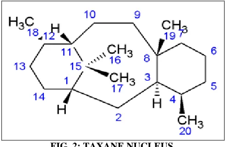
For anti-tumor activity, it is required that entire taxane molecule Fig. 2 be present since the ester and the tetraol formed by a low-temperature cleavage of paclitaxel are found to be essentially inactive 4.
Keywords:
Paclitaxel, Solubility, Compatibility study, Physicochemical
properties, Nanocrystals
Correspondence to Author: P. Sobhitarani
Associate Professor,
Vijaya College of Pharmacy, Munaganoor, Hyderabad - 501511, Telangana, India.
Paclitaxel was obtained in a pure form in 1969, and its structure was published in 1971, after many complexities due to its low concentration and structure complexities.
FIG. 2: TAXANE NUCLEUS.
The importance of paclitaxel was not recognized until the late 1970s, since, it is difficult to obtain and also due to its low solubility it has a formulation problem. It was in 1979 that Susan Horwitz discovered that paclitaxel has a unique mechanism of action and interest was further stimulated when impressive activity was demonstrated in NCI tumor screening. Unlike other microtubule agents, such as Vinca alkaloids, which induce the disassembly of microtubules, paclitaxel promotes the polymerization of tubulin 5-9. The microtubule formed in the presence of paclitaxel is extraordinarily stable and dysfunctional, thereby causing the death of the cell by disrupting the normal tubule dynamics required for cell division and vital interphase process 10.
Physicochemical properties of the drug that could influence the development of an effective dosage form should be investigated before formulation. Preformulation is the foremost stage for rational stable, safe and effective product development of an active pharmaceutical ingredient (API). It includes investigation of physicochemical characteristics of drug substance and drug-excipient compatibility 11-13. Preformulation studies of APIs and Polymers include the following course of events:
Identification:
Physical appearance. Melting point.
Fourier Transform Infrared Spectroscopy (FTIR).
Estimation Method:
UV spectroscopy.
Solid State Characterization:
X-Ray Diffraction Studies (XRD).
Differential Scanning Calorimetry (DSC). Solubility studies.
Partition coefficient. % Loss on drying
Stability Studies:
Drug-excipient compatibility studies. Solution stability.
MATERIALS AND METHODS:
Materials: Paclitaxel was a gift sample from Laurus labs, Hyderabad. Egg lecithin was purchased from Doosan Corporation, Korea. PVP (Plasdone C-12) was purchased from BASF, Germany. All other chemicals used in the study were of analytical grade.
Methods:
Identification of Drug and Excipients:
Melting Point Using Capillary Method: The Melting point is the temperature at which substance initiates to coalesce and gets completely melted. The melting point of Paclitaxel, PLGA, PVP and Egg lecithin was determined by using Thiel's tube method. The sufficient quantity of completely dried sample was introduced into a capillary glass tube to form a compact column of 2 to 4 mm height. Heat the melting-point apparatus to a temperature 5-10 °C below the expected temperature of melting. The sample tube was placed into a slot located on melting point apparatus (Perfit, India) for determination of melting point 14.
The confirmation of the identity of the compound was ascertained by comparison of the FTIR spectra of pharmacopoeial standard spectra.
Estimation Method: UV-Visible Spectroscopy:
Drug Scan for λmax and Standard Graph in IPA: Preparation of Stock Solution for IPA: Accurately weighed Paclitaxel (100 mg) was taken in 100 mL of volumetric flask and Isopropyl alcohol, was added up to 100 mL to get 1000 μg/ml (Stock solution –I). From the prepared stock solution, 10 mL taken and diluted to 100 mL with IPA to get 100 μg/mL (Stock solution-II). From the stock solution 4, 8, 16 µg/mL solution was prepared in IPA and scanned between 200-400 nm. Paclitaxel exhibits absorption peaks, at 227 nm & the other at 274 nm. The maximum absorbance is observed at 227 nm. The absorption maxima of 227 nm were selected and used for further studies.
Standard Calibration Curve of Paclitaxel: From the stock solution, serial dilutions were done to obtain solutions in the concentration ranging from 5-45 μg/ml. The absorbance of the solutions was measured against isopropyl alcohol (IPA) as blank at 227 nm using the UV spectrophotometer. The plot of absorbance versus concentration was plotted.
Drug Scan for λmax and Standard Graph in Phosphate Buffer Saline 7.4:
Preparation of Stock Solution for pH 7.4 Phosphate Buffer: Accurately weighed Paclitaxel (5 mg) was taken in 100 ml of volumetric flask and PBS pH 7.4 was added up to 100mL to get 50 μg/mL (stock solution). From the stock solution10, 20 and 30 µg /ml solution were prepared in PBS and scanned between 200-400 nm. Paclitaxel exhibits absorption peaks, at 229 nm & the other at 231 nm. The maximum absorbance is observed at 229 nm. The absorption maxima of 229 nm were selected and used for further studies.
Standard Calibration Curve of Paclitaxel: From the stock solution, serial dilutions were done to obtain solutions in the concentration ranging from 5-45 μg/ml. The absorbance of the solutions was measured against PBS 7.4 as blank at 229 nm using the UV spectrophotometer. The plot of absorbance versus concentration was plotted.
Solid State Characterization:
Solubility Study by Equilibrium Solubility Method: Solubility studies of Paclitaxel were performed by an equilibrating excess amount of drug in water, ethanol, acetone, and methanol. Assays were carried out in screw-capped vials and samples were kept in an orbital shaker (Remi, India) at 37 °C for 24 h (to achieve the equilibrium condition). After this interval, samples were filtered through 0.45 μm membrane filter and diluted in a volumetric flask with solvent followed by quantification using UV-Vis spectrophotometer at 227 nm.
Partition Coefficient (n-Octanol/Water) by Shake Flask Method: The partition coefficient of Paclitaxel was determined in octanol/water, n-hexane/water, oleyl alcohol/water, and dichloromethane/water systems at room temperature. 5 ml of the organic phase and 10 ml of aqueous phase were taken in a glass stopper graduated tube, and 100 mg of accurately weighed drug was added. The mixture was then shaken using mechanical shaker periodically for 24 h at room temperature. The mixture was transferred to a separating funnel and allowed to equilibrate for 6 h. The aqueous and organic phase was separated and filtered through a membrane filter, and drug content in each phase was analyzed by UV spectrophotometer. The apparent partition coefficient was obtained by the ratio of Paclitaxel concentration in the organic phase to the aqueous phase.
Ko/w = Co/Cw
Where, Co is the concentration of drug in the organic phase, and Cw is the concentration of drug in aqueous phase 15-17.
% Loss on drying = (W2 – W3) / (W2 – W1) × 100
Where W1 is the weight of empty weighing bottle; W2 is the weight of bottle with Paclitaxel before drying, and W3 is the weight of bottle with Paclitaxel after drying 19-21.
Drug–Excipient Compatibility Studies:
X-ray Diffraction (XRD) Analysis: Powder X-ray diffraction patterns were carried out on Paclitaxel and binary mixtures using a Bruker optics X-ray diffractometer (Bruker D8 Advance) using CuKα radiation. Scans were carried out at a rate of 5 ° /min in the 2θ range from 2.5° to 60°.
Differential Scanning Calorimetry (DSC) Analysis: DSC was carried out by using a Perkin Elmer DSC/7 differential scanning calorimeter (Perkin-Elmer, CT-USA) equipped with a TAC 7/DX instrument controller. The instrument was calibrated with indium for melting point and heat of fusion. A heating rate of 10 °C/min was employed in the 30-400 °C temperature range. Standard aluminum sample pans (Perkin-Elmer) were used; an empty pan was used as reference standard. Analyses were performed in triplicate on 5 mg samples under a nitrogen purge. DSC was performed with a rationale to determine the degree of crystallinity of paclitaxel alone and along with excipients.
FT-IR Spectroscopy: FTIR spectra of Paclitaxel and physical mixtures were obtained by KBr pellet method on an FTIR spectrometer (Jasco 6100, Tokyo, Japan) with a resolution of 2 cm-1. The data is presented as the average of 16 scans which were recorded over the range 4000–400 cm-1. The confirmation of the identity of the compound was ascertained by comparison of the FTIR spectra of all the samples with pharmacopoeial standard spectra.
Solution Stability: The solution stability of the drug and excipients is carried out in suitable solvents. The solvents selected for the study were evaluated for solution stability by preparing solutions of 10 μg/mL concentrations in different media like phosphate buffer with pH 4, 6 and 7.4, ethanol, methanol and stored it at 37 ± 0.5 °C for one week. The mixtures were kept in a thermostatically controlled oven and were analyzed for one week for drug content. The absence of
characteristic absorption maxima peaks or deviated from normal peaks indicates poor solution stability of the compound.
RESULTS AND DISCUSSION: Identification of Drug and Excipients
Melting Point Using Capillary Method: The melting point of Paclitaxel was determined and compared with the melting point as reported in pharmacopeia. Table 1 enumerates the average results of melting point studies conducted in triplicates and the comparison with a melting point as reported in official pharmacopeia.
TABLE 1: MELTING POINT OF DRUG/API
Drug / API Observed
Melting Point
Reference Melting Point
Paclitaxel 216 °C 216 - 217 °C
PVP 135 °C 150 °C
PLGA 42 °C (Tg) 40-44 °C
Egg lecithin 228 °C 231 °C
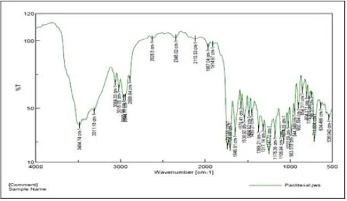
[image:4.612.314.566.383.529.2]FT-IR: The FTIR spectra of paclitaxel are as in Fig. 3. Paclitaxel characteristic peaks along with their description are tabulated in Table 2.
FIG. 3: FTIR SPECTRUM OF PACLITAXEL
From the data of IR analysis, the presence of the carbonyl (C=O), N-H/O-H and hydroxyl (-OH) groups have been confirmed, and all the identification data together confirmed the structure of supplied material as Paclitaxel.
TABLE 2: CHARACTERISTICS PEAKS OF
PACLITAXEL
Frequency (cm-1) Interpretation
3500-3200 NH stretching
1274 C-N stretching
1735-1720 C=O Stretching
941-803 C-H in-plane deformation 3300-2500 O-H Stretching (3311) 2973-2541 CH3 / C-H asymmetric Stretching
[image:4.612.314.565.627.745.2]Estimation Method: UV-Visible Spectroscopy:
Analytical Method Development for Paclitaxel: Drug Scan for λmax and Standard graph in IPA: λmax Determination in Isopropyl Alcohol:
FIG. 4: λmax SCAN OF PACLITAXEL IN ISOPROPYL
ALCOHOL
Standard Graph in Isopropyl Alcohol: The concentrations of Paclitaxel and the corresponding absorbance values are shown in Fig. 5. The solutions obeyed Beer-Lambert’s law over a concentration range of 4 to 20 µg/mL with a regression coefficient of 0.998.
FIG. 5: PACLITAXEL STANDARD GRAPH IN ISOPROPYL ALCOHOL
Drug Scan for λmax and Standard Graph in PBS 7.4:
λmax Determination in PBS:
FIG. 6: λmax DETERMINATION IN PBS
Standard Graph in Phosphate Buffered Saline 7.4: The concentrations of Paclitaxel and the corresponding absorbance values are shown in Fig. 7. The solutions obeyed Beer-Lambert’s law over a concentration range of 4 to 48 µg/ml with a regression coefficient of 0.999.
FIG. 7: STANDARD GRAPH OF PACLITAXEL IN PHOSPHATE BUFFERED SALINE 7.4
Solid State Characterization:
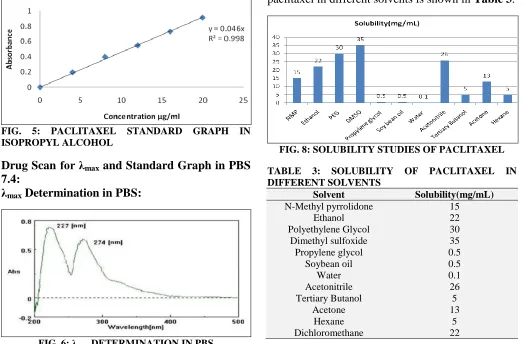
[image:5.612.49.299.132.276.2]Solubility Studies: Determination of solubility of the drug in different solvents is necessary as it helps in the selection of the solvent for the preparation of nanoformulations with controlled particle size. The qualitative solubility profile of paclitaxel in different solvents is shown in Table 3.
FIG. 8: SOLUBILITY STUDIES OF PACLITAXEL
TABLE 3: SOLUBILITY OF PACLITAXEL IN DIFFERENT SOLVENTS
Solvent Solubility(mg/mL)
N-Methyl pyrrolidone 15
Ethanol 22
Polyethylene Glycol 30
Dimethyl sulfoxide 35
Propylene glycol 0.5
Soybean oil 0.5
Water 0.1
Acetonitrile 26
Tertiary Butanol 5
Acetone 13
Hexane 5
[image:5.612.313.567.147.271.2] [image:5.612.48.302.397.539.2] [image:5.612.47.572.399.743.2] [image:5.612.314.567.414.542.2]Paclitaxel is insoluble in water, soluble in ethanol, dichloromethane and DMSO (dimethyl sulphoxide)
and the drug solubility in these solvents were found to be as follows.
[image:6.612.47.564.116.238.2]Partition Coefficient:
TABLE 4: PARTITION COEFFICIENT OF PACLITAXEL IN DIFFERENT SOLVENTS
Solvent system Solvent Qty of
Drug
Diluted with
Dilution Factor
Absorbance Conc
mg/mL
K Log p
Octanol-water Octanol 100mg IPA 3800 0.915 98.78 461.40 2.66
Water PBS 40 0.118 0.216
DCM -water DCM 300mg IPA 9000 0.160 320 612.66 2.91
Water PBS 100 0.090 0.428
Hexane-water Hexane 100mg IPA 5000 0.906 101 1057 3.02
Water PBS 100 0.040 0.190
Oleyl alcohol-water
Oleyl alcohol 100mg IPA 1000 0.50 111.1 2710 3.43
Water PBS 100 0.088 0.41
Loss on Drying (% LOD): % LOD of Paclitaxel was found 0.11 % of its weight after being dried at 105 °C for three hours which complied with the specification (% LOD ≤ 0.2%).
Drug - Excipient Compatibility Studies:
X-ray Diffraction (XRD) Analysis: The XRD studies were carried out for various experimental samples like paclitaxel, PLGA, PVP, and Egg lecithin. XRD diffraction patterns of paclitaxel, PLGA, PVP, Egg lecithin and all physical mixtures are presented in Fig. 9.
FIG. 9: X-RAY DIFFRACTION PATTERN FOR (A) PACLITAXEL (B) PVP (C) PACLITAXEL+PVP (D) PACLITAXEL + PLGA (E) PLGA (F) PACLITAXEL + EGG LECITHIN (G) EGG LECITHIN
It was clear that pure PTX showed partially sharp crystalline peaks, representative of the characteristics of a molecular compound with some crystallinity. The XRD graph of Paclitaxel exhibited strong peaks at 2θ values of 12.3921°, 13.7566° and 18.7720° with an intensity of 1442, 547 and 413 respectively. The resultant diffraction patterns represent the crystalline nature of the drug. All the crystalline drug signals were detectable in the physical mixture. A decrease in the intensity of the peaks of the physical mixture compared to pure
PTX was explained by the surface acting potential of selected surfactants. It should be inferred that there were no chemical interactions between PTX and selected surfactants because of no appearances of new functional groups.
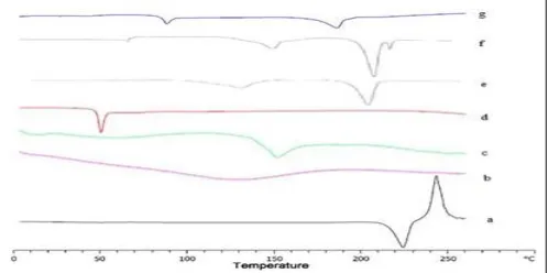
Differential Scanning Calorimetry: The DSC thermogram of paclitaxel, paclitaxel + PLGA, paclitaxel + PVP, paclitaxel + Tween-80 and paclitaxel + Egg lecithin was obtained to evaluate their thermal behavior. DSC diagrams of the seven samples are shown in Fig. 10. As illustrated in Fig. 10(a), the endothermic peak at 217.3 °C and exothermic peak at 244.5 °C were characteristic peaks of PTX in the crystalline state, while the endothermic peak at 125 °C in Fig. 10(b) represented the peak of PVP. Fig. 10(c) exhibited the endothermic peak of Egg lecithin at around 150 °C. Fig. 10(d) exhibited the endothermic peak of PLGA at around 50 °C. The Melting endothermic peak of paclitaxel lay at 217 °C in all the physical mixture samples indicating that there was no interaction between the selected surfactants and paclitaxel.
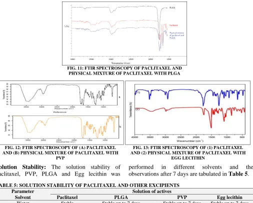
[image:6.612.49.300.419.567.2] [image:6.612.316.565.585.709.2]FTIR Spectroscopy: FTIR spectroscopy of physical mixtures of paclitaxel with three different surfactants is as shown in Fig. 11-13. The presence
of major characteristic peaks of paclitaxel in all the physical mixtures indicates the compatibility with the selected excipients.
FIG. 11: FTIR SPECTROSCOPY OF PACLITAXEL AND PHYSICAL MIXTURE OF PACLITAXEL WITH PLGA
Solution Stability: The solution stability of paclitaxel, PVP, PLGA and Egg lecithin was
[image:7.612.57.563.103.509.2]performed in different solvents and the observations after 7 days are tabulated in Table 5.
TABLE 5: SOLUTION STABILITY OF PACLITAXEL AND OTHER EXCIPIENTS
Parameter Solution of actives
Solvent Paclitaxel PLGA PVP Egg lecithin
Water Stable Stable up to 7 days Stable up to 7 days Stable up to 7 days
Phosphate buffer pH 7.4 Stable Stable up to 7 days Stable up to 7 days Stable up to 7 days Phosphate buffer pH of
4.0
Stable Stable up to 7 days Stable up to 7 days Stable up to 7 days
Phosphate buffer pH 6.0 Stable Stable up to 7 days Stable up to 7 days Stable up to 7 days
CONCLUSION: Before the formulation
development phase, preformulation studies were to be performed as they provide several key characteristics regarding the drug and the excipients. The solid-state characterization studies revealed the degree of crystallinity of the drug and polymer. The obtained sample of Paclitaxel was found crystalline by X-ray diffraction analysis and showed very close peaks at different 2θ values. The maximum intensity was observed at 2θ values 12.5 and microscopy also reveals the crystalline particles. Hence, the results of XRD studies of paclitaxel and other excipients unveil the
crystalline nature by the presence of sharp peaks indicating crystalline nature of the compound. The DSC studies were conducted for establishing the interaction between the drug and the polymers in solid state and also to determine the thermal behavior and melting point of drug and polymers. The results from DSC studies confirmed the identity and absence of interaction between drug and polymers in the solid state. The compatibility studies were further confirmed by conducting FTIR studies of different combinations of drug and polymer. The results indicated no significant interaction between 1:1 physical mixture of drug
FIG. 12: FTIR SPECTROSCOPY OF (A) PACLITAXEL AND (B) PHYSICAL MIXTURE OF PACLITAXEL WITH
PVP
FIG. 13: FTIR SPECTROSCOPY OF (1) PACLITAXEL AND (2) PHYSICAL MIXTURE OF PACLITAXEL WITH
and polymers. The solubility study of the drug in different solvents was performed, and from the results, paclitaxel exhibited good solubility in organic solvents like ethanol, dichloromethane and DMSO (dimethyl sulphoxide). The lipophilicity of paclitaxel was determined by calculating the log P or partition coefficient in different solvent systems like Octanol-Water, DCM-Water, Hexane-Water, Oleyl Alcohol-Water and from the results it was found that paclitaxel exhibited log P of 2.66, 2.91, 3.02 and 3.42 respectively which indicate the lipophilic nature of the compound. The solution stability of the drug in different solvents that are used in the preparation and characterization of nanoformulations was performed and was found to be stable in the resulted solvent system with negligible change in spectral properties. So, it was concluded that paclitaxel and all these excipients were suitable for further nanoformulation studies.
ACKNOWLEDGEMENT: The authors are very thankful to the management of Malla Reddy Pharmacy College & Vijaya College of Pharmacy, for providing necessary facilities to carry out the research work.
CONFLICT OF INTEREST: Declared none.
REFERENCES:
1. Nirmala CK, Mutsa TM, Orla MM, Michael Q and Magdalena P: Paclitaxel and its evolving role in the management of ovarian cancer. Bio Med Res Int 2015; 413076.
2. Jordan MA and Wilson L: Microtubules as a target for anticancer drugs. Nat Rev Cancer 2004; 4: 253-65. 3. Chih-Yang H, Da-Tong J, Chih-Fen C, Muralidhar-Reddy
P and Bharath Kumar V: A review on the effects of current chemotherapy drugs and natural agents in treating non– small cell lung cancer. Biomedicine 2017; 7(4): 23. 4. Mansukh CW: Campotthecin and Taxol: From nature to
bench to beside. Hamdan Med J 2015; 8: 69-82.
5. Chetan N, Sharad J, Ankit S and Vaibhav K: Paclitaxel formulations: challenges and novel delivery options. Current Drug Delivery 2014; 11(6): 666-86.
6. Iole C, Lucia F, Simona G, Mariamena A and Domenico C: Nanoparticle albumin bound paclitaxel in the treatment
of human cancer: nano delivery reaches prime-time. J Drug Del 2013; 905091.
7. Nirmala CK, Mutsa TM, Orla MM, Michael Q and Magdalena P: Paclitaxel and its evolving role in the management of ovarian cancer. BioMed Res Int 2015; 413076.
8. Richard AS, Kim MG, James HN and Ritu A: Drugs that target dynamic microtubules: a new molecular perspective. Med Res Rev. 2011; 31(3): 443-81.
9. Eiman M, Vaqar MA and Hasan M: Targeting microtubules by natural agents for cancer therapy. Mol Cancer Ther 2014; 13(2): 275-84.
10. Beth AW: How Taxol/paclitaxel kills cancer cells. Mol Biol Cell. 2014; 25(18): 2677-81.
11. Migoha CO, Ratansi M, Kaale E and Kagashe G: Preformulation studies for generic omeprazole magnesium enteric coated tablets. BioMed Res Int 2015; 2015: 307032-41.
12. Sandip MH, Yuvraj DD, Riyaz Ali MO and Dhanraj RJ: General considerations of design and development of dosage forms: the pre-formulation review. Asian Jour of Pharm 2017; 11(S3): S479-88.
13. Bharate SS and Vishwakarma RA: Impact of preformulation on drug development. Expert Opin Drug Deliv 2013; 10(9): 1239-57.
14. Killedar SG, Nale AB, More HN, Nadaf SJ, Pawar AA and Tamboli US: Isolation, characterization and evaluation of
C. fistula Linn. seed and pulp polymer for pharmaceutical
application. Int J Pharm Investig 2014; 4(4): 215-25. 15. Dezani AB, Pereira TM, Caffaro AM, Reis JM and Serra
CHR: Equilibrium solubility versus intrinsic dissolution: characterization of lamivudine, stavudine and zidovudine for BCS classification. Brazilian J Pharm Sci 2013; 49(4): 853-63.
16. The United Stated Pharmacopeia 2003; USP 26, NF 21. 17. Tiwari G: Biodegradable microspheres for controlled
delivery of metronidazole in the treatment of periodontal diseases: formulation consideration. Int J Pharm Bio Sci 2010; 1(2): 1-13.
18. Indian Pharmacopoeia, Ministry of Health and family welfare, Government of India, Published by the controller of publications, Delhi 1996.
19. Vercruysse J, Delaet U, Van Assche I, Cappuyns P and Arata F: Stability and repeatability of a continuous twin screw granulation and drying system. Euro Jour Pharm Biopham 2013; 85(3): 1031-38.
20. Mohamed JM, Bharathidasan P and Raffick MM: Preformulation and development of curcumin magnetic nanosuspension using magnetite (Fe3O4) and
methylcellulose. Int J Pharm Bio Sci 2012; 3(4): 419-32. 21. Vercruysse J, Peeters E, Fonteyne M, Cappuyns P and
Delaet U: Use of a continuous twin screw granulation and drying system during formulation development and process optimization. Euro Jour Pharm Biopham 2015; 89: 239-47.
All © 2013 are reserved by International Journal of Pharmaceutical Sciences and Research. This Journal licensed under a Creative Commons Attribution-NonCommercial-ShareAlike 3.0 Unported License.
This article can be downloaded to Android OS based mobile. Scan QR Code using Code/Bar Scanner from your mobile. (Scanners are available on Google Play store)
How to cite this article: