R E S E A R C H A R T I C L E
Open Access
An ex vivo continuous passive motion model in a
porcine knee for assessing primary stability of
cell-free collagen gel plugs
Turgay Efe
1*, Markus D Schofer
1, Alexander Füglein
2, Nina Timmesfeld
3, Susanne Fuchs-Winkelmann
1,
Thomas Stein
1, Bilal Farouk El-Zayat
1, Jürgen RJ Paletta
1, Thomas J Heyse
1Abstract
Background:Primary stability of cartilage repair constructs is of the utmost importance in the clinical setting but few continuous passive motion (CPM) models are available. Our study aimed to establish a novel ex vivo CPM animal model and to evaluate the required motion cycles for testing the mechanical properties of a new cell-free collagen type I gel plug (CaReS®-1S).
Methods:A novel ex vivo CPM device was developed. Full-thickness cartilage defects (11 mm diameter by 6 mm deep) were created on the medial femoral condyle of porcine knee specimens. CaReS®-1S was implanted in 16 animals and each knee underwent continuous passive motion. After 0, 2000, 4000, 6000, and 8000 motions, standardized digital pictures of the grafts were taken, focusing on the worn surfaces. The percentage of worn surface on the total CaReS®-1S surface was evaluated with image processing software.
Results:Significant differences in the worn surface were recorded between 0 and 2000 motion cycles (p < 0.0001). After 2000 motion cycles, there was no significant difference. No total delamination of CaReS®-1S with an empty defect site was recorded.
Conclusion:The ex vivo CPM animal model is appropriate in investigating CaReS®-1S durability under continuous passive motion. 2000 motion cycles appear adequate to assess the primary stability of type I collagen gels used to repair focal chondral defects.
Background
Cartilage defects of the knee are commonly encountered in orthopedic clinical practice [1,2]. Several well described repair strategies have been used in the man-agement of chondral lesions [3-6]. Debate persists about the best repair technique for symptomatic chondral defects of less than 2 cm² [7]. Currently, CaReS®-1S (Arthro Kinetics, Esslingen, Germany), a cell-free col-lagen type I gel plug was introduced to treat isolated cartilage defects. This graft is implanted in the debrided chondral lesion, in a single-stage procedure. The gel triggers chondrocyte migration and proliferation into
the plug, which has been proven both in vitro and ex vivo [8].
Most cartilage regenerative studies address the bio-chemical and histological composition of generated car-tilage [9,10]. However, successful carcar-tilage repair requires sufficient initial mechanical stability of the grafts [11]. The implanted grafts should withstand the forces in vivo in the early postoperative phase during joint movement. Secure primary stability is necessary to keep the grafts in place and to achieve sufficient joint surface congruity, on which the long-term results depend [12]. Partially or completely delaminated tissue engineered constructs can cause locking in the knee and poor clinical results [13,14].
Several authors describe the beneficial effects of con-tinuous passive motion (CPM) on neochondrogenesis and most postoperative protocols consider ex vivo * Correspondence: [email protected]
1
Department of Orthopedics and Rheumatology, University Hospital Marburg, Baldingerstrasse, 35043 Marburg, Germany
Full list of author information is available at the end of the article
non-weight bearing CPM in the first 6 weeks after sur-gery to be beneficial [15-17].
Drobnic et al. [18] reported a human cadaveric model of ex vivo CPM, testing four fixation techniques for fibrinogen and thrombin coated collagen fleece. They aimed to simulate the initial postoperative period by applying ex vivo CPM to specimens. However, the avail-ability of appropriate human specimens without advanced osteoarthritis is limited. This can be a chal-lenge for developing appropriate protocols to test the primary stability of collagen plugs; to date, there are no established animal models.
Moreover, little is known about the required motion cycles for testing the mechanical properties of cartilage repair constructs. The means to assess the early post-operative phase after cartilage regenerative procedures is lacking. Consequently, efforts to build a new ex vivo CPM device to assess the mechanical properties of tis-sue engineered scaffolds would be of great value. The aim of the present study is to construct a simplified, custom-made ex vivo CPM device and to investigate the number of motion cycles required for testing the mechanical properties of collagen plugs. We hypothe-sized that the new device would allow us to reliably evaluate the degree of wear to collagen plugs. We also aimed to determine the number of cycles required to assess wear appropriately using this model.
Methods
Sixteen porcine knees (age 9 months) were used to assess primary stability of cell-free collagen gel plugs. These were obtained from the local butcher, fresh fro-zen at -25°, and thawed for 16 hours at room tempera-ture (20°C) before testing. This project was performed in accordance with the Helsinki Declaration and with local legislation. CaReS®-1S was implanted in an identi-cal manner in each specimen. Right and left knees were not differentiated; both knees were used [19]. Some knee specimens were dissected to evaluate the anatomy of the porcine knee and to establish the surgical procedure.
CaReS®-1S
CaReS®-1S is a sterile cylindrical, three-dimensional col-lagen-based and cell-free gel plug, consisting of 4.8 mg/ mL rat tail collagen type I (CaReS® [20,21], Arthro Kinetics, Esslingen, Germany). It has been used for carti-lage defects in the knee and ankle in patients under 50 years of age. The plug is available in three different dia-meters (11, 22, and 34 mm) and thicknesses (4, 6, and 8 mm). The hydrogel consistency allows adequate cover-age and sufficient reconstruction of the articular surface. The basic concept of CaReS®-1S is to replenish the carti-lage defect with a matrix structure, facilitating
autologous chondrocytes to penetrate into the lesion zone. Low antigenicity and high biocompatibility of CaReS®-1S is achieved by the high conservation of pro-tein sequences of collagen type I within various species. The plugs used in this study measured 11 mm diameter by 6 mm deep and contained 4.27 mg collagen type I. CaReS®-1S were stored in phosphate-buffered saline solution and preserved at 4°C until use.
Ex vivo CPM device
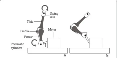
A custom-made, pneumatic ex vivo CPM device was constructed, on which extension-flexion motions were performed at a frequency of 1 Hz (Figure 1). There were no constrained forces and during passive motion the tibia was allowed to rotate freely. To simulate early postopera-tive rehabilitation, an additional axial load during testing was not performed. The extension and flexion motion cycles was performed via the coordination of two pneu-matic cylinders and detected by an integrated counter.
Surgical procedure
The tibia and the femur were transected approximately 10 cm proximal and distal to the knee joint, preserving muscles, articular capsule, ligaments, and tendons. A medial parapatellar arthrotomy was performed and the patella was dislocated laterally. A standardized full-thickness cartilage defect of 11 mm diameter by 6 mm deep was created in the weight-bearing area of the med-ial femoral condyle using a punch (Arthro Kinetics, Esslingen, Germany).
Ex vivo CPM protocol
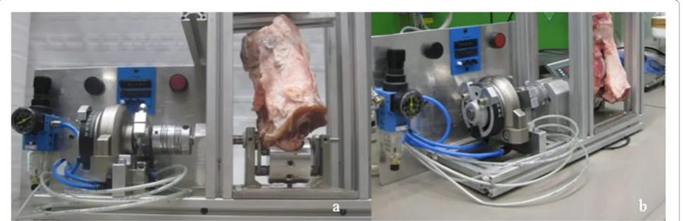
[image:2.595.305.539.584.700.2]Experiments were performed at 20°C. A constant ex vivo CPM protocol was established for the performance of experiments. After the preparation of the chondral defect, the proximal femur and distal tibia were fixed into a cylindric metal device and fastened in the vertical position (Figure 2). As the porcine hind leg could not be fully extended, specimens were flexed to and from an extension position, between 20°-120°-20°, encompassing
one complete motion cycle. After fixation, ex vivo CPM device test cycles through the whole range of motion were performed to align the mechanical axis of the stifle joint with the mechanical axis of the ex vivo CPM device. Both tibial and femoral fixation allowed adjust-ment of the stifle position. After adjustadjust-ment, 0.3 mL of a two-component fibrin sealant (Tissucol Duo, Baxter, Unterschleißheim, Germany) was applied to the pre-pared defect and the surrounding cartilage rim, prior to CaReS®-1S placement into the lesion. The gel plug was pressed into the defect according to standard manufac-turer guidelines and clinical practice. The graft was con-sidered to be sufficient when complete congruity with the surrounding cartilage rim was achieved. 5 mL of 0.9% sodium chloride solution was injected intraarticu-larly with a catheter to keep the articular surface moist and intraarticular friction low. After 2000, 4000, 6000, and 8000 motion cycles, the arthrotomy was reopened to evaluate the graft worn surface and to moisten the joint surface with 0.9% sodium chloride solution. Stan-dardized digital photographs were taken for analysis after CaReS®-1S implantation and after implantation and every 2000 motion cycles. During the entire procedure, test specimens were externally moistened with 0.9% sodium chloride solution every 10 minutes.
Graft Evaluation
The surface damage was defined as intact, marginally detached, partially detached, or completely displaced. Marginally detached only involved the junction between CaReS®-1S and adjacent cartilage, and appeared as a fis-sure. In partial detachment, the graft only covered a part of the defect. If the cartilage defect was completely empty, this was defined as displacement of the graft. Digital photographs obtained after 0, 2000, 4000, 6000, and 8000 motion cycles were transferred using image
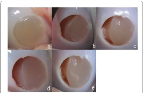
processing software (QUIPS, Leica, Wetzlar, Germany). The worn surface area and the total area of the defect were measured in pixels; the percentage of worn surface was calculated from the ratio of both measurements (Figure 3). The areas of interest were traced by an inde-pendent observer who was not involved in performing the experiments.
Statistical analysis
The repeated measured data at 2000, 4000, 6000, and 8000 motion cycles were assessed by analysis of variance (ANOVA). The increase in the worn surface within the first 2000 motion cycles and differences between 2000, 4000, 6000, and 8000 motion cycles were analyzed through the application of general linear hypothesis test-ing. P-value adjustment was performed by the method of Shaffer [22]. Additionally, for all contrasts, the corre-sponding 95% family-wise confidence intervals (CI) were calculated. Two-sided adjusted p-values less than 0.05 were considered statistically significant. All statistical analysis was performed with R software (Foundation for Statistical Computing, Vienna, Austria).
Results
[image:3.595.59.541.90.246.2]The ex vivo CPM device worked for 8000 motion cycles. The most time-consuming procedure was the mechanical axis of the stifle joint alignment with the mechanical axis of the testing device. Within the first 2000 motion cycles, the mean worn surface significantly increased by 20.2% (p < 0.0001, C.I. 10.2% - 30.2%). After 2000 cycles, no sig-nificant changes in the worn surface were observed (Table 1). Representative photos at 0, 2000, 4000, 6000, and 8000 motion cycles are shown in Figure 4. The eva-luation during ex vivo CPM showed that none of the CaReS®-1S grafts delaminated totally with an empty defect site.
Discussion
Information on ex vivo CPM devices and the number of motion cycles needed to investigate cartilage repair con-structs is lacking. The aim of our study was to construct a novel ex vivo CPM device and to create a small animal model to evaluate the required number of continuous passive motions for testing CaReS®-1S. Our hypothesis that this device would allow reproducible evaluation of wear to collagen plugs was proven to be correct. We also successfully determined number of cycles required to assess wear appropriately.
There are several limitations to our study. While por-cine anatomy closely approximates human anatomy [23], immanent differences between species are evident. Mechanical in vitro studies only simulate the physiologi-cal function and motion of the joint, since there is no force applied by the muscles. The presented ex vivo CPM device may only partially simulate its clinical application. In our study, analysis of the worn surface of the graft was performed by imaging processing software as a semiquantitative analysis. More objective quantita-tive measurements and absolute values may be prefer-able. However, analysis of cartilage repair constructs with the ex vivo CPM is difficult, raising the question of reliability. Other studies used qualitative scales [18,24], posing the problem of subjective bias. It is very likely that a potential malalignment of the joint within the testing device may have affected the results. In our setup, the tibia was allowed to rotate freely. It was also allowed to move freely in the frontal plane. Malalign-ment in the testing device resulted in impingeMalalign-ment of the specimen with the frame. Simulation in human cadaver knees would be the optimal standard for testing. However, human specimens are expensive, sometimes
difficult to obtain, and come from elderly people. There-fore, osteoarthritic changes are a common finding in these specimens, which may interfere with the evalua-tion of a procedure in an otherwise healthy joint of a young patient. The porcine model comes with the advantage of unlimited availability at a low cost.
[image:4.595.60.539.89.187.2]There are currently only two experimental ex vivo CPM models for testing cartilage implants. A recent study on human cadaver knees investigated the quality of PEOT/PBT scaffold fixation (transosseous fixation, fibrin glue, biodegradable pins, and continuous cartilage sutures) by ex vivo CPM in a vertical position [24]. The mechanical behavior was evaluated after 60 and 150 motion cycles with a 35N load, focusing on outline attachment, scaffold integrity, and area coverage. After 210 motion cycles, an endpoint fixation test was per-formed. Drobnic et al. [18] compared in a similar model four different fixation techniques (self-adhesion, fibrin glue, bone sutures, periosteal cover) of a collagen scaf-fold in the human cadaveric knee, horizontally oriented with and without loading. In both studies, the numbers of cycles remained small and did not correspond with the clinical situation after surgical management of carti-lage defects; whereas ex vivo CPM is often applied sev-eral hours a day. It is most likely that in the early postoperative phase the knee undergoes considerably more than 150 motion cycles. At our institution and in previous experience [25] patients usually use the ex vivo CPM machine for 6 to 8 hours daily after implantation of tissue engineered constructs. The rate is one cycle per minute but varies based on patient comfort and per-formance. This fact should be taken into consideration when performing mechanical testing. A study design with fewer motion cycles makes the transformation
Figure 3Worn surface analysis of CaReS®-1S. (a) initial image; calculation of percentage of worn surface from the ratio of (c) and (d).
Table 1 Analysis of surface wear in dependence of motion cycles (p < 0.05)
Motion Cycles Mean difference of worn surface (%) 95% Confidence Interval P-Value
0 to 2000 20.2 10.2 - 30.2 0.0001
2000 to 4000 1.9 -1.5 - 5.3 0.20
2000 to 6000 2.9 -0.6 - 6.3 0.12
[image:4.595.58.536.667.731.2]from in vitro to in vivo testing difficult. Consequently, long-term durability studies are necessary. However, it is possible to determine if such constructs were too fragile to be practical. Thus, shorter-term motion cycles studies have their place. In the present study, testing of CaReS®-1S was performed with 8000 motion cycles under ex vivo CPM. The question of how many motion cycles were necessary to evaluate wear to collagen plugs has not been answered satisfactorily as of yet. In our study, significant changes in the worn surface of the graft were only detected between 0 and 2000 motion cycles (p = 0.0001). Therefore, a protocol with 2000 motion cycles appears to be a sufficient number to investigate type I collagen gels used to repair focal chondral defects.
Delamination of tissue engineered constructs occurs mainly in the first six postoperative months [26]. A pos-sible explanation for this may include weak mechanical fixation and adherence. Since it is known that friction forces and shear forces may cause delamination of carti-lage implants [27], most authors allow only partial weight-bearing after cartilage regeneration techniques [28,29]. While Drobnic et al. [18] and Bekkers et al. [24] used additional load ex vivo CPM protocols, intraarticu-lar pressure measurements were not performed. It remains unclear how much load was applied on the spe-cimens intraarticularly. Although no additional load was applied in our study, we assume that the vertical orien-tation, the corresponding tibial cartilage, and the intraarticular liquid generates friction forces and shear forces on the gel that correspond with the clinical set-ting. Nevertheless, performing the experiments with an additional load might have been preferable.
Evaluation of tissue engineered constructs in the early postoperative rehabilitation phase is difficult. The ex vivo CPM device used in the present study is sim-plified and can be helpful to investigate the durability
of collagen gel constructs. Our findings may support research in new materials in a porcine knee model. Nevertheless, the ex vivo CPM device cannot be used in assessing biomechanical properties (viscoelasticity, permeability, tensile strength, and electromechanical properties).
Conclusion
The novel custom-made ex vivo CPM device allows the investigation of CaReS®-1S under a high number of motion cycles. Significant differences in the worn surface were detected between 0 and 2000 motion cycles, but not beyond 2000 cycles. This device and study model may be of great value in further investigations.
Acknowledgements
The authors wish to thank Dr. Karsten Gavenis, Department of Orthopaedic Surgery, University Hospital Aachen, Germany, and Professor Ulrich Schneider, Arthro Nova Clinic, Tegernsee, Germany for providing the laboratory facilities.
Author details
1Department of Orthopedics and Rheumatology, University Hospital
Marburg, Baldingerstrasse, 35043 Marburg, Germany.2Department of Oral and Maxillofacial Surgery, University Hospital Aachen, Pauwelsstrasse 30, 52074 Aachen, Germany.3Institute of Medical Biometry and Epidemiology, Philipps-University Marburg, Bunsenstrasse 3, 35037 Marburg, Germany.
Authors’contributions
TE participated in the study design, carried out the study, interpreted the results and drafted the manuscript. MDS, SFW, and TS participated in the study design and interpretation of the results. AF constructed the ex vivo CPM device and participated in the study design. NT performed the statistical analysis. JRJP and BFE designed the protocol and helped with data interpretation. TJH participated in the study design and the drafting of the manuscript. All authors read and approved the final manuscript.
Competing interests
TE, MDS, BFE and TJH are consultants to Smith&Nephew.
Received: 2 August 2010 Accepted: 15 December 2010 Published: 15 December 2010
References
1. Curl WW, Krome J, Gordon ES, Rushing J, Smith BP, Poehling GG:Cartilage injuries: a review of 31,516 knee arthroscopies.Arthroscopy1997, 13(4):456-460.
2. Hjelle K, Solheim E, Strand T, Muri R, Brittberg M:Articular cartilage defects in 1,000 knee arthroscopies.Arthroscopy2002,18(7):730-734.
3. Bert JM, Maschka K:The arthroscopic treatment of unicompartmental gonarthrosis: a five-year follow-up study of abrasion arthroplasty plus arthroscopic debridement and arthroscopic debridement alone. Arthroscopy1989,5(1):25-32.
4. Hangody L, Vasarhelyi G, Hangody LR, Sukosd Z, Tibay G, Bartha L, Bodo G: Autologous osteochondral grafting–technique and long-term results. Injury2008,39(Suppl 1):S32-39.
5. Mithoefer K, Williams RJ, Warren RF, Potter HG, Spock CR, Jones EC, Wickiewicz TL, Marx RG:The microfracture technique for the treatment of articular cartilage lesions in the knee. A prospective cohort study.J Bone Joint Surg Am2005,87(9):1911-1920.
6. Rae PJ, Noble J:Arthroscopic drilling of osteochondral lesions of the knee.J Bone Joint Surg Br1989,71(3):534.
[image:5.595.56.291.88.241.2]7. Smith GD, Knutsen G, Richardson JB:A clinical review of cartilage repair techniques.J Bone Joint Surg Br2005,87(4):445-449.
8. Morales TI:Chondrocyte moves: clever strategies?Osteoarthritis Cartilage 2007,15(8):861-871.
9. Getgood A, Brooks R, Fortier L, Rushton N:Articular cartilage tissue engineering: today’s research, tomorrow’s practice?J Bone Joint Surg Br 2009,91(5):565-576.
10. van Osch GJ, Brittberg M, Dennis JE, Bastiaansen-Jenniskens YM, Erben RG, Konttinen YT, Luyten FP:Cartilage repair: past and future–lessons for regenerative medicine.J Cell Mol Med2009,13(5):792-810.
11. Sittinger M, Hutmacher DW, Risbud MV:Current strategies for cell delivery in cartilage and bone regeneration.Curr Opin Biotechnol2004,
15(5):411-418.
12. Sittinger M, Perka C, Schultz O, Haupl T, Burmester GR:Joint cartilage regeneration by tissue engineering.Z Rheumatol1999,58(3):130-135. 13. Nehrer S, Spector M, Minas T:Histologic analysis of tissue after failed cartilage repair procedures.Clin Orthop Relat Res1999, ,365:149-162. 14. Peterson L, Minas T, Brittberg M, Nilsson A, Sjogren-Jansson E, Lindahl A:
Two- to 9-year outcome after autologous chondrocyte transplantation of the knee.Clin Orthop Relat Res2000, ,374:212-234.
15. Gikas PD, Bayliss L, Bentley G, Briggs TW:An overview of autologous chondrocyte implantation.J Bone Joint Surg Br2009,91(8):997-1006. 16. Moran ME, Kim HK, Salter RB:Biological resurfacing of full-thickness defects in patellar articular cartilage of the rabbit. Investigation of autogenous periosteal grafts subjected to continuous passive motion.J Bone Joint Surg Br1992,74(5):659-667.
17. O’Driscoll SW, Keeley FW, Salter RB:The chondrogenic potential of free autogenous periosteal grafts for biological resurfacing of major full-thickness defects in joint surfaces under the influence of continuous passive motion. An experimental investigation in the rabbit.J Bone Joint Surg Am1986,68(7):1017-1035.
18. Drobnic M, Radosavljevic D, Ravnik D, Pavlovcic V, Hribernik M:Comparison of four techniques for the fixation of a collagen scaffold in the human cadaveric knee.Osteoarthritis Cartilage2006,14(4):337-344.
19. Athanasiou KA, Rosenwasser MP, Buckwalter JA, Malinin TI, Mow VC: Interspecies comparisons of in situ intrinsic mechanical properties of distal femoral cartilage.J Orthop Res1991,9(3):330-340.
20. Andereya S, Maus U, Gavenis K, Muller-Rath R, Miltner O, Mumme T, Schneider U:[First clinical experiences with a novel 3D-collagen gel (CaReS) for the treatment of focal cartilage defects in the knee].Z Orthop Ihre Grenzgeb2006,144(3):272-280.
21. Schneider U, Andereya S:[First results of a prospective randomized clinical trial on traditional chondrocyte transplantation vs CaReS-Technology].Z Orthop Ihre Grenzgeb2003,141(5):496-497.
22. Shaffer J:Modified sequentially rejective multiple test procedures.Journal of the American Statistical Association1986,81:826-831.
23. Ahern BJ, Parvizi J, Boston R, Schaer TP:Preclinical animal models in single site cartilage defect testing: a systematic review.Osteoarthritis Cartilage 2009,17(6):705-713.
24. Bekkers JE, Tsuchida AI, Malda J, Creemers LB, Castelein RJ, Saris DB, Dhert WJ:Quality of scaffold fixation in a human cadaver knee model. Osteoarthritis Cartilage18(2):266-272.
25. Alford JW, Cole BJ:Cartilage restoration, part 2: techniques, outcomes, and future directions.Am J Sports Med2005,33(3):443-460.
26. Alparslan L, Minas T, Winalski CS:Magnetic resonance imaging of autologous chondrocyte implantation.Semin Ultrasound CT MR2001, 22(4):341-351.
27. Driesang IM, Hunziker EB:Delamination rates of tissue flaps used in articular cartilage repair.J Orthop Res2000,18(6):909-911.
28. Marcacci M, Zaffagnini S, Kon E, Visani A, Iacono F, Loreti I:Arthroscopic autologous chondrocyte transplantation: technical note.Knee Surg Sports Traumatol Arthrosc2002,10(3):154-159.
29. Peterson L, Brittberg M, Kiviranta I, Akerlund EL, Lindahl A:Autologous chondrocyte transplantation. Biomechanics and long-term durability.Am J Sports Med2002,30(1):2-12.
Pre-publication history
The pre-publication history for this paper can be accessed here: http://www.biomedcentral.com/1471-2474/11/283/prepub
doi:10.1186/1471-2474-11-283
Cite this article as:Efeet al.:An ex vivo continuous passive motion model in a porcine knee for assessing primary stability of cell-free collagen gel plugs.BMC Musculoskeletal Disorders201011:283.
Submit your next manuscript to BioMed Central and take full advantage of:
• Convenient online submission
• Thorough peer review
• No space constraints or color figure charges
• Immediate publication on acceptance
• Inclusion in PubMed, CAS, Scopus and Google Scholar
• Research which is freely available for redistribution