0095-1137/08/$08.00
⫹
0
doi:10.1128/JCM.01600-07
Copyright © 2008, American Society for Microbiology. All Rights Reserved.
Multiplex PCR for Rapid Detection of
Staphylococcus aureus
Isolates
Suspected to Represent Community-Acquired Strains
䌤
B. Strommenger,* C. Braulke, B. Pasemann, C. Schmidt, and W. Witte
National Reference Centre for Staphylococci, Robert Koch Institute, Wernigerode Branch, Wernigerode, Germany
Received 11 August 2007/Returned for modification 3 October 2007/Accepted 9 November 2007
The continuous spread of community-acquired methicillin-resistant
Staphylococcus aureus
(caMRSA) and
the introduction of these highly virulent isolates into hospitals represent increasing threats. The timely
recognition of caMRSA strains is crucial for infection control purposes. Thus, we developed a PCR-based assay
for the easy and rapid determination of those caMRSA clones that currently are the most prevalent in Germany
and Central Europe. This assay was able to correctly identify the majority of the isolates as caMRSA of
sequence type 80 (ST80), clonal complex 1 (USA400), and ST8 (USA300). In combination with
spa
typing-BURP (based upon repeat pattern) analysis and resistance typing, it provides a means for the extensive
characterization of suspicious isolates. Thus, this assay represents a reliable tool for monitoring the emergence
and spread of different caMRSA clones. The resulting information, in combination with careful interpretation
of the epidemiological records, might help to prevent the further spread of those highly virulent caMRSA
clones.
Staphylococcus aureus
is a facultative pathogenic
gram-pos-itive bacterium which is well known as colonizer of the human
skin, but it can also cause a variety of diseases, ranging from
minor skin and soft tissue infections to life-threatening disease
(20). Methicillin-resistant
S. aureus
(MRSA) and
multiresis-tant
S. aureus
strains are responsible for a large proportion of
nosocomial infections, making treatment difficult (34). Several
risk factors for MRSA infection or colonization have been
established and include previous hospitalization, residence in a
nursing home, antibiotic therapy, and the presence of
indwell-ing medical devices (20).
However, during the last decade an increasing number of
reports of MRSA cases among healthy community-dwelling
persons without classical risk factors for MRSA acquisition
were encountered worldwide (15). The majority of these
iso-lates, referred to as “community-acquired MRSA” (caMRSA)
strains (31), are genetically and phenotypically distinct from
representative “hospital-acquired” MRSA (haMRSA) strains,
as reflected by their narrow resistance patterns, as well as by
the distribution of their staphylococcal chromosomal cassette
(SCC
mec
) types (type IV or V) and resistance and toxin
de-terminants (15, 37). Different caMRSA clones were originally
shown to be continent specific (37), but recent studies
demon-strated their intercontinental spread, with the predominance of
particular clones in distinct geographic regions (3, 35, 36). The
caMRSA clones currently most predominant in Germany and
Central Europe are sequence type 80 (ST80;
spa
type t044,
European clone), ST1 (
spa
type t127, USA400), and ST8 (
spa
type t008, USA300) (17, 36, 40, 41).
A matter of particular concern is the threat of introducing
highly virulent caMRSA clones from the site of their origin in
the community into the hospital, where they meet considerably
more compromised patients, potentially leading to markedly
increased patient morbidity and mortality (39). Episodes of
health care-associated infections due to caMRSA have been
already reported in the United States and Europe (14, 18, 32).
Typing is a prerequisite to obtaining knowledge of the
epi-demiology of
S. aureus
strains in order to prevent the spread of
caMRSA within the community as well as from the community
into the hospitals. We have recently shown that
spa
typing in
combination with clustering by BURP (based upon repeat
pat-tern) analysis is a useful tool in
S. aureus
epidemiology,
espe-cially because of its reproducibility and the portability of the
typing data (33). However, recent studies (11, 32a) have
dem-onstrated that
spa
typing-BURP analysis is not always able to
discriminate unambiguously between different clones of
caMRSA (e.g., between ST80 and ST1 clones) and some
com-mon methicillin-susceptible S. aureus (MSSA) clones because
similar
spa
types occur in different clones, but this cannot be
explained by large chromosomal replacements (27). Moreover,
some
spa
types occur in different MRSA clones, including
caMRSA clones. This was demonstrated for
spa
type t008,
which was found in three different clones (characterized by the
possession of different SCC
mec
types) and which also included
caMRSA t008/ST8/USA300 (4).
Therefore, the aim of this study was the development of a
rapid and easy PCR assay that can be used to recognize the
predominant caMRSA clones based on lineage-specific genetic
markers.
MATERIALS AND METHODS
Bacterial strains.TheS. aureusisolates investigated in this study (n⫽125) were sent to the German Reference Centre for Staphylococci for further char-acterization and typing. Isolates originated from microbiological laboratories from throughout Germany, as well as from other Central European countries. The isolates included in this study were BURP analysis-defined relatives ofspa
types t044 (ST80) and t127 (ST1), as well as isolates of type t008/t024 (potentially ST8/USA300) and isolates from other clonal lineages (clonal complex 5 [CC5], CC22, CC30, CC45, CC121, ST59, ST152, and ST154), which were supposed to
* Corresponding author. Mailing address: Robert Koch Institute,
Wernigerode Branch, Burgstr. 37, D-38855 Wernigerode, Germany.
Phone: 0049/3943/679 260. Fax: 0049/3943/679 317. E-mail: strommengerb
@rki.de.
䌤
Published ahead of print on 21 November 2007.
582
on May 16, 2020 by guest
http://jcm.asm.org/
be of community origin on the basis of epidemiological information. Strains were isolated from patients with skin and soft tissue infections (n⫽55), conjunctivitis (n⫽2), and otitis (n⫽1); 9 isolates were collected from patients with invasive infections (bacteremia, pneumonia, urinary tract infections). Twenty-one strains were isolated from nasal swabs. For 39 isolates no information concerning the medical history of the patient was available (most of them were selected because of their relatedness tospatypes t044 and t127).
All isolates were cultured on sheep blood agar and were confirmed to beS. aureusby colony morphology and a positive plasma coagulase reaction. They were subjected to susceptibility testing by the broth microdilution method, as described by DIN (5). Previously characterized reference isolates for the most prevalent caMRSA clones in Germany and Central Europe were the following: 05-01290, ST1/t127 (USA400),seh lukPV; 06-01172, ST8/t008 (USA300),arcA lukPV; and 06-00300, ST80/t044 (European caMRSA),etd lukPV.
DNA extraction.Genomic DNA was isolated from 2 ml overnight culture with a DNeasy tissue kit (Qiagen, Hilden, Germany) by using lysostaphin (100 mg/ liter; Sigma, Taufkirchen, Germany) to achieve bacterial lysis.
Selection of lineage-specific loci.The determinants most likely to be specific for particular caMRSA clones were selected from the literature as well as from published genomes (23, 36). The following determinants were chosen for ampli-fication by a multiplex approach: the enterotoxin H gene (seh) as a marker for caMRSA of clonal lineage ST1/USA400 (12, 30), the arginine deiminase gene (arcA) as part of the ACME (arginine catabolic mobile element) cluster for ST8/t008/USA300 (6, 9), and the gene for exfoliative toxin D (etd) for European caMRSA clones of ST80 (42, 43). In addition, the Panton-Valentine leukocidin gene (lukPV) was selected as a marker as it is often epidemiologically associated with the caMRSA clones prevalent in Central Europe and the United States (1).
caMRSA-MP.The primers used for the multiplex PCR (MP) for caMRSA detection (caMRSA-MP) were designed to facilitate the concomitant amplifica-tion of all putative PCR products in a single reacamplifica-tion. Therefore, all oligonucle-otides had similar melting temperatures of approximately 60°C and yielded PCR products of 200 to 600 bp (Table 1). All primers were selected from public databases by using the freely available software Primer 3 (29) and were synthe-sized by Metabion (Munich, Germany). Single PCR amplifications as well as MP amplifications were performed with Ready-to-Go-PCR beads (GE Healthcare, Munich, Germany) in a 25-l reaction mixture containing approximately 10 ng of template DNA and 2.5 pmol of each primer. Initial denaturation at 94°C for 3 min was followed by 30 cycles of amplification with 94°C for 30 s, annealing at 55°C for 30 s, and extension at 72°C for 30 s (except for the final cycle, which had an extension step of 4 min). The PCR products were analyzed on a 2% agarose gel. Initially, the PCR products were confirmed by sequencing. Sequencing re-actions were carried out with an ABI Prism BigDye Terminator cycle sequencing ready reaction kit (Applied Biosystems, Foster City, CA), as specified by the manufacturer. Comparison of the sequences to the published sequence data was performed with the DNAStar software package (DNAStar Inc., Madison, WI).
Molecular typing.Spatyping and BURP analysis, as well as multilocus se-quence typing (MLST) and eBURST analysis, were conducted as described elsewhere (32a).
RESULTS
Molecular characterization of isolates and validation of MP
results.
Individual primer pairs as well as the combination of
optimized primer sets were tested with previously
character-ized isolates representing caMRSA clones ST1/USA400, ST8/
USA300, and ST80 before they were used for the molecular
characterization of the study isolates (Fig. 1). A total of 125
isolates were examined by caMRSA-MP. Additionally, all
iso-lates were characterized by
spa
typing. A subset of isolates was
also typed by MLST analysis. The resulting types were grouped
by BURP and eBURST analyses. The results of the strain
characterization are summarized in Table 2. As demonstrated
in other studies (11, 32a), grouping by BURP analysis was not
always sufficient to group the isolates unambiguously into
def-inite groups associated with caMRSA, e.g., into CC1 and ST80.
Thus, BURP group A contains a mixture of isolates belonging
to CC1, CC7, CC15, CC80, and CC97 (Table 2, BURP group
A c6). Adjustment of the default parameters of the BURP
algorithm to a more stringent group definition, as proposed in
a recent study by Mellmann et al. (21), only partly solved this
problem (Table 2, BURP group A c4).
[image:2.585.46.542.81.210.2]Our caMRSA-MP approach facilitated the unambiguous
as-signment of isolates to caMRSA/caMSSA clones CC1, ST8,
and ST80 in the majority of cases. All ST80 isolates examined
in this study (
n
⫽
22; seven different
spa
types) were positive
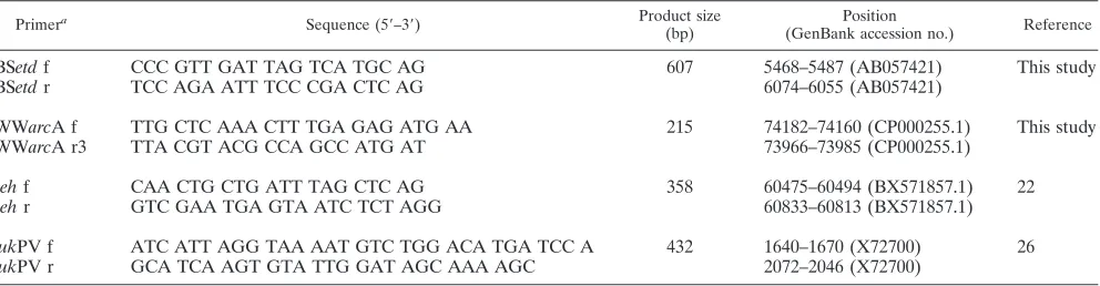
TABLE 1. Primers used in this study
Primera Sequence (5⬘–3⬘) Product size
(bp)
Position
(GenBank accession no.) Reference
BS
etd
f
CCC GTT GAT TAG TCA TGC AG
607
5468–5487 (AB057421)
This study
BS
etd
r
TCC AGA ATT TCC CGA CTC AG
6074–6055 (AB057421)
WW
arc
A f
TTG CTC AAA CTT TGA GAG ATG AA
215
74182–74160 (CP000255.1)
This study
WW
arc
A r3
TTA CGT ACG CCA GCC ATG AT
73966–73985 (CP000255.1)
seh
f
CAA CTG CTG ATT TAG CTC AG
358
60475–60494 (BX571857.1)
22
seh
r
GTC GAA TGA GTA ATC TCT AGG
60833–60813 (BX571857.1)
luk
PV f
ATC ATT AGG TAA AAT GTC TGG ACA TGA TCC A
432
1640–1670 (X72700)
26
luk
PV r
GCA TCA AGT GTA TTG GAT AGC AAA AGC
2072–2046 (X72700)
a
f, forward; r, reverse.
FIG. 1. caMRSA-MP characterization of clinical
S. aureus
isolates.
A 2% agarose gel stained with ethidium bromide is shown. Lanes: M,
marker, 100-bp ladder; 1, 07-00812, t127/ST1/
seh
; 2, 07-00821, t127/
ST1/
seh lukPV
; 3, 05-01197-2, t008/ST8/
arcA lukPV
; 4, 06-00468, t311/
ST5/
lukPV
; 5, 04-02349, t131/ST80/
etd lukPV
; 6, 05-02914, t044/ST80/
etd lukPV
; A, control isolate 06-01172, t008/ST8/
arcA lukPV
; B, control
isolate 06-00300, t044/ST80/
etd lukPV
; C, control isolate 05-01290,
t127/ST1/
seh lukPV
.
on May 16, 2020 by guest
http://jcm.asm.org/
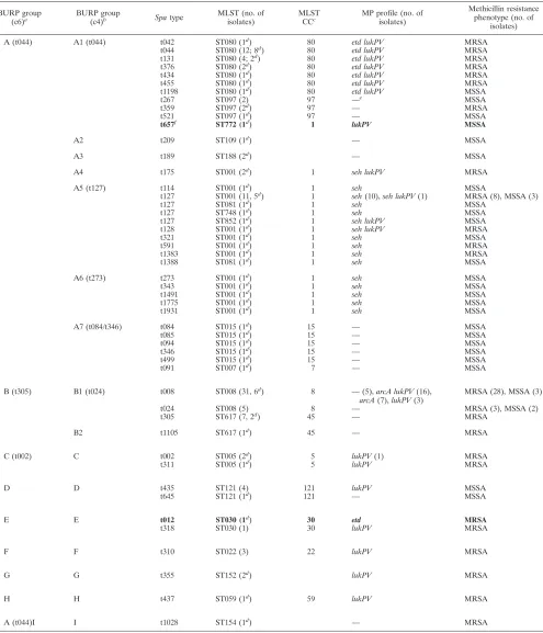
[image:2.585.302.542.507.651.2]TABLE 2. Characteristics of isolates in this study
BURP group
(c6)a BURP group
(c4)b Spatype MLST (no. of
isolates)
MLST
CCc MP profile (no. of
isolates)
Methicillin resistance phenotype (no. of
isolates)
A (t044) A1 (t044) t042 ST080 (1d
) 80 etd lukPV MRSA
t044 ST080 (12; 8d
) 80 etd lukPV MRSA
t131 ST080 (4; 2d) 80 etd lukPV MRSA
t376 ST080 (2d) 80 etd lukPV MRSA
t434 ST080 (1d
) 80 etd lukPV MRSA
t455 ST080 (1d
) 80 etd lukPV MRSA
t1198 ST080 (1d) 80 etd lukPV MSSA
t267 ST097 (2) 97 —e MSSA
t359 ST097 (2d
) 97 — MRSA
t521 ST097 (1d
) 97 — MSSA
t657f ST772 (1d) 1 lukPV MSSA
A2 t209 ST109 (1d
) — MSSA
A3 t189 ST188 (2d
) — MSSA
A4 t175 ST001 (2d) 1 seh lukPV MRSA
A5 (t127) t114 ST001 (1d) 1 seh MSSA
t127 ST001 (11, 5d) 1 seh(10),seh lukPV(1) MRSA (8), MSSA (3)
t127 ST081 (1d
) 1 seh MSSA
t127 ST748 (1d
) 1 seh MSSA
t127 ST852 (1d) 1 seh lukPV MSSA
t128 ST001 (1d) 1 seh lukPV MRSA
t321 ST001 (1d
) 1 seh MSSA
t591 ST001 (1d
) 1 seh MRSA
t1383 ST001 (1d) 1 seh MRSA
t1388 ST081 (1d) 1 seh MSSA
A6 (t273) t273 ST001 (1d) 1 seh MSSA
t343 ST001 (1d) 1 seh MSSA
t1491 ST001 (1d
) 1 seh MSSA
t1775 ST001 (1d
) 1 seh MSSA
t1931 ST001 (1d) 1 seh MSSA
A7 (t084/t346) t084 ST015 (1d
) 15 — MSSA
t085 ST015 (1d) 15 — MSSA
t094 ST015 (1d) 15 — MSSA
t346 ST015 (1d
) 15 — MSSA
t499 ST015 (1d
) 15 — MSSA
t091 ST007 (1d) 7 — MSSA
B (t305) B1 (t024) t008 ST008 (31, 6d) 8 —(5),arcA lukPV(16),
arcA(7),lukPV(3)
MRSA (28), MSSA (3)
t024 ST008 (5) 8 — MRSA (3), MSSA (2)
t305 ST617 (7, 2d
) 45 — MRSA
B2 t1105 ST617 (1d
) 45 — MRSA
C (t002) C t002 ST005 (2d
) 5 lukPV(1) MRSA
t311 ST005 (1d) 5 lukPV MRSA
D D t435 ST121 (4) 121 lukPV MSSA
t645 ST121 (1d
) 121 — MSSA
E E t012 ST030 (1d
) 30 etd MRSA
t318 ST030 (1) 30 lukPV MRSA
F F t310 ST022 (3) 22 lukPV MRSA
G G t355 ST152 (2d
) lukPV MRSA
H H t437 ST059 (1d
) 59 lukPV MRSA
A (t044)I I t1028 ST154 (1d) — MRSA
a
Results of BURP grouping with calculated cost between members of a group less than or equal to 6. Types in parentheses indicate the putative ancestor of the group as defined by BURP analysis.
b
Results of BURP grouping with calculated cost between members of a group less than or equal to 4. Types in parentheses indicate the putative ancestor of the group as defined by BURP analysis.
c
CCs as defined by eBURST analysis with a stringent group definition with six of seven loci.
d
Number of isolates with confirmed MLST types; all other MLST data were inferred on the basis of thespa-MLST mappings done previously at our institute.
e
—, negative for the relevant determinants.
f
Boldface indicates a false classification of the isolates on the basis of the MP profile.
on May 16, 2020 by guest
http://jcm.asm.org/
for
etd
and carried
lukPV
; all but one of the isolates were
MRSA. In contrast, isolates of CC1 were more heterogeneous
(
n
⫽
28; 14 different
spa
types, 4 different MLST types, 15
MSSA and 13 MRSA isolates) and only 5 isolates carried
lukPV
; However, all isolates except one (ST772, single-locus
variant ST1) were positive for
seh.
Two more distantly related
isolates with
spa
type t189 (ST188, double-locus variant ST1)
were also negative for
seh.
All other isolates clustered into
BURP group A but did not belong to CC1 or ST80 (ST97,
ST15, ST7, ST109, ST188) and so were negative for the
deter-minants
etd
and
seh
, thus indicating their affiliation to
alterna-tive clonal lineages not associated with caMRSA.
The caMRSA-MP profiles of isolates of type t008/ST8 (
n
⫽
31; 3 MSSA and 28 MRSA isolates) were found to be the most
heterogeneous. While the majority of isolates tested (
n
⫽
16)
contained
arcA
as well as
lukPV
, indicating their affiliation with
the USA300 caMRSA clone, we also found isolates that
car-ried only
arcA
(
n
⫽
7) or
lukPV
(
n
⫽
3). Five isolates of
spa
type t008, as well as all
spa
type t024 isolates, lacked any
determinant for which analyses were carried out. Interestingly,
six of seven type t008 isolates that carried
arcA
only were
isolated within the same hospital.
Except for one isolate (t012, ST30,
etd
positive), all isolates
not belonging to the caMRSA clones of interest were negative
for the respective determinants (
etd
,
seh
,
arcA
). In contrast,
lukPV
was detected in isolates of all clonal lineages examined
except ST45 and ST154 (Table 2). Thus, on the basis of
caMRSA-MP alone, one isolate of ST30 would have been
classified as a
lukPV
-negative ST80 isolate; on the other hand,
one isolate of CC1 (ST772) would have been classified as a
lukPV
-positive caMRSA isolate not belonging to the
predom-inant caMRSA lineages, leading to two falsely classified
iso-lates among a total of 127 (1.6%) isoiso-lates tested.
DISCUSSION
Monitoring the epidemiology of caMRSA clones is crucial to
prevent their spread within the community as well as their
introduction into hospitals. In particular, the emergence of
“new” clones and the acquisition of additional resistance
de-terminants by already circulating clones pose an ongoing
chal-lenge for infection control authorities (10, 36).
Recent studies (11, 32a) demonstrated that
spa
typing and
BURP analysis, which are, in general, accepted helpful tools in
studies of the short-term as well as the long-term epidemiology
of
S. aureus
, are not able to discriminate unambiguously
be-tween particular clones of caMRSA and common MSSA
clones. In addition, the emergence and spread of “new”
caMRSA clones in Central Europe (especially the spread of
caMRSA t008/ST8/USA300) cannot be monitored efficiently,
because this caMRSA lineage exhibits ambiguous
spa
type
t008, which also occurs in other clones, especially in
ST8-haMRSA-SCC
mec
type IV (epidemic MRSA clones 2 and 6)
and ST8-haMRSA-SCC
mec
type II (“Irish-1”), as well as
MSSA strains.
Therefore, the aim of this study was the development and
validation of a rapid and easy PCR tool for the detection of the
caMRSA clones currently most prevalent in Germany and
Central Europe. The genetic determinants most likely specific
for the three clones were selected on the basis of previous
studies, with a focus on the distribution of particular virulence
determinants within the
S. aureus
population (
seh
,
etd
, and
arcA
). In addition, we selected
lukPV
as a determinant, as it is
often epidemiologically associated with caMRSA.
LukPV
oc-curs in both MRSA and MSSA strains (24), but its role in the
virulence of
S. aureus
is currently controversial (1, 8, 38).
Al-though
lukPV
is not generally associated with caMRSA isolates
(25, 28), it seems to be widespread among European and
American caMRSA populations (36). In the present study we
detected
lukPV
in all ST80 isolates, while the determinant was
variably present in community-acquired
S. aureus
isolates of
ST1 and ST8. We also found
lukPV
in caMRSA isolates of
ST5, ST30, ST22, ST152, and ST59 but not in isolates of CC45
(ST617) and ST154, which is in agreement with the findings of
other studies published previously (36). Thus, we consider
lukPV
to be a useful marker for the detection of caMRSA in
this multiplex approach, in particular, because of its putative
role in invasive infections like necrotizing pneumonia (1, 16);
however, it cannot replace the careful interpretation of
epide-miological records for the classification of isolates as
commu-nity or hospital acquired.
caMRSA isolates of ST8 (USA300) were previously shown
to be positive for
arcA
and
lukPV
in the majority of cases (9);
however, we found a high degree of variability of multiplex
profiles within
spa
type t008, once again highlighting the
di-versity of clones exhibiting this
spa
type. Seven isolates carried
arcA
but not
lukPV
. Interestingly, six of them were collected
within a single hospital, indicating the local spread of a new
caMRSA clone descending from USA300 by the loss of the
lukPV
determinant.
The characterization of community-acquired
S. aureus
iso-lates of CC1 also revealed a high degree of heterogeneity with
a high number of MSSA isolates and only a few
lukPV
-positive
isolates, which is in agreement with the findings of previous
studies (2, 24). However, the large number of
lukPV
-negative
MSSA isolates might represent a putative community reservoir,
in which isolates are waiting for the acquisition of SCC
mec
,
lukPV
, and other virulence or resistance determinants to
be-come caMRSA clones in the future.
The determinants included in our assay are located on
mo-bile genetic elements and thus are subject to putative
horizon-tal transfer (19). Although Holtfreter et al. (13) demonstrated
a strong association of mobile genetic elements with a clonal
background, they found remarkable variations in gene profiles,
indicating horizontal gene transfer within clonal lineages as
well as between isolates of different lineages, finally leading to
the occurrence of particular markers within unrelated lineages.
This was demonstrated for
etd
, which was found in MSSA
CC25 lineages in that study as well as for
seh
, which was found
in MSSA isolates of ST1 and a second genetic background
(ST34,
spa
type t089). Other studies demonstrated the rare
detection of
arcA
in genetic backgrounds different from t008/
ST8/USA300 (7, 9). In this study, we unexpectedly found one
isolate of ST30 carrying
etd
. However, since these “different”
genetic backgrounds are characterized by clearly distinct
spa
types (13) they can be distinguished unambiguously in most
instances. This is also the case for caMRSA isolates of clonal
lineages different from ST80, CC1, and ST8. Thus, we advise
the use of a combination of
spa
typing and caMRSA-MP to
detect suspicious isolates rapidly.
on May 16, 2020 by guest
http://jcm.asm.org/
In conclusion, we present the development of an easy MP
assay for the rapid detection of the most common caMRSA
clones in Central Europe. Our assay facilitated the
unambig-uous assignment of caMRSA/caMSSA isolates to the currently
most prevalent clones in the majority of cases. In combination
with
spa
typing-BURP analysis and resistance testing, this
as-say facilitates the rapid detection of isolates suspected of being
community-acquired
S. aureus
and MRSA isolates. Monitoring
of the emergence and spread of caMRSA clones might assist
with the prevention of the further introduction of virulent
caMRSA strains into hospitals.
ACKNOWLEDGMENTS
We are grateful to all the laboratories that delivered isolates to the
National Reference Centre for Staphylococci. We highly appreciate
strain donations from Hendrik Westh, Hviodore Hospital,
Copenha-gen, Denmark; Alexander W. Friedrich, University of Muenster,
Muenster, Germany; Stephan Harbarth, Hopital Cantonal
Universita-ire, Geneva, Switzerland; Werner Ruppitsch, AGES, Vienna, Austria,
Karina Krziwanek, Krankenhaus der Elisabethinen, Linz, Austria; and
Michal A. Borg, St. Luke’s Hospital, G’Mangia, Malta. We highly
appreciate the excellent technical assistance of our sequencing unit at
the Robert Koch Institute in Berlin, Germany.
REFERENCES
1.Boyle-Vavra, S., and R. S. Daum.2006. Community-acquired methicillin-resistantStaphylococcus aureus: the role of Panton-Valentine leukocidin. Lab. Investig.87:3–9.
2.Coombs, G. W., J. C. Pearson, F. G. O’Brien, R. J. Murray, W. B. Grubb, and K. J. Christiansen.2006. Methicillin-resistantStaphylococcus aureusclones, Western Australia. Emerg. Infect. Dis.12:241–247.
3.Denis, O., A. Deplano, H. De Beenhouwer, M. Hallin, G. Huysmans, M. G. Garrino, Y. Glupczynski, X. Malaviolle, A. Vergison, and M. J. Struelens.
2005. Polyclonal emergence and importation of community-acquired methi-cillin-resistantStaphylococcus aureusstrains harbouring Panton-Valentine leucocidin genes in Belgium. J. Antimicrob. Chemother.56:1103–1106. 4.Deurenberg, R. H., C. Vink, S. Kalenic, A. W. Friedrich, C. A. Bruggeman,
and E. E. Stobberingh.2007. The molecular evolution of methicillin-resistant
Staphylococcus aureus. Clin. Microbiol. Infect.13:222–235.
5.Deutsches Institut fu¨r Normung.2004. DIN 58940. Medical microbiology— susceptibility testing of pathogens to antimicrobial agents. Part 8. Microdi-lution. General method specific requirements, p. 342–353. InDeutsches Institut fu¨r Normung eV (ed.), DIN-Taschenbuch 222: medizinische Mikro-biologie und Immunologie—diagnostische Verfahren. Beuth-Verlag, Berlin, Germany.
6.Diep, B. A., S. R. Gill, R. F. Chang, T. H. Phan, J. H. Chen, M. G. Davidson, F. Lin, J. Lin, H. A. Carleton, E. F. Mongodin, G. F. Sensabaugh, and F. Perdreau-Remington.2006. Complete genome sequence of USA300, an epidemic clone of community-acquired meticillin-resistantStaphylococcus aureus. Lancet367:731–739.
7.Ellington, M. J., L. Yearwood, M. Ganner, C. East, and A. M. Kearns.2007. Distribution of the ACME-arcA gene among methicillin-resistant Staphylo-coccus aureusfrom England and Wales. J. Antimicrob. Chemother. doi: 10.1093/jac/dkm422.
8.Ellington, M. J., R. Hope, M. Ganner, M. Ganner, C. East, G. Brick, and A. M. Kearns.2007. Is Panton-Valentine leucocidin associated with the pathogenesis ofStaphylococcus aureusbacteraemia in the UK? J. Antimi-crob. Chemother.60:402–405.
9.Goering, R. V., L. K. McDougal, G. E. Fosheim, K. K. Bonnstetter, D. J. Wolter, and F. C. Tenover.2007. Epidemiologic distribution of the arginine catabolic mobile element among selected methicillin-resistant and methicil-lin-susceptibleStaphylococcus aureusisolates. J. Clin. Microbiol.45:1981– 1984.
10.Graber, C. J., M. K. Wong, H. A. Carleton, F. Perdreau-Remington, B. L. Haller, and H. F. Chambers.2007. Intermediate vancomycin susceptibility in a community-associated MRSA clone. Emerg. Infect. Dis.13:491–493. 11.Hallin, M., A. Deplano, O. Denis, R. De Mendonca, R. de Ryck, and M. J.
Struelens.2007. Validation of pulsed-field gel electrophoresis andspatyping for long-term, nationwide epidemiological surveillance studies of Staphylo-coccus aureusinfections. J. Clin. Microbiol.45:127–133.
12.Holden, M. T. G., E. J. Feil, J. A. Lindsay, S. J. Peacock, N. P. J. Day, M. C. Enright, T. J. Foster, C. E. Moore, L. Hurst, R. Atkin, A. Barron, N. Bason, S. D. Bentley, C. Chillingworth, T. Chillingworth, C. Churcher, L. Clark, C. Corton, A. Cronin, J. Doggett, L. Dowd, T. Feltwell, Z. Hance, B. Harris, H. Hauser, S. Holroyd, K. Jagels, K. D. James, N. Lennard, A. Line, R. Mayes,
S. Moule, K. Mungall, D. Ormond, M. A. Quail, E. Rabbinowitsch, K. Rutherford, M. Sanders, S. Sharp, M. Simmonds, K. Stevens, S. Whitehead, B. G. Barrell, B. G. Spratt, and J. Parkhill.2004. Complete genomes of two clinicalStaphylococcus aureusstrains: evidence for the rapid evolution of virulence and drug resistance. Proc. Natl. Acad. Sci. USA101:9786–9791. 13.Holtfreter, S., D. Grumann, M. Schmudde, H. T. T. Nguyen, P. Eichler, B.
Strommenger, K. Kopron, J. Kolata, S. Giedrys-Kalemba, I. Steinmetz, W. Witte, and B. M. Broker.2007. Clonal distribution of superantigen genes in clinicalStaphylococcus aureusisolates. J. Clin. Microbiol.45:2669–2680. 14.Klevens, R. M., M. A. Morrison, S. K. Fridkin, A. Reingold, S. Petit, K.
Gershman, S. Ray, L. H. Harrison, R. Lynfield, G. Dumyati, J. M. Townes, A. S. Craig, G. Fosheim, L. K. McDougal, and F. C. Tenover.2006. Com-munity-associated methicillin-resistantStaphylococcus aureusand healthcare risk factors. Emerg. Infect. Dis.12:1991–1993.
15.Kluytmans-Vandenbergh, M. F., and J. A. Kluytmans.2006. Community-acquired methicillin-resistantStaphylococcus aureus: current perspectives. Clin. Microbiol. Infect.12(Suppl. 1):9–15.
16.Labandeira-Rey, M., F. Couzon, S. Boisset, E. L. Brown, M. Bes, Y. Benito, E. M. Barbu, V. Vazquez, M. Hook, J. Etienne, F. Vandenesch, and M. G. Bowden.2007.Staphylococcus aureusPanton-Valentine leukocidin causes necrotizing pneumonia. Science315:1130–1133.
17.Larsen, A., M. Stegger, R. Goering, M. Sorum, and R. Skov.2007. Emergence and dissemination of the methicillin resistantStaphylococcus aureusUSA300 clone in Denmark (2000-2005). Euro Surveill. http://www.eurosurveillance.org /em/v12n02/1202-222.asp.
18.Linde, H., F. Wagenlehner, B. Strommenger, I. Drubel, J. Tanzer, U. Reis-chl, U. Raab, C. Holler, K. G. Naber, W. Witte, F. Hanses, B. Salzberger, and N. Lehn.2005. Healthcare-associated outbreaks and community-acquired infections due to MRSA carrying the Panton-Valentine leucocidin gene in southeastern Germany. Eur. J. Clin. Microbiol. Infect. Dis.24:419–422. 19.Lindsay, J. A., C. E. Moore, N. P. Day, S. J. Peacock, A. A. Witney, R. A.
Stabler, S. E. Husain, P. D. Butcher, and J. Hinds.2006. Microarrays reveal that each of the ten dominant lineages ofStaphylococcus aureushas a unique combination of surface-associated and regulatory genes. J. Bacteriol.188:
669–676.
20.Lowy, F. D.1998.Staphylococcus aureusinfections. N. Engl. J. Med.339:
520–532.
21.Mellmann, A., T. Weniger, C. Berssenbruegge, M. Sammeth, J. Stoye, A. W. Friedrich, H. Grundmann, and D. Harmsen.2007. Determination of the clonal relatedness of the natural population ofStaphylococcus aureususing a calibrated BURP (based upon repeat patterns) algorithm based onspa
typing data, abstr. C-322. Abstr. 107th Gen. Meet. Am. Soc. Microbiol. American Society for Microbiology, Washington, DC.
22.Monday, S. R., and G. A. Bohach.1999. Use of multiplex PCR to detect classical and newly described pyrogenic toxin genes in staphylococcal iso-lates. J. Clin. Microbiol.37:3411–3414.
23.Monecke, S., P. Slickers, H. Hotzel, G. Richter-Huhn, M. Pohle, S. Weber, W. Witte, and R. Ehricht. 2006. Microarray-based characterisation of a Panton-Valentine leukocidin-positive community-acquired strain of methi-cillin-resistantStaphylococcus aureus. Clin. Microbiol. Infect.12:718–728. 24.Mongkolrattanothai, K., S. Boyle, M. D. Kahana, and R. S. Daum.2003.
SevereStaphylococcus aureusinfections caused by clonally related commu-nity-acquired methicillin-susceptible and methicillin-resistant isolates. Clin. Infect. Dis.37:1050–1058.
25.O’Brien, F. G., T. T. Lim, F. N. Chong, G. W. Coombs, M. C. Enright, D. A. Robinson, A. Monk, B. Said-Salim, B. N. Kreiswirth, and W. B. Grubb.2004. Diversity among community isolates of methicillin-resistantStaphylococcus aureusin Australia. J. Clin. Microbiol.42:3185–3190.
26.Prevost, G., B. Cribier, P. Couppie, P. Petiau, G. Supersac, V. Finck-Bar-bancon, H. Monteil, and Y. Piemont.1995. Panton-Valentine leucocidin and gamma-hemolysin fromStaphylococcus aureusATCC 49775 are encoded by distinct genetic loci and have different biological activities. Infect. Immun.
63:4121–4129.
27.Robinson, D. A., and M. C. Enright. 2004. Evolution ofStaphylococcus aureusby large chromosomal replacements. J. Bacteriol.186:1060–1064. 28.Rossney, A. S., A. C. Shore, P. M. Morgan, M. M. Fitzgibbon, B. O’Connell,
and D. C. Coleman.2007. The emergence and importation of diverse geno-types of MRSA harboring the Panton-Valentine leukocidin genepvlreveals thatpvlis a poor marker for community-acquired MRSA in Ireland. J. Clin. Microbiol.45:2554–2563.
29.Rozen, S., and H. Skaletsky.2000. Primer3 on the WWW for general users and for biologist programmers. Methods Mol. Biol.132:365–386. 30.Said-Salim, B., B. Mathema, and B. N. Kreiswirth.2003.
Community-ac-quired methicillin-resistantStaphylococcus aureus: an emerging pathogen. Infect. Control Hosp. Epidemiol.24:451–455.
31.Salgado, C. D., B. M. Farr, and D. P. Calfee.2003. Community-acquired methicillin-resistantStaphylococcus aureus: a meta-analysis of prevalence and risk factors. Clin. Infect. Dis.36:131–139.
32.Stam-Bolink, E. M., D. Mithoe, W. H. Baas, J. P. Arends, and A. V. Moller.
2007. Spread of a methicillin-resistantStaphylococcus aureusST80 strain in the community of the northern Netherlands. Eur. J. Clin. Microbiol. Infect. Dis.26:723–727.
on May 16, 2020 by guest
http://jcm.asm.org/
32a.Strommenger, B., C. Braulke, D. Heuck, C. Schmidt, B. Pasemann, U. Nu¨bel, and W. Witte.2008.Spatyping ofStaphylococcus aureusas a frontline tool in epidemiological typing. J. Clin. Microbiol.46:574–581.
33.Strommenger, B., C. Kettlitz, T. Weniger, D. Harmsen, A. W. Friedrich, and W. Witte.2006. Assignment ofStaphylococcus isolates to groups byspa
typing, SmaI macrorestriction analysis, and multilocus sequence typing. J. Clin. Microbiol.44:2533–2540.
34.Tiemersma, E. W., S. L. Bronzwaer, O. Lyytikainen, J. E. Degener, P. Schrijnemakers, N. Bruinsma, J. Monen, W. Witte, and H. Grundman.2004. Methicillin-resistantStaphylococcus aureusin Europe, 1999–2002. Emerg. Infect. Dis.10:1627–1634.
35.Tietz, A., R. Frei, and A. F. Widmer. 2005. Transatlantic spread of the USA300 clone of MRSA. N. Engl. J. Med.353:532–533.
36.Tristan, A., M. Bes, H. Meugnier, G. Lina, B. Bozdogan, P. Courvalin, M. E. Reverdy, M. C. Enright, F. Vandenesch, and J. Etienne.2007. Global dis-tribution of Panton-Valentine leukocidin-positive methicillin-resistant
Staphylococcus aureus, 2006. Emerg. Infect. Dis.13:594–600.
37.Vandenesch, F., T. Naimi, M. C. Enright, G. Lina, G. R. Nimmo, H. Heffer-nan, N. Liassine, M. Bes, T. Greenland, M. E. Reverdy, and J. Etienne.2003. Community-acquired methicillin-resistant Staphylococcus aureus carrying Panton-Valentine leukocidin genes: worldwide emergence. Emerg. Infect. Dis.9:978–984.
38.Voyich, J. M., M. Otto, B. Mathema, K. R. Braughton, A. R. Whitney, D.
Welty, R. D. Long, D. W. Dorward, D. J. Gardner, G. Lina, B. N. Kreiswirth, and F. R. DeLeo.2006. Is Panton-Valentine leukocidin the major virulence determinant in community-associated methicillin-resistantStaphylococcus aureusdisease? J. Infect. Dis.194:1761–1770.
39.Wenzel, R. P., G. Bearman, and M. B. Edmond.2007. Community-acquired methicillin-resistantStaphylococcus aureus(MRSA): new issues for infection control. Int. J. Antimicrob. Agents30:210–212.
40.Witte, W., C. Braulke, C. Cuny, B. Strommenger, G. Werner, D. Heuck, U. Jappe, C. Wendt, H. J. Linde, and D. Harmsen.2005. Emergence of methi-cillin-resistant Staphylococcus aureus with Panton-Valentine leukocidin genes in central Europe. Eur. J. Clin. Microbiol. Infect. Dis.24:1–5. 41.Witte, W., C. Cuny, B. Strommenger, C. Braulke, and D. Heuck.2004.
Emergence of a new community acquired MRSA strain in Germany. Euro. Surveill.9:1–2.
42.Yamaguchi, T., K. Nishifuji, M. Sasaki, Y. Fudaba, M. Aepfelbacher, T. Takata, M. Ohara, H. Komatsuzawa, M. Amagai, and M. Sugai.2002. Identification of theStaphylococcus aureus etdpathogenicity island which encodes a novel exfoliative toxin, ETD, and EDIN-B. Infect. Immun.70:
5835–5845.
43.Yamasaki, O., A. Tristan, T. Yamaguchi, M. Sugai, G. Lina, M. Bes, F. Vandenesch, and J. Etienne.2006. Distribution of the exfoliative toxin D gene in clinicalStaphylococcus aureusisolates in France. Clin. Microbiol. Infect.12:585–588.