R E S E A R C H A R T I C L E
Open Access
Phenolic compounds isolated from
fermented blueberry juice decrease
hepatocellular glucose output and enhance
muscle glucose uptake in cultured murine
and human cells
Abir Nachar
1,2, Hoda M. Eid
1,2,3, Melinda Vinqvist-Tymchuk
4, Tri Vuong
5, Wilhelmina Kalt
4, Chantal Matar
5and Pierre S. Haddad
1,2*Abstract
Background:We recently reported that blueberry juice fermented (FJ) withSerratia vacciniibacterium has antidiabetic activities both in vivo and in vitro. The purpose of this project was to elucidate the effect of FJ on glucose homeostasis in liver and skeletal muscle cells and to identify active fractions/compounds responsible for this effect.
Methods:FJ was fractionated using standard chromatography procedures. Hepatic (H4IIE, HepG2) and skeletal muscle cells (C2C12) were treated with maximum non-toxic concentrations of FJ, fractions and isolated compounds thereof. Glucose-6-phosphatase (G6Pase) activity was measured using glucose oxidase method. To measure glucose uptake and glycogen synthase (GS) activity, radioactive assays were used.
Results:Fractionation of FJ yielded seven fractions. FJ and its phenolic fractions F2, F3-1 and F3-2 respectively inhibited G-6Pase by 31, 45, 51 and 26%; activated GS by 2.3-, 2.3-, 2.2- and 2-fold; and stimulated glucose uptake by 19, 25, 18 and 15%, as compared to DMSO vehicle control. Subfractionation of the active fractions yielded 4 compounds (catechol, chlorogenic, gallic and protocatechuic acid). Catechol, yielding the greatest bioactivity in G6Pase and glucose uptake assays, decreased G6Pase activity by 54%, increased GS by 2-fold and stimulated glucose uptake by 44% at 45.5μM.
Conclusions:This study identifies novel potential antidiabetic compounds that can help standardize FJ.
Keywords:Diabetes, Glucose homeostasis, Glucose-6-phosphatase, Glycogen Synthase, Glucose transport
Background
Type 2 diabetes (T2D) is a chronic metabolic disease that affects 382 million people worldwide and is associ-ated with many complications, especially cardiovascular diseases [1]. Insulin resistance plays a major role in the physiopathology of type 2 diabetes. It is associated with
an impaired insulin stimulation of glucose transport in muscle and fat as well as an impaired suppression of hepatic glucose production [2]. Nowadays, several people are using natural health products alone or in combination with their hypoglycemic drugs to manage their T2D. Currently, more than one third of Canadian diabetic patients are using alternative medicine [3, 4].
Members of the Vaccinium genus (family Ericaceae), notablyVaccinium angustifoliumAit (Canadian lowbush blueberry), are well known for their antidiabetic activ-ities and have been used in the traditional medicine of many populations to treat T2D [5–8]. Previous studies * Correspondence:[email protected]
1Natural Health Products and Metabolic Diseases Laboratory, Department of
Pharmacology and Physiology, Université de Montréal, Station Centre-Ville, P.O. Box 6128, Montréal, Québec H3C 3J7, Canada
2Canadian Institutes of Health Research Team in Aboriginal Antidiabetic
Medicines and Montreal Diabetes Research Center, Montreal, Canada Full list of author information is available at the end of the article
have shown that different parts of the V. angustifolium plant reduces insulin resistance in obese rats [9] and possess insulin-like, glitazone-like and cytoprotective ef-fects [10]. Blueberry fruits are rich in phenolic com-pounds with antidiabetic properties [8, 11]. Interestingly, the biotransformation of blueberry juice by a bacterium called Serratia vacciniiwas found to greatly increase its content in phenolic compounds and its antioxidant activity [12].
This process also had an impact on the antidiabetic potential of blueberry juice. Indeed, fermented blueberry juice (FJ), in contrast to normal juice, stimulated glucose uptake in muscle cells and adipocytes using an insulin-independent pathway implicating the phosphorylation of AMP-activated protein kinase (AMPK) [13]. This antidi-abetic effect was validated in an animal model using KK-Ay hyperphagic mice [14].
Glucose homeostasis results from equilibrium between the intestinal absorption of glucose, its production by the liver and its utilization by peripheral tissues such as muscle and fat [15]. Insulin regulates hepatic glucose production and storage. It inhibits some transcription factors like the forkhead family and the Peroxisome proliferator-activated receptor-gamma coactivator-1α (PGC-1α) leading to a decrease in the activity of Glucose-6-phosphatase (G6Pase), a key enzyme impli-cated in hepatic glucose production [16, 17]. On the other hand, insulin signaling phosphorylates glycogen synthase kinase-3 (GSK-3) leading to the activation of glycogen synthase (GS), a key enzyme implicated in glu-cose storage [18]. In addition, insulin regulates gluglu-cose uptake and utilization in muscle through the stimulation of glucose transporter 4 (GLUT4) translocation to the plasma membrane in order to mediate facilitative glu-cose diffusion [19].
In continuity with aforementioned studies on FJ in muscle cells and adipocytes, the aim of this project is to elucidate the antidiabetic action of FJ at the level of glu-cose homeostasis in cultured hepatocytes and muscle cells. Importantly, our purpose is to identify active frtions and compounds responsible of the antidiabetic ac-tivity of FJ.
Methods Cell culture
All cell lines used–H4IIE (rat hepatoma), HepG2 (human hepatoma) and C2C12 (murine skeletal myoblasts)–were purchased from American Type Culture Collection (ATCC; Manassas, VA, USA). H4IIE cells were grown in a high glucose Dubelcco’s Modified Eagle Medium (DMEM) containing 10% Fetal Bovin Serum (FBS) and 0.5% antibi-otics (PS: Penicillin 100 U/mL, Streptomycin 100μg/mL). HepG2 cells were grown in DMEM/F12 (50/50) medium, containing 10 FBS and 0.5% PS. On the other hand,
C2C12 myocytes were cultured in DMEM medium con-taining 10 FBS, 10 HS and 0.5% PS then switched to DMEM medium containing 2% HS to initiate ation. Glucose uptake assay was performed on differenti-ated myotubes at the 7th day of differentiation. All cells were cultured and incubated at 37 °C with 5% CO2in
12-well plates for glucose uptake and G6Pase experiments and in 6-well plates for GS experiments. Overnight treat-ment (16–18 h) with the different samples was initiated prior to the determinations.
Preparation of fermented blueberry juice
Mature lowbush blueberry fruits were purchased from Cherryfield Foods Inc. (Cherryfield, Maine, USA). The species was identified by the plant taxonomist Dr. Alain Cuerrier (Montreal Botanical Garden). The frozen fruit preparation represents a homogeneous mixture of sev-eral genotypes provided by many wild blueberry pro-ducers from Canada and Northeastern United States. We selected this starting material because it appropri-ately represents what is regularly used in the industry to prepare commercial wild blueberry juice. For the pur-poses of this study, blueberry fruit mixture (hereafter called juice) was prepared by blending the fruits (100 g) with an equivalent quantity (100 g) of Minimal Broth Davis without dextrose (MM) (Difco Laboratories, Detroit, MI, USA). The fruit mixture was then centri-fuged to remove insoluble particles. The resulting juice was sterilized using 0.22μm Express Millipore filters (Millipore, Etobicoke, ON, Canada).
Serratia vacciniiwere cultured as previously described [12]. The juice was inoculated with a saturated culture of Serratia vaccinii at (7.5 ± 0.3) log CFU ml−1 corre-sponding to 2% of the total juice volume. A control flask was prepared under the same conditions but without in-oculation. The blueberry preparations were incubated in a Lab-Line low-temperature benchtop incubated shaker (Lab-Line Instruments, Inc, Melrose Park, IL, USA) in 250-ml flasks at 22 °C, 120 rev min−1, under aerobic conditions. After a 4 day fermentation period, the FJ was sterilized by 0.22μm filtration as detailed elsewhere [12]. In order to facilitate the handling and insure the stabil-ity of the blueberry preparations, they were freeze-dried to produce powdered material that was kept at −20 °C until use.
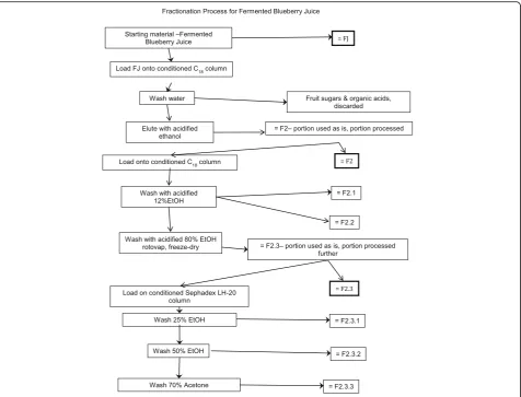
Fractionation of FJ and identification of active compounds
scheme that was carried out to identify active com-pounds present in fermented blueberry juice (FJ). The starting material for the fractionation process was either normal blueberry juice prepared from wild blueberries (the control Juice, CJ), or the FJ in batches of approxi-mately 500 ml. These were placed onto 29.5 cm × 5 cm chromatography columns (pre-conditioned with 1 col-umn volume methanol then 2 colcol-umn volumes water) containing Waters preparative C18 resin (125 Å, 55– 105μ) then washed with 2 column volumes of water, which was sufficient to remove sugars and organic acids (discarded). The phenolic compounds were eluted from the column using 1.2 column volumes of 13 mM tri-fluoroacetic acid (Sigma Aldrich, Oakville, ON) in etha-nol. The ethanol eluent was dried using rotary evaporation and lyophilized to give the Phenolic fraction (F2). The CJ underwent no further fractionation after this initial step. On the other hand, a portion of F2 was dissolved in water and further fractionated on C18 col-umn as outlined above. The first subfraction (F2.1) was eluted using 4 column volumes of 0.16 M HCl (Ricca
Chemical Company, Texas, USA) and 2.06 M ethanol in water. This subfraction was composed mainly of gallic acid, protocatechuic acid, and catechol, as confirmed by HPLC (comparing retention times and UV-vis profiles of the peaks to standards). The next subfraction (F2.2) was eluted using an additional 2 column volumes of 0.16 M HCl and 2.06 M ethanol in water and was rich in chloro-genic acid (as confirmed by HPLC). The remaining bound materials, mainly flavonoids, were eluted using 0.16 M HCl and 13.7 M ethanol in water and were called the flavonoid fraction (F2.3). The three fractions were dried using rotary evaporation and lyophilisation. Next, a portion of the F2.3 fraction was dissolved in 4.28 M ethanol and applied to a 34.5 cm × 5 cm lipophilic Sephadex column (LH-20, 25–100μ, Sigma Aldrich, Oakville, ON, Canada). HCl was neutralized and three fractions were collected. The first fraction enriched in anthocyanins (F2.3.1) was eluted using 7 column vol-umes of 4.28 M ethanol. The second fraction enriched in heteropolymers (F2.3.2) was then eluted using 3 col-umn volumes of 8.56 M ethanol. Finally, a third fraction
[image:3.595.62.539.87.450.2]enriched in proanthocyanidins (F2.3.3) was eluted using 3 column volumes of 9.53 M acetone. All three fractions were dried using rotary evaporation and lyophilisation.
HPLC analysis of normal and fermented blueberry juice The HPLC system used was an Agilent 1100 binary pump system (Agilent Technologies, Mississauga, ON, Canada) with thermostatted column chamber, refriger-ated autosampler, degasser, and DAD detection. Samples were separated on an Agilent Zorbax SB-C18 2.1×50mm 1.8μcolumn held at 26 °C. Samples were held at 4 °C in the autosampler. Elution was done using a two solvent gradient (solvent A was 8 ml trifluoroacetic acid (TFA) per litre water (pH 1.35), solvent B was 6.8 ml TFA per liter acetonitrile) at a flow rate of 0.4 mL/min as follows: 5-10% B (0–12.5 min), 10–20% B (12.5–43.75 min), 20– 100% B (43.75–45 min), hold 100% B 5 min, 100–5% B (50–51 min), re-equilibrating 9 min before the next in-jection. The following wavelengths were monitored: 280 nm (phenolics), 360 nm (flavonols), and 520 nm (anthocyanins), and as well full spectra were recorded from 190 to 600 nm. Designated pure compounds were quantified using authentic standards (Sigma Aldrich, Oakville, ON, Canada).
Cytotoxicity assay (LDH)
The cytotoxicity assay was based on a lactate dehydro-genase (LDH) release kit (LDH Colorimetric Kit; Roche, Mannheim, Germany) and served to determine max-imum non-toxic concentration of each sample in H4IIE, HepG2 and C2C12 cells. As was described previously [22], the cells were treated overnight (16–18 h) with CJ, FJ, fractions thereof, or pure compounds at different concentrations. The culture media were collected separ-ately for each condition and kept on ice (representing released LDH from cells).
Then the cells were lysed by adding culture medium with 1% Triton, for 10 min at 37 °C, 5% CO2
(represent-ing cellular LDH). All the samples were centrifuged at 250×g, 4 °C for 10 min and kept on ice in Eppendorf tubes. The ratio of released LDH to total LDH (total LDH = released LDH + cellular LDH) was calculated for each condition and results normalized to the values ob-tained from cells treated with the vehicle control (DMSO), always present at a final concentration of 0.1%. Maximum non-toxic concentrations for each sample were the highest ones that yielded LDH release compar-able to that of DMSO controls.
Glusose-6-phosphatase (G6Pase) activity
H4IIE cell line was used to measure the activity of G6Pase. Confluent cells were treated overnight (16– 18 h) with DMSO 0.1% (vehicle control), insulin 100 nM (positive control), CJ, FJ, each of the seven fractions (at
5μg/mL) or each of the four pure compounds, all at their maximal non-toxic concentrations (Additional file 1: Table S1). After the treatment, cells were rinsed with PBS then lysed using a 15 mM phosphate buffer containing 0.05% Triton and 1.3 mM Phenol (pH = 6.5). A glucose-6-phosphate-containing buffer (200 mM) was then added to the cell lysates for 40 min at 37 °C; G-6-P contained in this buffer served as a substrate for endogenous G6Pase to yield glucose. A Wako AutoKit Glucose colorimetric assay (Wako Chemicals, Richmond, VA, USA) was used to de-termine the quantity of glucose generated in this reaction according to manufacturers’recommendations. The BCA method was used to determine the protein content for each condition. Results were expressed relative to vehicle control (DMSO 0.1%).
Glycogen Synthase (GS) activity
Glucose uptake bioassay
C2C12 myocytes were grown in 12-well plates to 60% confluence then differentiated into myotubes over a 7-day period. On 7-day 6 of differentiation, C2C12 cells were treated overnight (16–18 h) with either 0.1% DMSO (ve-hicle control), the maximal non-toxic concentrations of CJ, FJ, each of the seven fractions (12.5μg/mL) or each the four pure compounds (Additional file 1: Table S1). Metformin (400μM) was used as a positive control in similar conditions. After treatment, cells were rinsed twice with a warm Krebs phosphate buffer (KPB: 20 mM Hepes, 4.05 mM Na2HPO4, 0.95 mM
NaH2PO4, 136 mM NaCl, 5 mM glucose, 4.7 mM
KCl, 1 mM CaCl2, 1 mM MgSO4, pH 7.4) then
incu-bated with KPB for 30 min at 37 °C. At this point, insulin (100 nM) was added to specific wells to act as another positive control (incubation in KPB buffer for 30 min). Cells were then rinsed twice with warm glucose-free KPB, then incubated in glucose-free KPB containing 0.5μCi/mL 2-deoxy-D-3H-glucose (TRK-383, Amersham Biosciences, Baie d’Urfé, Canada) for 10 min at 37 °C. After incubation, cells were kept on ice and rinsed 3 times with ice-cold glucose-free KPB then lysed in 0.5μL of NaOH (0.1 M) for 30 min. The lysates were transferred with 1 mL of water to scintillation vials, then 4 mL of liquid scintillation cocktail (Beckman Coulter, Fullerton, USA) was added to each vial and incorporated radioactivity was mea-sured using a liquid scintillation counter (LKB Wallac 1219; Perkin Elmer, Woodbridge, ON, Canada).
Statistical analysis
All data were reported as the mean ± SEM of 3 different experiments with triplicates for each sample. Results were analyzed by one-way analysis of variance (ANOVA) (post-hoc pairwise comparisons were carried out with Bonferroni correction). In case the requirement of homogeneity of variances was not fulfilled, Games-Howell test for post-hoc was used. All statistical analyses were carried out using SPSS software, version 24 (IBM Corporation, NY, USA). A pvalue below 0.05 was con-sidered statistically significant.
Results
Cytotoxicity assay (LDH)
After overnight treatment of H4IIE, HepG2 and differ-entiated C2C12 cells with CJ, FJ, each of the seven frac-tions or each of the four pure compounds at different concentrations, LDH released and total LDH were mea-sured for each condition. Additional file 1: Table S1 shows the maximum non-toxic concentration deter-mined for each sample and each cell line based on the results of LDH test.
Fermented blueberry juice and its fractions decrease G6Pase activity and increase GS activation
[image:5.595.304.539.309.634.2]As illustrated in Fig. 2a, FJ significantly decreased G6Pase activity (31% reduction; p< 0.05) as compared to vehicle control (0.1% DMSO). This effect repre-sented roughly half of the effect of the insulin posi-tive control (67% decrease). In contrast, CJ did not induce a significant change in the activity of G6Pase. After fractionation of FJ, the seven fractions were tested in the G6Pase bioassay and all of them showed varied, yet statistically significant, inhibitory effect on the enzyme’s activity that ranged from 20 to 61% when compared to the DMSO (0% inhibition refer-ence; Fig. 2a).
Figure 2b presents results concerning GS activity. FJ induced a significant increase in the enzyme’s activity (2.3 fold increase, p< 0.001) identical to that of the
positive control, insulin (2.3 fold activation). As observed with G6Pase activity, CJ was without any significant ef-fect. In addition, all seven fractions derived from FJ were able to significantly increase the activity of GS and ranged from 1.9- to 2.7-fold when compared to the DMSO vehicle control (100% activation; Fig. 2b).
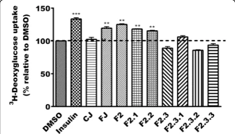
Phenolic fractions enhance glucose uptake in C2C12 muscle cells
When tested for their ability to enhance glucose up-take in C2C12 muscle cells, FJ showed a 20% stimula-tion of this uptake (p< 0.01) whereas CJ was without any effect (similar to vehicle control, DMSO 0.1%; Fig. 3). Insulin used as the positive control stimulated glucose transport into cells by 33% (p< 0.001). The phenolic fraction (F2) along with its subfractions F2.1 and F2.2, F2.3.2 were able to enhance glucose uptake into muscle cells by 25, 18 and 15% respectively (p< 0.01). Bonferroni post hoc test did not reveal signifi-cant differences between FJ, F2, F2.1, and F2.2. The other fractions were without effect when compared to vehicle control (100% activation; Fig. 3).
Identification and isolation of phenolic compounds from the active fractions
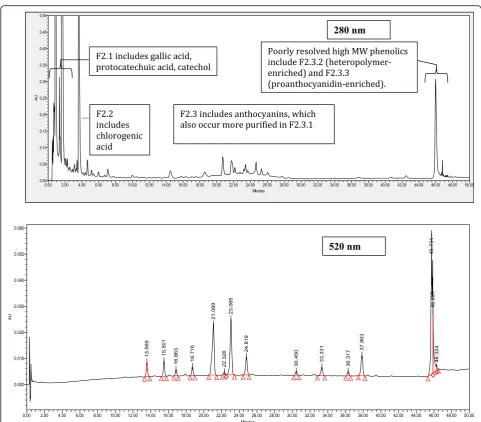
Results of G6Pase and GS activity as well as glucose up-take bioassays were used to guide the identification of compounds found in the active fractions. HPLC analysis confirmed that the three fractions F2, F2.1 and F2.2 were composed mainly of four phenolic compounds: chloro-genic acid (CA), gallic acid (GA), protocatechuic acid (PA) and catechol (Cat). Compound identification was confirmed by comparing retention times and UV-vis
profiles of each peak with respective standards (Fig. 4 and Additional file 1: Table S2). Calculation of phenolic compounds concentrations was based on the external standard method. The concentrations of individual phenolic compounds in FJ were as follows: CA (3.0 mg/ g DW), GA (3.2 mg/g DW), PA (0.3 mg/g DW) and GA (3.2 mg/g DW).
Identification of GA and Cat as the most active
compounds acting on key enzymes implicated in hepatic glucose output
As mentioned above, the most active fractions (F2, F2.1 and F2.2) were rich in phenolic compounds mainly CA, GA, PA and Cat. These compounds were tested at their maximal non-toxic concentration in the G6Pase and the GS assays. Two of the four compounds, namely CA and PA, did not significantly decrease G6Pase activity as compared to DMSO. In contrast, GA was able to reduce the enzyme activity by 25% (p< 0.05) whereas Cat yielded a 54% decrease in G6Pase activity (similar to the positive control, insulin) compared to the vehicle control DMSO 0.1% (p< 0.001, Fig. 5a). Games-Howell test showed that difference between Cat and GA was statisti-cally significant (p< 0.05). In the GS assay, all of the four compounds were able to significantly increase the activ-ity of the enzyme by about 2 fold (CA) or slightly less (GA, PA and Cat) when compared to DMSO 0.1% ve-hicle control (p< 0.05, Fig. 5b). These effects compared favorably with the effect of insulin (2.3 fold increase). No statistically significant differences were detected between the compounds.
Enhancement of glucose uptake by CA, GA and Cat Along with their ability to regulate key enzymes impli-cated in hepatic glucose output, CA, GA and Cat signifi-cantly enhanced glucose uptake in C2C12 cells by 15% (p< 0,01), 16% (p< 0.05) and 43% (p< 0.001) respect-ively. In contrast, PA had no effect when compared to the vehicle control DMSO 0.1% (Fig. 6). Games-Howell test revealed that Cat yielded the greatest activity, it dif-fered significantly from CA (p< 0.01) and GA (p< 0.05). CA and GA were not statistically different.
Discussion
Insulin resistance is an important risk factor for the de-velopment of T2D. Along with the decrease in the phos-phorylation of insulin receptor substrates (IRS-1 and IRS-2) and in PI3-K activity observed in insulin resist-ance, the translocation of glucose transporters and the activity of key enzymes implicated in glucose homeosta-sis are also reduced [24]. Despite the presence of many hypoglycemic drugs in the market, the control of gly-caemia is sometimes hard to achieve. Moreover, popula-tions in Canada and the USA use natural health
[image:6.595.57.290.500.633.2]products alone or in combination with their oral hypoglycemic in order to better manage diabetes [3, 4].
Several parts of the Canadian “lowbush blueberry” (Vaccinium Angustifolium Ait) plant were previously shown by our group to exert antidiabetic activities [10]. A recent study by Klimis-Zakas et al. has reported the ability of a wild blueberry-enriched diet to reduce meta-bolic syndrome risk factors such as chronic inflamma-tion and endothelial dysfuncinflamma-tion in obese Zucker rats, a rat model for metabolic dysfunction [25]. Meanwhile, Matar and colleagues demonstrated that biotransform-ation of blueberry juice by theSerratia vaccinii bacteria conferred a new phytochemical profile to the juice [12]. HPLC evaluation of the phenolic profiles of the normal and fermented juices detected differences in the con-tents of flavonoids and organic acids. Notably, gallic
acid, which was not detected in normal juice, reached concentrations that varied from 26.7 ± 0.9 to 64.6 ± 0.5 mg/kg FW in FJ. On the other hand, chlorogenic acid was rather abundant in FJ, being present at a con-centration of 852.7 ± 2.8 mg/kg FW. In general, total phenolic content nearly tripled after 3 days of fermenta-tion (from 1251.3 ± 278.7 to 3640.3 ± 201.1 mg of gallic acid equivalent (GAE)/kg) and continued to increase modestly thereafter to reach 3926.3 ± 194.3 mg of GAE/ kg after 7 days of fermentation [12]. Moreover, fermen-ted blueberry juice (FJ) not only exhibifermen-ted an increase in phenolic content but was also characterized by more pronounced antioxidant activities compared to the nor-mal juice (CJ) [12, 13, 21]. Our laboratories therefore combined efforts to examine the impact of fermentation on the antidiabetic potential of wild blueberry
[image:7.595.56.538.83.505.2]preparations. We found that fermentation conferred to blueberry juice the capacity to enhance glucose uptake in muscle cells and adipocytes via phosphorylation of AMPK [13].
In the current studies, we first chose to further evalu-ate the antidiabetic potential of FJ by assessing revalu-ate- rate-limiting enzymes of gluconeogenesis (G6Pase) and glycogen synthesis implicated in hepatic glucose output. Indeed, the regulation of hepatic glucose production, along with glucose utilization by peripheral tissues like muscle and fat, are key regulators of systemic glycaemia [17, 26]. We first compared effects of crude preparations of control and fermented blueberry juice. Consistent with our previous studies showing that fermentation conferred antidiabetic potential, we found that fermen-ted blueberry juice significantly affecfermen-ted hepatocellular
glucose homeostasis while control blueberry juice was without effect. Indeed, FJ significantly inhibited G6Pase (to a level roughly half as potent as insulin) and en-hanced GS activity (to a level similar to insulin) in cul-tured murine and human hepatocytes, respectively. These results thus indicate that fermentation of blue-berry juice also confers it the potential to control hepatic glucose output by reducing glucose production and in-creasing glucose storage. We also confirmed our previ-ous finding in C2C12 cells [13] by demonstrating that our fermented blueberry preparation increased glucose transport whereas normal juice extract did not. This ascertained that the current lyophilized preparations be-haved similarly to the actual juice that was used in these previous experiments.
Secondly and importantly, the current studies sought to isolate active fractions/compounds participating in the antidiabetic activity of the FJ in the muscle and liver cell bioassays of glucose homeostasis by using a phyto-chemical fractionation approach that we carried out pre-viously to examine the protective effects of CJ on cardiomyocytes [27]. This approach yielded seven poly-phenolic fractions of FJ, which were tested in the G6Pase, GS and glucose uptake bioassays. FJ fractions were able to significantly decrease G6Pase activity to levels ranging from 20 to 61%. The heteropolymer-enriched fraction, F2.3.2, gave the highest effect, which was very close to that of the positive control insulin. Un-fortunately, the complexity of this fraction precluded further sub-fractionation. Next in apparent potency were the F2 and F2.1 fractions, the latter being enriched in
Fig. 5Effect of CA, GA, PA and Cat isolated from the active fractions of FJ on the activity of G6Pase (a) and GS (b). Results shown represent the change in G6Pase (a) and GS (b) activities observed after overnight treatment of H4IIE and HepG2 cells with maximum non-toxic concentration of isolated compounds: CA (70.5μM), GA (147μM), PA (162.2μM) and Cat (45.5μM). Results are expressed relative to DMSO (0.1%) vehicle control (a: 0% inhibition;b: 100 % activity). Insulin (100 nM) was used as a positive control. **:p<0.01 and ***:p<0.001 significantly different from DMSO (n=3
in triplicates)
[image:8.595.302.538.86.263.2] [image:8.595.57.290.87.418.2]GA, PA and Cat. In contrast, all FJ phenolic fractions in-creased the activity of GS within a relatively narrow range (1.9- to 2.7- fold increase), which was comparable to the action of the insulin positive control.
In terms of glucose uptake in skeletal muscle cells, on the other hand, significant glucose transport activity was associated only with three phenolic-enriched fractions, namely F2, F2.1 and F2.2. Since all fractions were active to varying degrees in both G6Pase and GS assays but only these three phenolic fractions showed a significant effect on glucose uptake, we examined in greater detail the constituents of the latter fractions. HPLC analysis re-vealed that the phenolic fractions contained mainly four compounds, namely chlorogenic acid (CA), gallic acid (GA), protochatecuic acid (PA) and catechol (Cat). These compounds are known for their antioxidant activ-ity. Many studies also showed additional beneficial ef-fects of CA, GA and PA. Indeed, CA has demonstrated anti-inflammatory, anti-diabetic, neuro-protective and cardio-protective properties [28–31]. GA exerts anti-diabetic activities [32], improves hyperglycaemia and glucose tolerance [33], while offering protection against diabetes complications, notably through cardio-protective properties [34, 35]. On the other hand, PA possesses anti-inflammatory [36, 37] and anti-apoptotic properties [38]. It also improves angiogenesis [39] and protects against hepatotoxicity and nephrotoxicity [40]. In contrast, and to our knowledge, no recent studies have addressed effects of catechol on glucose metabol-ism. Thus, our study is the first to reveal the effect of these phenolic compounds on hepatic glucose homeo-stasis and glucose transport in muscle.
The four compounds were able to increase GS activity to levels similar to those observed for FJ and its active fractions. GA and Cat showed stronger effects than the two others on the reduction of G6Pase activity. This ef-fect was close to that observed for FJ and its active frac-tions. In terms of glucose transport, PA was inactive; GA and CA had similar effects, whereas Cat had the most pronounced activity. Interestingly, Cat stands out in this study as having the best activity profile in almost all bio-assays, yielding equivalent if not stronger effects than FJ and its phenolic active fractions themselves.
Conclusion
Altogether, the results of this study confirmed that fer-mentation of blueberry juice confers it antidiabetic po-tential in liver and skeletal muscle cells through the regulation of key hepatic enzymes implicated in gluco-neogenesis and glycogen synthesis and the enhancement of skeletal muscle glucose uptake. Using a phytochemical fractionation approach, we now demonstrate that this activity resides principally in phenolic fractions and can be attributed, at least in part, to CA, GA, PA and Cat.
This is congruent with studies showing antidiabetic properties for CA and GA, whereas it uncovers novel beneficial actions for PA. As mentioned, however, of all pure compounds in our study, Cat stood out as the most promising constituent, showing activity similar to or higher than the parent FJ (and even the insulin control) in all three bioassays. Future studies will be needed to further examine effects of flavonoids, anthocyanins, het-eropolymers and proanthocyanins present in FJ.
Our study thus provides important insights into novel potential antidiabetic molecules that are produced when blueberry juice is fermented with Serratia vaccinii. Im-portantly, the identified compounds represent quality control tools that can be used to ensure the efficacy of FJ and hence standardize FJ preparations.
Additional file
Additional file 1: Table S1.A. Optimum non-toxic concentrations of CJ, FJ and corresponding fractions used for bioassays in H4IIE, HepG2 and C2C12 cells. B. Maximum non-toxic concentrations of pure compounds used for bioassays in H4IIE, HepG2 and C2C12 cells.Table S2.Fractionation of fermented blueberry extract, indicating the major component(s) in each fraction (F). (DOCX 19 kb)
Abbreviations
AMPK:AMP- activated protein kinase; Antho: Anthocyanins fraction; CA: Chlorogenic acid; Cat: Catechol; CJ: Control blueberry juice; FJ: Fermented blueberry juice; Flv: Flavonoids fraction; G6Pase: Glucose-6-Phosphatase; GA: Gallic acid; GLUT4: Glucose transporter 4; GS: Glycogen Synthase; GSK-3: Glycogen Synthase kinase-3; Hetero: Heteropolymers fraction; IRS: Insulin Receptor Substrate; PA: Protocatechuic acid; PAC: Proanthocyanidins fraction; PGC-1α: Peroxisome proliferator-activated receptor gamma coactivator 1-alpha; Phe: Phenolic fraction
Acknowledgments
The project was carried out in collaboration with BioAtlantech and Vaccinium Technologies, Fredericton, New Brunswick, Canada; special thanks are expressed to John Argall and Denise Philpott. We also wish to thank Dr John Thor Arnason of the University of Ottawa for valuable discussions and advice.
Funding
This study was funded by Agriculture and AgriFood Canada through its “Developing Innovative Agrifood Products”(DIAP) program (no 5025).
Availability of data and materials
The datasets supporting the conclusions of this article are included within the manuscript and its supplementary information files.
Authors’contributions
AN performed the biological activities, data analysis and wrote the manuscript. HME performed the statistical analysis, interpreted data, wrote and critically revised the manuscript. MVT performed the fractionation and HPLC analysis. WK and CM collaborated by providing precious professional advice for the phytochemical analysis, fermentation and fractionation of the juice. TV prepared the fermented juice. PSH is the supervisor and the corresponding author. All co-authors read, commented and approved the final manuscript.
Competing interests
The authors declare that they have no competing interests.
Consent for publication
Ethics approval and consent to participate
Not applicable.
Author details
1Natural Health Products and Metabolic Diseases Laboratory, Department of
Pharmacology and Physiology, Université de Montréal, Station Centre-Ville, P.O. Box 6128, Montréal, Québec H3C 3J7, Canada.2Canadian Institutes of
Health Research Team in Aboriginal Antidiabetic Medicines and Montreal Diabetes Research Center, Montreal, Canada.3Department of
Pharmacognosy, University of Beni-Suef, Beni-Suef, Egypt.4Food chemistry,
Agriculture and Agri-Food Canada, Government of Canada, Kentville, Nova Scotia, Canada.5Department of Nutritional Sciences, Faculty of Health Sciences, University of Ottawa, Ottawa, Canada.
Received: 10 September 2016 Accepted: 23 February 2017
References
1. Riaz M, Zia-Ul-Haq M, Saad B. The role of anthocyanins in obesity and diabetes. In: Anthocyanins and human health: biomolecular and therapeutic aspects. Cham: Springer International Publishing; 2016.
2. Fandriks L. Roles of the gut in the metabolic syndrome: an overview. J Intern Med. 2016. doi:10.1111/joim.12584. [Epub ahead of print] 3. Nahas R, Moher M. Complementary and alternative medicine for the
treatment of type 2 diabetes. Can Fam Physician. 2009;55(6):591–6. 4. Esmail N. Complementary and AlternativeMedicine in Canada: Trends in Use
and Public Attitudes, 1997–2006. In: Public Policy Sources, vol. 87. Vancouver: The Fraser Institute; 2007.
5. Koupy D, Kotolova H, Kucerova J. Effectiveness of phytotherapy in supportive treatment of type 2 diabetes mellitus Billberry (Vaccinium myrtillus). Ceska Slov Farm. 2015;64(1–2):3–6.
6. Haddad PS, Depot M, Settaf A, Cherrah Y. Use of antidiabetic plants in Morocco and Quebec. Diabetes Care. 2001;24(3):608–9.
7. Stull AJ, Cash KC, Johnson WD, Champagne CM, Cefalu WT. Bioactives in blueberries improve insulin sensitivity in obese, insulin-resistant men and women. J Nutr. 2010;140(10):1764–8.
8. Edirisinghe I, Burton-Freeman B. Anti-diabetic actions of Berry polyphenols– Review on proposed mechanisms of action. J Berry Res. 2016;6:237–50. 9. Seymour EM, Tanone II, Urcuyo-Llanes DE, Lewis SK, Kirakosyan A,
Kondoleon MG, Kaufman PB, Bolling SF. Blueberry intake alters skeletal muscle and adipose tissue peroxisome proliferator-activated receptor activity and reduces insulin resistance in obese rats. J Med Food. 2011; 14(12):1511–8.
10. Martineau LC, Couture A, Spoor D, Benhaddou-Andaloussi A, Harris C, Meddah B, Leduc C, Burt A, Vuong T, Le Mai P, et al. Anti-diabetic properties of the Canadian lowbush blueberry Vaccinium angustifolium Ait.
Phytomedicine. 2006;13(9–10):612–23.
11. Song Y, Huang L, Yu J. Effects of blueberry anthocyanins on retinal oxidative stress and inflammation in diabetes through Nrf2/HO-1 signaling. J Neuroimmunol. 2016;301:1–6.
12. Martin LJ, Matar C. Increase of antioxidant capacity of the lowbush blueberry (Vaccinium angustifolium) during fermentation by a novel bacterium from the fruit microflora. J Sci Food Agric. 2005;85(9):1477–84. 13. Vuong T, Martineau LC, Ramassamy C, Matar C, Haddad PS. Fermented
Canadian lowbush blueberry juice stimulates glucose uptake and AMP-activated protein kinase in insulin-sensitive cultured muscle cells and adipocytes. Can J Physiol Pharmacol. 2007;85(9):956–65.
14. Vuong T, Benhaddou-Andaloussi A, Brault A, Harbilas D, Martineau LC, Vallerand D, Ramassamy C, Matar C, Haddad PS. Antiobesity and antidiabetic effects of biotransformed blueberry juice in KKA(y) mice. Int J Obes (Lond). 2009;33(10):1166–73.
15. Han HS, Kang G, Kim JS, Choi BH, Koo SH. Regulation of glucose metabolism from a liver-centric perspective. Exp Mol Med. 2016;48:e218. 16. Oh KJ, Han HS, Kim MJ, Koo SH. CREB and FoxO1: two transcription factors
for the regulation of hepatic gluconeogenesis. BMB Rep. 2013;46(12):567–74. 17. Anyamaneeratch K, Rojvirat P, Sukjoi W, Jitrapakdee S. Insights into
transcriptional regulation of hepatic glucose production. Int Rev Cell Mol Biol. 2015;318:203–53.
18. Dieni CA, Bouffard MC, Storey KB. Glycogen synthase kinase-3:
cryoprotection and glycogen metabolism in the freeze-tolerant wood frog. J Exp Biol. 2012;215(3):543–51.
19. Leto D, Saltiel AR. Regulation of glucose transport by insulin: traffic control of GLUT4. Nat Rev Mol Cell Biol. 2012;13(6):383–96.
20. Matchett MD, MacKinnon SL, Sweeney MI, Gottschall-Pass KT, Hurta RA. Inhibition of matrix metalloproteinase activity in DU145 human prostate cancer cells by flavonoids from lowbush blueberry (Vaccinium angustifolium): possible roles for protein kinase C and mitogen-activated protein-kinase-mediated events. J Nutr Biochem. 2006;17(2):117–25. 21. Vuong T, Matar C, Ramassamy C, Haddad PS. Biotransformed blueberry juice
protects neurons from hydrogen peroxide-induced oxidative stress and mitogen-activated protein kinase pathway alterations. Br J Nutr. 2010;104(5):656–63. 22. Nachar A, Vallerand D, Musallam L, Lavoie L, Badawi A, Arnason J, Haddad
PS. The action of antidiabetic plants of the canadian james bay cree traditional pharmacopeia on key enzymes of hepatic glucose homeostasis. Evid Based Complement Alternat Med. 2013;2013:189819.
23. Thomas JA, Schlender KK, Larner J. A rapid filter paper assay for UDPglucose-glycogen glucosyltransferase, including an improved biosynthesis of UDP-14C-glucose. Anal Biochem. 1968;25(1):486–99. 24. Abdul-Ghani MA, DeFronzo RA. Pathogenesis of Insulin Resistance in
Skeletal Muscle. J Biomed Biotechnol. 2010;2010:19. Article ID 476279. doi: 10.1155/2010/476279.
25. Klimis-Zacas D, Vendrame S, Kristo AS. Wild blueberries attenuate risk factors of the metabolic syndrome. J Berry Res. 2016;6(2):225–36.
26. Mues C, Zhou J, Manolopoulos KN, Korsten P, Schmoll D, Klotz LO, Bornstein SR, Klein HH, Barthel A. Regulation of glucose-6-phosphatase gene expression by insulin and metformin. Horm Metab Res. 2009;41(10):730–5. 27. Louis XL, Thandapilly SJ, Kalt W, Vinqvist-Tymchuk M, Aloud BM, Raj P, Yu L, Le H,
Netticadan T. Blueberry polyphenols prevent cardiomyocyte death by preventing calpain activation and oxidative stress. Food Funct. 2014;5(8):1785–94. 28. Cheng DM, Pogrebnyak N, Kuhn P, Krueger CG, Johnson WD, Raskin I.
Development and phytochemical characterization of high polyphenol red lettuce with anti-diabetic properties. PLoS One. 2014;9(3):e91571. 29. Shen W, Qi R, Zhang J, Wang Z, Wang H, Hu C, Zhao Y, Bie M, Wang
Y, Fu Y, et al. Chlorogenic acid inhibits LPS-induced microglial activation and improves survival of dopaminergic neurons. Brain Res Bull. 2012;88(5):487–94.
30. Hwang SJ, Kim YW, Park Y, Lee HJ, Kim KW. Anti-inflammatory effects of chlorogenic acid in lipopolysaccharide-stimulated RAW 264.7 cells. Inflamm Res. 2014;63(1):81–90.
31. Li Y, Shen D, Tang X, Li X, Wo D, Yan H, Song R, Feng J, Li P, Zhang J, et al. Chlorogenic acid prevents isoproterenol-induced hypertrophy in neonatal rat myocytes. Toxicol Lett. 2014;226(3):257–63.
32. Kade IJ, Ogunbolude Y, Kamdem JP, Rocha JB. Influence of gallic acid on oxidative stress-linked streptozotocin-induced pancreatic dysfunction in diabetic rats. J Basic Clin Physiol Pharmacol. 2014;25(1):35–45. 33. Bak EJ, Kim J, Jang S, Woo GH, Yoon HG, Yoo YJ, Cha JH. Gallic acid
improves glucose tolerance and triglyceride concentration in diet-induced obesity mice. Scand J Clin Lab Invest. 2013;73(8):607–14.
34. Umadevi S, Gopi V, Vellaichamy E. Inhibitory effect of gallic acid on advanced glycation end products induced up-regulation of inflammatory cytokines and matrix proteins in H9C2 (2–1) cells. Cardiovasc Toxicol. 2013;13(4):396–405. 35. Ramkumar KM, Vijayakumar RS, Vanitha P, Suganya N, Manjula C, Rajaguru P,
Sivasubramanian S, Gunasekaran P. Protective effect of gallic acid on alloxan-induced oxidative stress and osmotic fragility in rats. Hum Exp Toxicol. 2014;33(6):638–49.
36. Del Corno M, Varano B, Scazzocchio B, Filesi C, Masella R, Gessani S. Protocatechuic acid inhibits human dendritic cell functional activation: role of PPARgamma up-modulation. Immunobiology. 2014;219(6):416–24. 37. Wei M, Chu X, Guan M, Yang X, Xie X, Liu F, Chen C, Deng X.
Protocatechuic acid suppresses ovalbumin-induced airway inflammation in a mouse allergic asthma model. Int Immunopharmacol. 2013;15(4):780–8. 38. Deng JS, Lee SD, Kuo WW, Fan MJ, Lin YM, Hu WS, Huang YC, Velmurugan BK,
Tsai FJ, Tsai CH, et al. Anti-apoptotic and pro-survival effect of protocatechuic acid on hypertensive hearts. Chem Biol Interact. 2014;209:77–84.
39. Kang Z, Zhu H, Jiang W, Zhang S. Protocatechuic acid induces angiogenesis through PI3K-Akt-eNOS-VEGF signalling pathway. Basic Clin Pharmacol Toxicol. 2013;113(4):221–7.