Copyright © 1999, American Society for Microbiology. All Rights Reserved.
Development of Amplified 16S Ribosomal DNA Restriction Analysis
for Identification of
Actinomyces
Species and Comparison
with Pyrolysis-Mass Spectrometry and Conventional
Biochemical Tests
VAL HALL,* G. L. O’NEILL,† J. T. MAGEE,ANDB. I. DUERDEN
Anaerobe Reference Unit, Department of Medical Microbiology and Public Health Laboratory, University Hospital of Wales, Cardiff CF4 4XW, United Kingdom
Received 19 January 1999/Returned for modification 4 March 1999/Accepted 8 April 1999
Identification ofActinomycesspp. by conventional phenotypic methods is notoriously difficult and unreliable. Recently, the application of chemotaxonomic and molecular methods has clarified the taxonomy of the group and has led to the recognition of several new species. A practical and discriminatory identification method is now needed for routine identification of clinical isolates. Amplified 16S ribosomal DNA restriction analysis (ARDRA) was applied to reference strains (n527) and clinical isolates (n536) ofActinomycesspp. and other gram-positive rods. Clinical strains were identified initially to the species level by conventional biochemical tests. However, given the low degree of confidence in conventional methods, the findings obtained by ARDRA were also compared with those obtained by pyrolysis-mass spectrometry. The ARDRA profiles generated by the combination ofHaeIII andHpaII endonuclease digestion differentiated all reference strains to the species or subspecies level. The profiles correlated well with the findings obtained by pyrolysis-mass spectrometry and by conventional tests and enabled the identification of 31 of 36 clinical isolates to the species level. ARDRA was shown to be a simple, rapid, cost-effective, and highly discriminatory method for routine identification of
Actinomycesspp. of clinical origin.
The genusActinomycescomprises a heterogeneous group of anaerobic and facultatively anaerobic, asporogenous, nonmo-tile, non-acid-fast, gram-positive rods with a G1C content of 55 to 71 mol% (2). ManyActinomycesspp. are known to be indigenous to mucous membranes, particularly those in the oral cavity, in humans and other animals. Members of the genus are known to cause actinomycosis and may be found in polymicrobial infections arising from tissue invasion by oral anaerobes (16). Some species are significant in periodontal disease (16).
Detection of the presence of Actinomyces spp. in clinical specimens may affect the prognosis and patient management, but identification by conventional phenotypic methods is no-toriously difficult and unreliable (6). Problems arise from the slow and granular growth of some isolates (particularly Acti-nomyces israelii), the poor reproducibilities of biochemical tests, and the lack of discriminatory power of biochemical tests. The lack of discriminatory power may indicate heterogeneity within species. Indeed, for some species, subdivisions based on biochemical or serological differences have been described (17, 18), and combination in a single genospecies of some serotypes ofActinomyces naeslundiiandActinomyces viscosus has been proposed (8).
Recently, the application of chemotaxonomic and molecular methods has clarified the taxonomy of the group and has led to the recognition of several new species. These include
Actino-myces europaeus(4),Actinomyces graevenitzii(12),Actinomyces neuii(5),Actinomyces radingae(22), andActinomyces turicensis
(22). Other new species have been assigned to other, similar genera: Arcanobacterium bernardiae (13), Arcanobacterium phocae (13), Arcanobacterium pyogenes (13), Actinobaculum schaalii (9), and Actinobaculum suis (9). Currently, little is known of the natural habitats, clinical prevalence, and patho-genic potential of these species. A discriminatory, reproduc-ible, and practical method for characterization of clinical iso-lates may help elucidate their occurrence and significance.
Pyrolysis mass-spectrometry (PMS) has been shown to be a useful tool in differentiating groups of organisms to the species or subspecies level (10) and represents a whole-cell analysis approach independent of conventional biochemical tests. However, PMS is a fingerprinting method and is best suited to examination of large batches of organisms. Barsotti et al. (1) demonstrated the potential of rRNA gene (rDNA) restriction patterns as taxonomic tools forActinomyces. This approach may be enhanced by initial amplification of rDNA by PCR. Amplified rDNA restriction analysis (ARDRA) has been used to identify various fungi (21) and bacteria (19, 20).
In this study, ARDRA was applied to reference strains and clinical isolates of Actinomyces spp. and some other gram-positive rods. Clinical isolates were identified initially to the species level by conventional biochemical tests and comparison of the results with those for reference strains. However, given the low degree of confidence in this method, the findings were also compared with those obtained by PMS.
MATERIALS AND METHODS
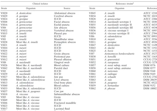
Bacterial strains.A total of 63 strains were examined (Table 1). Reference
strains (n527) represented 17 species ofActinomycesand 5 species of other, similar genera. Clinical strains comprised 35 human isolates and 1 animal isolate from the collection held by the Anaerobe Reference Unit, Public Health Labo-ratory Service. Strains were selected to represent the range of species and sites
* Corresponding author. Mailing address: Anaerobe Reference Unit, Department of Medical Microbiology and Public Health Labo-ratory, University Hospital of Wales, Cardiff CF4 4XW, United King-dom. Phone: 44(0)1222 742171. Fax: 44(0)1222 744123. E-mail: hallv @cardiff.ac.uk.
† Present address: Laboratory of Hospital Infection, Central Public Health Laboratory, London NW9 5HT, United Kingdom.
2255
on May 15, 2020 by guest
http://jcm.asm.org/
of isolation of strains referred for identification from laboratories throughout England and Wales. Strains were stored at280°C on Microbank beads (Pro-lab Diagnostics, Wirral, United Kingdom) and were recovered on Fastidious Anaer-obe Agar (FAA; LabM, Bury, United Kingdom) incubated anaerobically at 37°C for 48 h.
Conventional tests.Strains were determined to be members of the genus
Actinomyceson the basis of volatile and nonvolatile fatty acid end products of glucose metabolism, as detected by gas-liquid chromatography as described else-where (7).
Cell and colonial morphologies, pigment production, fluorescence under long-wave UV illumination, ability to grow in air and in air plus 5% CO2, and
production of catalase and indole were recorded. Hydrolysis of esculin and starch and production of acid from amygdalin, arabinose, cellobiose, glucose, mannitol, raffinose, ribose, salicin, sucrose, trehalose, and xylose were tested by the method of Phillips (14). Production of nitrate reductase, urease, pyrazinamidase,b -ga-lactosidase, a-glucosidase, andb-N-acetyl-glucosaminidase was detected after incubation for 18 to 24 h with Rosco diagnostic tablets (BioConnections, Leeds, United Kingdom). Identifications were made initially by reference to the scheme of Brazier and Hall (3) and subsequently by reference to the publications of Funke et al. (5), Pascual Ramos et al. (12), Funke et al. (4) and Lawson et al. (9).
PMS.Pyrolysis was performed with colonies from 48-h anaerobic FAA cul-tures, and the mass spectra were analyzed as described elsewhere (11).
ARDRA.All strains were tested in triplicate. Strains were cultured
anaerobi-cally on FAA for 48 h.
For DNA extraction, a 1-ml loopful of bacteria was suspended in 100ml of Chelex resin (5%; Bio-Rad Laboratories, Richmond, Calif.) in sterile distilled water, boiled for 8 min, and centrifuged at 17,0003gfor 10 min. The superna-tant was decanted.
For PCR, DNA extract (5ml) was added to 45ml of a reaction mixture containing 1 U of Taqpolymerase (Pharmacia Biotech, St. Albans, United Kingdom), each deoxynucleoside triphosphate at a concentration of 200 mM, 20 pmol of each primer (pA and pH9), 2.25 mM magnesium chloride, 10 mM Tris HCl (pH 9), 50 mM KCl, and 0.1% Triton X-100. The primer sequences were AGAGTTTGATCCTGGCTCAG (pA) and AAGGAGGTGATCCAGCCGCA (pH9). Amplification was accomplished by 31 cycles of denaturation at 92°C for 2 min, annealing at 55°C for 1 min, and extension at 72°C for 1.5 min, with a final extension period of 5 min. The specific PCR product (approximately 1,600 bp) was detected by electrophoresis of a 5-ml sample for 40 min at 5 V/cm in 1% agarose in TAE (Tris-acetate-EDTA) buffer with ethidium bromide (0.5mg/ml) and was visualized under UV illumination.
Restriction endonuclease digestion was performed withHaeIII andHpaII endonucleases in separate reaction mixtures containing 1ml of endonuclease (10 U/ml), 1.5ml of matched incubation buffer, and 12.5ml of PCR product; the mixture was incubated for 1.5 h at 37°C. For electrophoresis, 3ml of 63loading buffer (Advanced Biotechnologies, Epsom, United Kingdom) was added to each sample, and 15ml was electrophoresed for 1.75 h at 5 V/cm in Metaphor agarose (3.5%; FMC, Rockland, Maine) in TAE buffer with ethidium bromide (0.5
mg/ml). Molecular size markers (2 kb; Sigma, Poole, United Kingdom) were run in duplicate or triplicate alongside the samples. The gels were destained in deionized water for 10 min and photographed under UV illumination, and the images were stored on a floppy disk.
[image:2.612.54.563.83.443.2]Gel data were analyzed with GelCompar software (Applied Maths, Kortrijk, Belgium). Distinct banding patterns were assigned three-digit codes, arbitrarily numbered in the order encountered, and were stored as types in HAE and HPA libraries. The combined results for each strain were recorded as, for example, 001/003, which represent theHaeIII andHpaII profiles, respectively.
TABLE 1. Sources of bacterial strainsa
Clinical isolates Reference strainsb
Strain Organism Source Strain Organism Reference
VH20 A. denticolens Abdominal abscess VH45 A. israelii ATCC 12102
VH29 A. georgiae Cerebral abscess VH34 A. israelii NCTC 10236
VH24 A. georgiae IUCD VH28 A. gerencseriae ATCC 23860
VH48 A. gerencseriae Facial abscess VH14 A. naeslundiiserotype I NCTC 10301
VH16 A. gerencseriae Eyelid puncta VH55 A. naeslundiiserotype II ATCC 44339
VH4 A. gerencseriae Jaw abscess VH56 A. naeslundiiserotype III ATCC 44340
VH32 A. gerencseriae Vertebral abscess VH36 A. viscosusserotype I NCTC 10951
VH15 A. israelii Pleural pus VH54 A. viscosusserotype II ATCC 27044
VH5 A. israelii IUCD VH6 A. odontolyticus NCTC 09935
VH1 A. israelii Mandibular sinus VH10 A. meyeri ATCC 35568
VH38 Most likeA. israelii Perinephric abscess VH11 A. georgiae ATCC 49285
VH21 A. israelii IUCD VH47 A. denticolens NCTC 11490
VH44 A. meyeri IUCD VH43 A. slackii NCTC 11923
VH35 A. meyeri IUCD VH25 A. bovis NCTC 11535
VH22 A. meyeri Groin abscess VH7 Actinomyces hordeovulneris ATCC 35275
VH41 A. meyeri Peritoneal pus VH30 A. howellii NCTC 11636
VH12 A. meyeri Pleural effusion VH51 A. graevenitzii CCUG 27294
VH8 A. naeslundii Gingival swab VH52 A. europaeus CCUG 32789A
VH26 Most likeA. naeslundii Blood culture VH57 A. neuiisubsp.neuii DSM 8576
VH42 Most likeA. naeslundii Parotid duct VH58 A. neuiisubsp.anitratus DSM 8577
VH2 A. naeslundii IUCD VH60 A. turicensis DSM 9168
VH39 A. naeslundii IUCD VH61 A. radingae DSM 9169
VH27 Most likeA. odontolyticus Jaw pus VH53 A. schaalii CCUG 27420
VH50 Most likeA. odontolyticus Liver abscess VH63 A. suis DSM 20639
VH13 A. odontolyticus Oral bone plate VH19 A. pyogenes NCTC 05224
VH40 A. odontolyticus Blood culture VH59 A. bernardiae DSM 9152
VH33 Most likeA. odontolyticus IUCD VH62 A. phocae DSM 10002
VH37 Most likeA. pyogenes Cow jaw
VH18 A. viscosus Submandibular abscess
VH3 Most likeA. viscosus Groin VH49 Most likeA. viscosus IUCD
VH46 Most likeA. viscosus Fractured mandible VH23 Most likeA. viscosus IUCD
VH17 Most likeA. viscosus Lacrimal fluid
VH9 Actinomycesspecies Dental abscess
VH31 Actinomycesspecies Osteomyelitis
aAbbreviations: IUCD, intrauterine contraceptive device; ATCC, American Type Culture Collection, Rockville, Md.; NCTC, National Collection of Type Cultures,
London, United Kingdom; CCUG, Culture Collection, University of Goteborg, Goteborg, Sweden; DSM, Deutsche Sammlung von Mikroorganismen, Braunschweig, Germany.
bReference strains other thanA. israeliiNCTC 10236 represent type strains for species or subspecies.
on May 15, 2020 by guest
http://jcm.asm.org/
RESULTS
Conventional biochemical tests. For all strains, minor amounts of acetic acid and major amounts of lactic and suc-cinic acids were detected as products of glucose metabolism.
(i) Reference strains.The identities of 18 of the 27 strains were confirmed by conventional tests. Strains that gave anom-alous results are listed in Table 2.
(ii) Clinical strains. Identifications obtained by conven-tional tests are listed in Table 1. Interpretation of results was sometimes difficult due to insufficient growth or the poor re-producibility of reactions. When results were inconsistent with those described for recognized species, strains are designated “most like” followed by the species name.
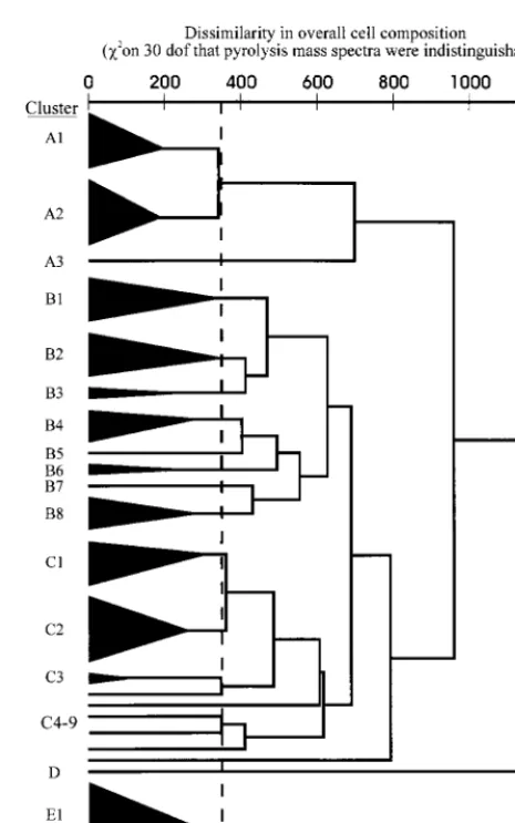
PMS.By PMS, strains VH20, VH21, and VH28 were con-taminated and thus were excluded from analysis. The remain-ing strains formed four superclusters (superclusters A, B, C, and E) plus cluster with a single strain (cluster D) (Fig. 1; Table 3). The majority of strains clustered with members of the same species. However, five clinical strains identified by con-ventional tests asActinomyces meyericlustered closely with the type strain ofA. turicensisand remotely from the type strain of
A. meyeri. Also, two strains conventionally identified as Acti-nomyces georgiaeclustered with fourActinomyces gerencseriae
strains; one strain (strain VH29) was subsequently reidentified asA. gerencseriae; the identity of the other strain (strain VH24) remains uncertain. A reference strain ofA. israelii(strain VH34) inexplicably clustered with this A. gerencseriae group. One strain (strain VH23), conventionally identified as most likeA. viscosus, clustered with the type strain ofA. neuiisubsp.neuii
and was subsequently reidentified as A. neuiisubsp.neuiiby virtue of its abilities to produce catalase, ferment mannitol, and reduce nitrate. Strains VH7, VH9, VH19, VH27, VH31, VH43, VH47, VH52, VH58, VH59, and VH61 each formed single-strain clusters.
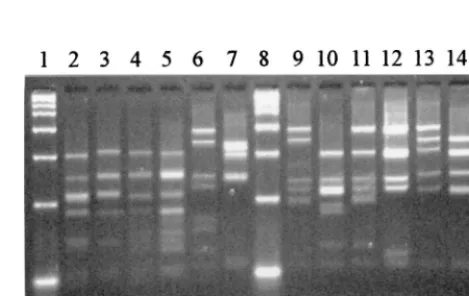
ARDRA. All strains were cleaved by both endonucleases, yielding 6 to 10 bands byHaeIII and 5 to 12 bands byHpaII typing (Fig. 2 and 3, respectively). The resulting HaeIII and
HpaII profiles were found to be highly reproducible, allowing the assignation of permanent types to each strain (Table 3). Clinical strains yielding HaeIII and HpaII profiles indistin-guishable from those of a reference strain were assigned to that species. The types obtained for A. naeslundii reference sero-vars indicated subspecies variation inHaeIII profiles and the possible species specificity of the HpaII profiles. Therefore, clinical strains were assigned to species for which the HpaII profile was indistinguishable from that for a reference strain and identification by conventional biochemical tests concurred. When the latter did not concur or when strains yielded distinct profiles with both enzymes, the identity was considered to be
uncertain. This approach was supported by the findings ob-tained by conventional biochemical tests and PMS.
(i) Reference strains.ByHaeIII typing, 17 of the 22 species represented and the three serotypes ofA. naeslundiiand two of
[image:3.612.53.552.83.185.2]A. viscosuswere clearly differentiated.Actinomyces odontolyti-cus, A. meyeri, and A. georgiae strains were indistinguishable TABLE 2. Reference strains demonstrating results anomalous to those published elsewherea
Strain Species and strain designation Conventional identification Anomaly
VH28 A. gerencseriaeATCC 23860 A. georgiae Raffinose negative
VH36 A. viscosusser. I NCTC 10951 A. naeslundii Catalase negative
VH47 A. denticolensNCTC 11490 No identification Trehalose positive
VH52 A. europaeusCCUG 32789A No identification Nitrate positive
VH59 A. bernardiaeDSM 9152 No identification b-N-Acetyl-glucosaminidase positive
VH60 A. turicensisDSM 9168 Poor differentiation Pyrazinamidase positive
VH61 A. radingaeDSM 9169 No identification Esculin negativeb
VH62 A. phocaeDSM 10002 A. georgiae Xylose positive
VH63 A. suisDSM 20639 A. meyeriorA. naeslundii b-Galactosidase negative
aThe previous publications are references 4, 5, 9, and 12.
[image:3.612.314.547.327.699.2]bReaction listed as positive in the previous studies but negative or slowly positive in the original description of Wust et al. (22).
FIG. 1. Dendrogram of similarities between strains on the basis of PMS, which reflects whole-cell composition. Strain membership of the clusters is out-lined in Table 3. dof, degrees of freedom.
on May 15, 2020 by guest
http://jcm.asm.org/
(type 001), as wereA. pyogenesandA. schaalii(type 004). Both
A. israeliistrains were type 008. The twoA. neuiisubspecies were type 022.
ByHpaII typing, 20 species were clearly differentiated; but
[image:4.612.55.550.83.659.2]A. naeslundii serotypes I, II, and III,A. viscosus serotype II, and A. howelliiwere indistinguishable (type 005). BothA. is-raeliistrains were type 009. The twoA. neuiisubspecies were type 019. The combination ofHaeIII andHpaII types allowed TABLE 3. Results by ARDRA, PMS, and conventional biochemical tests, arranged byHpaII profile
ARDRA profile PMS cluster Strain Conventional identification True identification
001/001 B2 VH6 A. odontolyticus A. odontolyticusNCTC 09935
001/001 B2 VH13 A. odontolyticus A. odontolyticus
001/001 B2 VH33 Most likeA. odontolyticus A. odontolyticus
001/001 B2 VH50 Most likeA. odontolyticus A. odontolyticus
025/001 B2 VH40 A. odontolyticus A. odontolyticus
002/002 C9 VH7 A. hordeovulneris A. hordeovulnerisATCC 35275
001/003 B3 VH10 A. meyeri A. meyeriATCC 35568
001/004 B3 VH11 A. georgiae A. georgiaeATCC 49285
003/005 B1 VH2 A. naeslundii A. naeslundii
003/005 C1 VH14 A. naeslundii A. naeslundiiserotype I NCTC 10301
003/005 C1 VH26 Most likeA. naeslundii A. naeslundii
007/005 B4 VH30 A. howellii A. howelliiNCTC 11636
009/005 C1 VH39 A. naeslundii A. naeslundii
014/005 C1 VH8 A. naeslundii A. naeslundii
014/005 B4 VH56 A. naeslundii A. naeslundiiserotype III ATCC 44340
014/005 B4 VH42 Most likeA. naeslundii A. naeslundii
017/005 C2 VH55 A. naeslundii A. naeslundiiserotype II ATCC 44339
017/005 C2 VH18 A. viscosus A. viscosus
021/005 C2 VH54 A. viscosus A. viscosusserotype II ATCC 27044
021/005 C2 VH17 Most likeA. viscosus A. viscosus
021/005 C2 VH46 Most likeA. viscosus A. viscosus
021/005 C2 VH49 Most likeA. viscosus A. viscosus
021/005 C1 VH3 Most likeA. viscosus A. viscosus
004/006 D VH19 A. pyogenes A. pyogenesNCTC 05224
005/007 B4 VH25 A. bovis A. bovisNCTC 11535
006/008 A2 VH4 A. gerencseriae A. gerencseriae
006/008 A2 VH16 A. gerencseriae A. gerencseriae
006/008 A2 VH29 A. georgiae A. gerencseriae
006/008 A2 VH32 A. gerencseriae A. gerencseriae
006/008 A2 VH48 A. gerencseriae A. gerencseriae
006/008 NDa VH28 A. georgiae A. gerencseriaeATCC 23860
008/009 A1 VH45 A. israelii A. israeliiATCC 12102
008/009 A2 VH34 A. israelii A. israeliiNCTC 10236
008/009 ND VH21 A. israelii A. israelii
018/009 A1 VH5 A. israelii A. israelii
018/009 ND VH20 A. denticolens A. israelii
020/009 A1 VH1 A. israelii A. israelii
020/009 A1 VH15 A. israelii A. israelii
020/009 A1 VH38 Most likeA. israelii A. israelii
009/010 B1 VH36 A. naeslundii A. viscosusserotype I NCTC 10951
010/011 C5 VH43 A. slackii A. slackiiNCTC 11923
024/011 B7 VH31 Actinomycesspecies Uncertain
011/012 B5 VH47 Uncertain A. denticolensNCTC 11490
013/013 B1 VH51 A. graevenitzii A. graevenitziiCCUG 27294
012/014 C6 VH52 Uncertain A. europaeusCCUG 35789A
004/015 B8 VH53 A. schaalii A. schaaliiCCUG 27420
001/016 E2 VH27 Most likeA. odontolyticus Uncertain
015/016 E1 VH60 A. turicensisorA. meyeri A. turicensisDSM 9168
015/016 E1 VH12 A. meyeri A. turicensis
015/016 E1 VH22 A. meyeri A. turicensis
015/016 E1 VH35 A. meyeri A. turicensis
015/016 E1 VH41 A. meyeri A. turicensis
015/016 E1 VH44 A. meyeri A. turicensis
019/017 A3 VH9 Actinomycesspecies Uncertain
022/019 C3 VH57 A. neuiisubsp.neuii A. neuiisubsp.neuiiDSM 8576
022/019 C3 VH23 A. viscosus A. neuiisubsp.neuii
022/019 C4 VH58 A. neuiisubsp.anitratus A. neuiisubsp.anitratusDSM 8577
023/020 A2 VH24 Most likeA. georgiae Uncertain
026/021 C8 VH59 Uncertain A. bernardiaeDSM 9152
027/022 B6 VH37 Most likeA. pyogenes Uncertain
028/023 C7 VH61 Uncertain A. radingaeDSM 9169
029/024 B6 VH62 A. georgiae A. phocaeDSM 10002
030/025 B8 VH63 A. meyeriorA. naeslundii A. suisDSM 20639
aND, no data available.
on May 15, 2020 by guest
http://jcm.asm.org/
differentiation of all 22 species represented by reference strains.
(ii) Clinical strains.In reactions with both endonucleases, 16 of the 36 strains were indistinguishable from reference strains of the same species, as determined by conventional tests, and were assigned to species with confidence. Eight strains were indistinguishable by ARDRA from the reference strains of a species other than that identified by conventional tests. These comprised the five strains conventionally identified asA. meyeri, which were indistinguishable fromA. turicensis; VH23, which was originally most likeA. viscosusbut which was redesignatedA. neuii subsp. neuii and which gave the same profile as the A. neuii strains; VH29, which was originally considered to beA. georgiae but which was indistinguishable fromA. gerencseriae; and VH18 (A. viscosus), which gave the same profile asA. naeslundiiserotype II. These were assigned to the species determined by ARDRA, with strain VH18 being deemed a member ofA. naeslundiigenospecies 2, which in-cludesA. viscosusserotype II.
Seven strains had distinct HaeIII profiles but gave HpaII profiles indistinguishable from those of reference strains of the species determined by conventional tests and were assigned to that species. One strain (strain VH31), which had a distinct
HaeIII profile and which was indistinguishable from the refer-ence strain ofActinomyces slackii by HpaII typing, was not clearly identified by conventional tests and was deemed to be of uncertain identity.
One strain (strain VH27) that was identified by conventional tests as most likeA. odontolyticusgave profiles indistinguish-able from those of reference strains of A. odontolyticus by
HaeIII typing andA. turicensisbyHpaII typing. The remaining three strains (strains VH9, VH24, and VH37) gave unique profiles in both reactions. The true identities of these four strains remain uncertain.
DISCUSSION
The application of chemotaxonomic and molecular methods has clarified the taxonomy of the genus Actinomyces, but the description of novel species has rendered previously published identification schemes obsolete. When few strains of a species have been studied, the reliability of identification by
conven-tional tests is questionable. Furthermore, given the diversity within the genus, further modification of the taxonomic status of strains may be warranted.
Currently, the Public Health Laboratory Service Anaerobe Reference Unit receives over 100 referrals each year from clinical laboratories throughout England and Wales for con-firmation of isolates presumptively identified as Actinomyces
spp. There is a need for a practical and discriminatory method for the identification of clinical isolates.
The identification of Actinomyces to the species level by conventional biochemical tests is beset with problems. These include technical difficulties, poorly discriminatory tests, and heterogeneity within described species. In this study, nine ref-erence strains demonstrated reactions anomalous from those published elsewhere, and this resulted in mis- or nonidentifi-cation (Table 2). Notably,A. europaeus was found to reduce nitrate to nitrite, andA. bernardiae producedb-N -acetyl-glu-cosaminidase. Anomalies, particularly in enzyme reactions, may be due to differences in test methodologies.
PMS has been shown to be highly discriminatory for a wide range of organisms but is best suited to the testing of large batches of isolates. Direct comparisons between batches are not readily made. Thus, PMS would be impractical for exam-ination of occasional clinical isolates. However, given the low degree of confidence in results obtained by conventional tests for the identification ofActinomyces, evaluation of the efficacy of novel methods is difficult. In this study, PMS provided a valuable independent approach with which to compare find-ings. It is noteworthy and reassuring that the dendrogram derived from PMS analysis reflects the taxonomic relationships generated by 16S rDNA sequencing (4, 9, 13).A. israeliiandA. gerencseriaestrains formed a supercluster (supercluster A) re-mote from other species. Superclusters B and C were linked and contained strains identified asA. odontolyticus,A. naeslun-dii,A. viscosus, and several other species, largely represented by single reference strains. Within this group, the type strains of A. meyeri and A. georgiae formed a cluster (cluster B3) closely linked toA. odontolyticusstrains (cluster B2). The re-moteness of cluster E (A. turicensis strains and strain VH27) from other species raises doubts as to their place in the genus
Actinomyces.
ARDRA has proven to be useful for discrimination of
var-FIG. 3.HpaII restriction profiles for some reference strains. Lanes 1, 8, and 15, 2-kb marker; lane 2,A. israeliiATCC 12102; lane 3,A. gerencseriaeATCC 23860; lane 4,A. naeslundiiserotype I NCTC 10301; lane 5,A. viscosusserotype II ATCC 27044; lane 6,A. neuiisubsp.neuiiDSM 8576; lane 7,A. graevenitzii
CCUG 27294; lane 9,A. odontolyticusNCTC 09935; lane 10,A. georgiaeATCC 49285; lane 11,A. meyeriATCC 35568; lane 12,A. turicensisDSM 9168; lane 13,
[image:5.612.56.290.73.229.2]A. radingaeDSM 9169; lane 14,A. europaeusCCUG 32789A. FIG. 2.HaeIII restriction profiles for some reference strains. Lanes 1, 8, and
15, 2-kb marker; lane 2,A. israeliiATCC 12102; lane 3,A. gerencseriaeATCC 23860; lane 4,A. naeslundiiserotype I NCTC 10301; lane 5,A. viscosusserotype II ATCC 27044; lane 6,A. neuiisubsp.neuiiDSM 8576; lane 7,A. graevenitzii
CCUG 27294; lane 9,A. odontolyticusNCTC 09935; lane 10,A. georgiaeATCC 49285; lane 11,A. meyeriATCC 35568; lane 12,A. turicensisDSM 9168; lane 13,
A. radingaeDSM 9169; lane 14,A. europaeusCCUG 32789A.
on May 15, 2020 by guest
http://jcm.asm.org/
[image:5.612.314.549.521.669.2]ious bacterial species. In this study, the types generated by the combination ofHaeIII andHpaII endonuclease digestion pro-files correlated well with the findings obtained by PMS and conventional tests and enabled the identification of 31 of 36 clinical strains to the species level. The remaining five strains were not clearly identified by conventional tests, and three strains formed distinct clusters by PMS; the latter strains may well represent novel species or other genera.
Seven strains were identified by ARDRA and PMS as spe-cies other than those initially assigned by conventional tests. Five of these seven strains initially identified asA. meyeri clus-tered tightly with theA. turicensistype strain by PMS and were indistinguishable fromA. turicensis by ARDRA. Review of conventional test results showed that the two species are poorly differentiated by the range of biochemical tests per-formed. The identity of one of the five strains (strain VH12) was confirmed asA. turicensisby 16S rDNA sequence analysis (data not shown). The strain that was apparently misidentified by PMS and ARDRA asA. gerencseriaehad been designated
A. georgiae by conventional tests by virtue of its inability to ferment raffinose; the morphology and other reactions were consistent withA. gerencseriae. This property was also demon-strated by the A. gerencseriae type strain; thus, raffinose fer-mentation appears to be an unreliable determinant in the iden-tification of this species. The strain originally designated most likeA. viscosusand identified asA. neuiiby PMS and ARDRA was subsequently redesignatedA. neuiisubsp.neuiiby virtue of catalase production, nitrate reduction, and fermentation of mannitol.
A. gerencseriae and A. turicensis showed homogeneous, distinct ARDRA profiles. The subdivisions observed within
A. israelii, A. odontolyticus, A. naeslundii, and A. viscosus
may correspond to those previously recognized by other work-ers (8, 17, 18). One strain ofA. viscosuswas indistinguishable fromA. naeslundiiserotype II by PMS and ARDRA, adding to the body of evidence thatA. naeslundiiandA. viscosusisolates form a heterogeneous complex that requires further investiga-tion.
When species were represented by single reference strains, all strains gave distinct profiles, and none was misidentified. However, no further conclusions regarding the efficacy of ARDRA for identification of these species can be drawn with-out data derived from additional strains.
Current consumable costs, based on a batch of 10 isolates, were £2.80 (as of April 1999, £1 is equal to $1.60) per isolate. Each batch required approximately 3.5 h of labor spread over 1.5 days.
In conclusion, ARDRA was shown to be a simple, rapid, cost-effective, and highly discriminatory method for identifica-tion ofActinomycesspp. of clinical origin. Application of the method to further clinical and veterinary isolates may confirm its usefulness, and an extensive investigation of strains referred to the Anaerobe Reference Unit is in progress. Within the limitations of the current study, identification of a strain as a member of the genusActinomycesby gas-liquid chromatogra-phy is a prerequisite. However, preliminary investigations of strains of Propionibacterium spp.,Lactobacillus spp., and Bi-fidobacteriumspp. have demonstrated the clear differentiation of strains by ARDRA (unpublished data), indicating potential for identification to the species level of members of these genera and obviating the need for gas-liquid chromatography. Identification of clinical isolates ofActinomycesto the spe-cies level may be important for patient management. Addition-ally, a reliable identification system is essential for the discov-ery of the natural habitats, prevalence, and pathogenicity of recently described species. With increasing knowledge of these
aspects, species-level identification of clinical isolates may be-come more relevant to patient management. Some species, e.g., A. turicensis, have been shown to be identifiable in com-mercial biochemical systems (15). However, genotypic meth-ods may be advantageous in their ability to detect novel species as well as those listed in commercial databases. A practical, highly discriminatory, and cost-effective method such as ARDRA applied to many strains may greatly aid in the eluci-dation of the ecology and clinical spectra ofActinomyces spe-cies and may further clarify the taxonomy of the genus.
ACKNOWLEDGMENTS
We thank Margaret Heginbotham and Paul Talbot for technical assistance with PMS and conventional tests.
REFERENCES
1.Barsotti, O., D. Decoret, G. Benay, A. Carlotti, J. Freney, V. Guerin-Faublee,
and J. Morrier.1994. rRNA gene restriction patterns as possible taxonomic
tools for the genusActinomyces. Zentbl. Bakteriol. Parasitenkd. Infektion-skr. Hyg. Abt. 1 Orig.281:433–441.
2.Bowden, G. H. W.1998.Actinomyces, p. 445–462.InA. Balows and B. I.
Duerden (ed.), Topley and Wilson’s microbiology and microbial infections, 9th ed., vol. 2. Edward Arnold, London, United Kingdom.
3.Brazier, J. S., and V. Hall.1997.Actinomyces, p. 625–639.InA. M.
Emmer-son, P. M. Hawkey, and S. H. Gillespie (ed.), Principles and practice of clinical bacteriology. John Wiley & Sons, Chichester, United Kingdom.
4.Funke, G., N. Alvarez, C. Pascual, E. Falsen, E. Akervall, L. Sabbe, L.
Schouls, N. Weiss, and M. D. Collins.1997.Actinomyces europaeussp. nov.,
isolated from human clinical specimens. Int. J. Syst. Bacteriol.47:687– 692.
5.Funke, G., S. Stubbs, A. von Graevenitz, and M. D. Collins.1994.
Assign-ment of human-derived CDC group 1 coryneform bacteria and CDC 1-like coryneform bacteria to the genusActinomycesasActinomyces neuiisubsp.
neuiisp. nov., subsp. nov., andActinomyces neuiisubsp.anitratussubsp. nov. Int. J. Syst. Bacteriol.44:167–171.
6.Hall, V., and J. S. Brazier.1997. Identification of actinomyces—what are the
major problems?, p. 187–192. InA. R. Eley and K. W. Bennett (ed.), Anaerobic pathogens. Sheffield Academic Press, Sheffield, United Kingdom.
7.Holdeman, L. V., E. P. Cato, and W. E. C. Moore.1977. Chromatographic
procedures for analysis of acid and alcohol products, p. 134–136.In Anaer-obe laboratory manual, 4th ed. Virginia Polytechnic Institute and State University, Blacksburg.
8.Johnson, J. L., L. V. H. Moore, B. Kaneko, and W. E. C. Moore.1990.
Actinomyces georgiaesp. nov.,Actinomyces gerencseriaesp. nov., designation of two genospecies ofActinomyces naeslundii, and inclusion ofA. naeslundii
serotypes II and III andActinomyces viscosusserotype II inA. naeslundii
genospecies 2. Int. J. Syst. Bacteriol.40:273–286.
9.Lawson, P., E. Falsen, E. Åkervall, P. Vandamme, and M. D. Collins.1997.
Characterization of someActinomyces-like isolates from human clinical spec-imens: reclassification ofActinomyces suis(Soltys and Spratling) as Acti-nobaculum suiscomb. nov. and description ofActinobaculum schaaliisp. nov. Int. J. Syst. Bacteriol.47:899–903.
10. Magee, J.1993. Whole-organism fingerprinting, p. 383–427.InM.
Goodfel-low and A. G. O’Donnell (ed.), Handbook of new bacterial systematics. Academic Press, London, United Kingdom.
11. Magee, J. T., M. J. Hindmarch, K. W. Bennett, B. I. Duerden, and R. E.
Aries.1989. A pyrolysis mass spectrometry study of fusobacteria. J. Med.
Microbiol.28:227–236.
12. Pascual Ramos, C., E. Falsen, N. Alvarez, E. Åkervall, B. Sjo¨de´n, and M. D.
Collins.1997.Actinomyces graevenitziisp. nov. isolated from human clinical
specimens. Int. J. Syst. Bacteriol.47:885–888.
13. Pascual Ramos, C., G. Foster, and M. D. Collins.1997. Phylogenetic analysis
of the genusActinomycesbased on 16S rRNA gene sequences: description of
Arcanobacterium phocaesp. nov.,Arcanobacterium bernardiaecomb. nov., andArcanobacterium pyogenescomb. nov. Int. J. Syst. Bacteriol.47:46–53.
14. Phillips, K. D.1976. A simple and sensitive technique for determining the
fermentation reactions of non-sporing anaerobes. J. Appl. Bacteriol.41:325– 328.
15. Sabbe, L. J. M., D. Van de Merwe, L. Schoulls, A. Bergmans, M.
Vanee-choutte, and P. Vandamme.1999. Clinical spectrum of infections due to the
newly describedActinomycesspeciesA. turicensis,A. radingaeandA. euro-paeus. J. Clin. Microbiol.37:8–13.
16. Schaal, K. P.1997. Actinomycoses, actinobacillosis and related diseases,
p. 777–798.InW. J. Hausler and M. Sussman (ed.), Topley and Wilson’s microbiology and microbial infections, 9th ed., vol. 3. Edward Arnold, Lon-don, United Kingdom.
on May 15, 2020 by guest
http://jcm.asm.org/
17.Schofield, G. M., and K. P. Schaal.1981. A numerical taxonomic study of members of theActinomycetaceaeand related taxa. J. Gen. Microbiol.127:
237–259.
18.Slack, J. M., and M. A. Gerencser.1975.Actinomyces, filamentous bacteria.
Biology and pathogenicity, p. 57–64. Burgess, Minneapolis, Minn.
19.Vaneechoutte, M., P. Riegel, D. de Briel, H. Monteil, G. Verschraegen, A. De
Rouck, and G. Claeys.1995. Evaluation of the applicability of amplified
rDNA-restriction analysis (ARDRA) to identification of species of the genus
Corynebacterium. Res. Microbiol.146:633–641.
20.Vaneechoutte, M., C. P. Cartwright, E. C. Williams, B. Ja¨ger, H.-V. Tichy, T.
De Baere, A. De Rouck, and G. Verschraegen.1996. Evaluation of 16S rRNA
gene restriction analysis for the identification of cultured organisms of clin-ically importantClostridiumspecies. Anaerobe2:249–256.
21.Vilgalys, R., and M. Hester.1990. Rapid genetic identification and mapping
of enzymatically amplified ribosomal DNA from severalCryptococcus spe-cies. J. Bacteriol.172:4238–4246.
22.Wust, J., S. Stubbs, N. Weiss, G. Funke, and M. D. Collins.1995. Assignment
ofActinomyces pyogenes-like (CDC coryneform group E) bacteria to the genusActinomycesasActinomyces radingaesp. nov. andActinomyces turicen-sissp. nov. Lett. Appl. Microbiol.20:76–81.