Correcting temperature-sensitive protein folding
defects.
C R Brown, … , L Q Hong-Brown, W J Welch
J Clin Invest.
1997;
99(6)
:1432-1444.
https://doi.org/10.1172/JCI119302
.
Recently, we found that different low molecular weight compounds, all known to stabilize
proteins in their native conformation, are effective in correcting the temperature-sensitive
protein folding defect associated with the deltaF508 cystic fibrosis transmembrane regulator
(CFTR) protein. Here we examined whether the folding of other proteins which exhibit
temperature-sensitive folding defects also could be corrected via a similar strategy. Cell
lines expressing temperature-sensitive mutants of the tumor suppressor protein p53, the
viral oncogene protein pp60src, or a ubiquitin activating enzyme E1, were incubated at the
nonpermissive temperature (39.5 degrees C) in the presence of glycerol, trimethylamine
N-oxide or deuterated water. In each case, the cells exhibited phenotypes similar to those
observed when the cells were incubated at the permissive temperature (32.5 degrees C),
indicative that the particular protein folding defect had been corrected. These observations,
coupled with our earlier work and much older studies in yeast and bacteria, indicate that
protein stabilizing agents are effective in vivo for correcting protein folding abnormalities.
We suggest that this type of approach may prove to be useful for correcting certain protein
folding abnormalities associated with human diseases.
Research Article
Find the latest version:
http://jci.me/119302/pdf
1432 Brown et al. J. Clin. Invest.
© The American Society for Clinical Investigation, Inc. 0021-9738/97/03/1432/13 $2.00
Volume 99, Number 6, March 1997, 1432–1444
Correcting Temperature-sensitive Protein Folding Defects
C. Randell Brown, Ly Q. Hong-Brown,* and William J. Welch*‡
*Department of Medicine and ‡Department of Physiology, The University of California, San Francisco, California 94143
Abstract
Recently, we found that different low molecular weight compounds, all known to stabilize proteins in their native conformation, are effective in correcting the temperature-sensitive protein folding defect associated with the DF508 cystic fibrosis transmembrane regulator (CFTR) protein. Here we examined whether the folding of other proteins which exhibit temperature-sensitive folding defects also could be corrected via a similar strategy. Cell lines express-ing temperature-sensitive mutants of the tumor suppressor protein p53, the viral oncogene protein pp60src, or a
ubiq-uitin activating enzyme E1, were incubated at the nonper-missive temperature (39.58C) in the presence of glycerol, tri-methylamine N-oxide or deuterated water. In each case, the
cells exhibited phenotypes similar to those observed when the cells were incubated at the permissive temperature (32.58C), indicative that the particular protein folding de-fect had been corrected. These observations, coupled with our earlier work and much older studies in yeast and bacte-ria, indicate that protein stabilizing agents are effective in vivo for correcting protein folding abnormalities. We sug-gest that this type of approach may prove to be useful for correcting certain protein folding abnormalities associated with human diseases. (J. Clin. Invest. 1997. 99:1432–1444.)
Key words: temperature-sensitive mutations• protein
fold-ing• molecular chaperones• chemical chaperones• genetic disease
Introduction
A number of low molecular weight compounds have been shown to be effective in stabilizing proteins in vitro against thermally induced denaturation (1–3). Representative com-pounds include polyols such as glycerol, solvents such as DMSO, and deuterated water (D2O)1. In addition to their ef-fects in vitro, some of these compounds appear to influence protein folding and/or stability in vivo (4–6). Animal cells
incu-bated in the presence of either deuterated water or glycerol, for example, can withstand severe heat shock treatments that would otherwise be lethal to the cells. Here, addition of the compounds to the cells helps to reduce the overall extent of thermal denaturation of intracellular proteins. In yeast and bacteria, the addition of glycerol into the growth medium not only protects the cells against thermal treatments, but in some cases also is effective in correcting protein folding abnormali-ties due to specific mutations (7). This type of osmotic remedi-ation has been shown to be the most effective for those mutant proteins that exhibit a temperature-sensitive protein folding defect.
Based on these earlier observations, we have been examin-ing whether different protein stabilizexamin-ing agents might be effec-tive in correcting protein folding abnormalities associated with particular diseases (8). For example, in the majority of patients with cystic fibrosis, a mutation in the gene encoding the cystic fibrosis transmembrane conductance regulator (CFTR) pro-tein results in the deletion of a phenylalanine residue at posi-tion 508 (DF508 CFTR). As a consequence, the newly synthe-sized DF508 CFTR protein fails to fold properly, and does not move to the plasma membrane where it normally functions as a cAMP-regulated chloride channel (9). Instead, the newly synthesized protein becomes trapped in the endoplasmic retic-ulum, likely in a complex with one or more members of the molecular chaperone family (10, 11). This folding defect of the DF508 CFTR protein is temperature dependent. Lowering the growth temperature (e.g., 308C or less) of animal cells ex-pressing the DF508 CFTR protein causes a portion of the mu-tant protein to move to the plasma membrane. These cells now appear competent for cAMP-stimulated chloride exchange, in-dicative of a functional CFTR protein (12–14).
We have found that the temperature-sensitive folding de-fect of the DF508 CFTR protein can be corrected when cells are cultured in the presence of different protein-stabilizing agents. For example, incubation of mouse fibroblasts express-ing DF508 CFTR at 378C in the presence of glycerol, trimethyl-amine N-oxide (TMAO), or deuterated water (D2O), resulted in the proper processing of the DF508 CFTR protein and its subsequent transport to the cell surface. Importantly, these cells then exhibited cAMP-dependent chloride transport, simi-lar in rate and magnitude as that observed for the cells express-ing the wild-type CFTR protein (8).
Cystic fibrosis is but one of a number of genetic diseases which arise because of specific mutations that ultimately lead to protein folding errors (for reviews see reference 15 and 16). Prompted by our success with the mutant CFTR protein, we examined whether protein-stabilizing agents also might be ef-fective for correcting other temperature-sensitive protein fold-ing mutants. For these studies, cell lines expressfold-ing three dif-ferent temperature-sensitive folding mutants were examined: (a) the tumor suppressor protein p53, (b) pp60src, the trans-forming protein encoded by Rous sarcoma, and (c) an enzyme (E1) which catalyzes the first step in the pathway of protein ubiquitination. We show that treatment with the different
pro-The first two authors made equal contributions to this manuscript. Address correspondence to C. Randell Brown or William J. Welch, UC Box 0854, UCSF, San Francisco, CA 94143. Phone: 415-206-5909; FAX: 415-206-4123; E-mail: [email protected]
Received for publication 22 October 1996 and accepted in revised form 3 January 1997.
1. Abbreviations used in this paper: CFTR, cystic fibrosis transmem-brane conductance regulator; D2O, deuterated water; TMAO,
tein-stabilizing agents resulted in the cells adopting a wild-type phenotype at the nonpermissive temperature, indicative that the particular protein folding defect had been corrected. Thus, we suspect that mutations which result in temperature-sensi-tive protein folding defects are amenable in general to correc-tion in vivo via the use of protein-stabilizing agents. Further-more, we suggest that our results may have broad implications as they relate to the correction of protein folding defects asso-ciated with certain genetic diseases.
Methods
Cell culture and indirect immunofluorescence.A1-5 cells (a generous gift from A. Levine), a cell line expressing a temperature-sensitive (ts) mutant p53 protein (ala to val change at amino acid 135), were cultured and maintained at 378C in DMEM containing 10% fetal bo-vine serum. Cells expressing a ts pp60src protein (kindly provided by
D. T. Aftab) were routinely maintained under the same conditions. ts20 cells, which express a temperature-sensitive mutant of the E1 en-zyme (a component of the ubiquitin pathway) (kindly provided by H. Ozer), were maintained at 32.58C (permissive temperature) in DMEM medium containing 10% fetal bovine serum. For indirect im-munofluorescence, cells were grown on glass coverslips. After the particular experimental treatment (described in the figure legends),
cells were fixed with 100% methanol, and then subsequently rehy-drated in PBS. The intracellular localization of p53 was determined by incubation of the fixed cells with the antibody 421 (Oncogene Sci-ence Inc., Mahasset, NY), which recognizes both the mutant and wild-type forms of p53. Primary antibody was visualized by subse-quent staining with a rhodamine conjugated goat anti–mouse anti-body. All antibodies were diluted in 5 mg/ml BSA in PBS.
Cell morphology analysis. Cells expressing the ts E1 enzyme or the ts pp60src protein were incubated at either 32.58C (permissive
tem-perature) or at 39.58C (nonpermissive temperature) in the presence or absence of protein-stabilization agents. Following 2 d of treat-ments, cells were examined by phase-contrast microscopy.
Effects of chemical treatments on cell growth.A1-5 cells were plated in 60-mm dishes at an initial concentration of 5 3 104 cells/plate.
[image:3.612.59.538.339.682.2]Af-ter allowing them to attach at 378C for 16 h, the cells were shifted to the appropriate temperature in the presence or absence of the vari-ous protein stabilizing agents. A few plates of the cells were collected at the time of chemical addition, and cell number was determined us-ing a hemocytometer. Thereafter, the cells were counted at daily in-tervals. All time points were examined in duplicate with the values given representing the mean for replicate counts. For ts20 experi-ments, cells growing at 32.58C were plated on 60-mm dishes and maintained at this temperature for 16 h. Some of the cells were incu-bated with the chemicals, while the others were maintained in normal growth media. The cells were then shifted to the appropriate temper-atures in the presence or absence of the chemicals.
1434 Brown et al.
Chemical treatments. Various concentrations of protein-stabiliz-ing agents were added to DMEM supplemented with 10% fetal bo-vine serum. For D2O experiments, powdered DMEM was
reconsti-tuted using 100% D2O (Sigma Chemical Co., St. Louis, MO) and
supplemented with 10% fetal bovine serum. For each experiment, the culture medium was removed and replaced with fresh medium con-taining the chemical of interest. For recovery experiments (see Fig. 4) cells were incubated with the various chemicals for 2 d. The medium was then removed, and the cells were washed with and further incu-bated in normal growth medium not containing the particular protein stabilizing agent.
Western blotting. Following the particular experimental treat-ment, cells were lysed in Laemmli sample buffer and heated at 1008C for 5 min. Lysates were clarified, and the proteins were separated by SDS-PAGE. The resolved proteins were transferred to nitrocellulose and subsequently immunoblotted using the mouse monoclonal anti– p53 antibody, 421 (Oncogene Science) or the hsp73/hsp 72-specific antibody, N27 (StressGen Biotech. Corp., Victoria, Canada).
Results
Correction of a p53 protein folding mutant. A cell line that ex-presses a mutant form of the tumor suppressor protein p53 was chosen for our initial studies (17). This cell line (A1-5) was
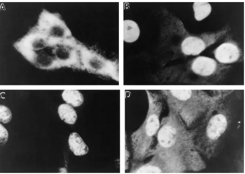
produced by transfection of primary rat fibroblasts with a p53 gene containing a missense mutation (ala to val at amino acid 135). As was shown previously, this mutation results in a tem-perature-sensitive protein folding defect (18–20). At tempera-tures around 32.58C or less, the p53 protein adopts a wild-type conformation and localizes within the cell nucleus (Fig. 1 D). In contrast, at temperatures of 39.58C or greater, the p53 pro-tein fails to fold properly and does not accumulate within the nucleus. Instead, the vast majority of the mutant protein is found predominantly within the cytoplasm (Fig. 1 B).
[image:4.612.60.538.334.673.2]We examined whether treatment of the A1-5 cells with dif-ferent protein stabilizing agents would correct the tempera-ture-sensitive protein folding defect of mutant p53. Three dif-ferent reagents were examined: (a) the carbohydrate (or polyol) glycerol; (b) a methylamine, trimethylamine N-oxide; and finally, (c) deuterated water (D2O). Cells were plated in normal culture medium and then incubated at 39.5 C where the p53 protein adopted the mutant conformation. The next day the culture medium was removed, and the cells were incu-bated in medium supplemented with either 0.6 M glycerol or 75 mM trimethylamine N-oxide (TMAO). In the case of the deuterated water, powdered DMEM was reconstituted with 100% D2O, supplemented with serum, and added to the cells.
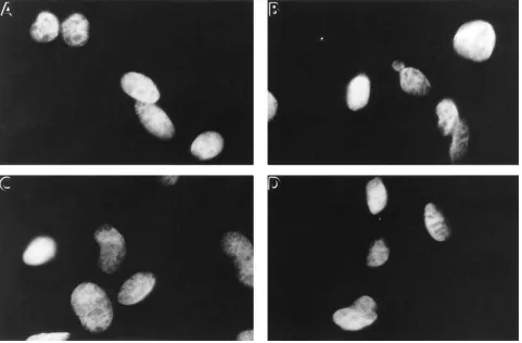
Figure 2. A1-5 cells incubated at the nonpermissive temperature in the presence of protein-stabilizing agents exhibit nuclear p53 staining. A1-5 cells were grown on glass coverslips and incubated for 1 d at 39.58C. Some of the coverslips then were transferred into medium supplemented with either D2O (100%), TMAO (75 mM), or glycerol (0.6 M), and the cells were further incubated at 39.58C. After 2 d of incubation at 39.58C,
the cells were analyzed for the intracellular distribution of p53 by indirect immunofluorescence as described in Fig. 1. Fig. 2 shows only the im-munofluorescent micrographs. (A) Control cells incubated in normal medium; (B) cells incubated in medium prepared with 100% D2O; (C) cells
After 2 d of incubation at 39.58C, the cells were analyzed for their distribution of p53. In the control cells maintained at 39.58C in normal growth medium, p53 was predominantly cy-toplasmic (Fig. 2 A). In contrast, cells incubated at 39.58C in the presence of D2O (Fig. 2 B), TMAO (Fig. 2 C), or glycerol (Fig. 2 D) now exhibited a nuclear locale of p53. Thus, incuba-tion of the cells in the presence of the different protein-stabi-lizing agents appeared to result in the mutant p53 protein adopting a wild-type conformation at 39.58C.
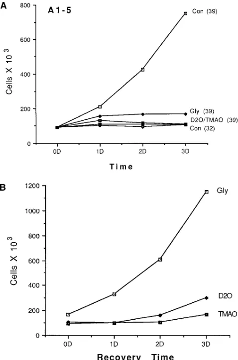
As an alternative method to screen for the potential benefi-cial effects of these compounds on p53 function, the prolifera-tive capacity of the A1-5 cells incubated at the nonpermissive temperature (in either the absence or presence of the different chemicals) was determined. As has been shown previously (19), the A1-5 cells become growth-arrested when maintained at the permissive temperature (32.58C), ostensibly because of the high levels of the wild-type p53 tumor suppressor protein (Fig. 3 A). In contrast, at 39.58C where the overexpressed p53 protein folds incorrectly (and therefore is biologically inac-tive), the cells continue to proliferate normally. Hence, if treat-ment with the various protein-stabilizing agents corrects the folding of the mutant p53 protein, the cells now should exhibit cell cycle arrest, even when incubated at the nonpermissive temperature. Indeed, treatment of the A1-5 cells maintained at 39.58C with any one of the three different compounds now resulted in a growth-arrested phenotype, similar to that ob-served for the cells incubated at the permissive temperature where the p53 protein is functional (Fig. 3 A).
The effects of the different protein-stabilizing agents on the phenotype of the A1-5 cells were reversible (Fig. 4). As was shown earlier, cells incubated in the presence of the different protein-stabilizing agents for 2 d at 39.58C exhibited a nuclear locale of p53, indicative of the protein being in its wild-type conformation. When the culture medium containing the pro-tein-stabilizing agents was removed and replaced with fresh culture medium, however, p53 again began to accumulate within the cytoplasm. For example, after only 1 d following the removal of glycerol, the cells maintained at 39.58C no longer displayed a nuclear p53 distribution. In the case of either D2O or TMAO, after 2 d following their removal, all of p53 now was found within the cytoplasm. Similar results were obtained when the analysis was performed by cell counts. As was shown in Fig. 3 A, cells incubated at 39.58C in the presence of the three different protein-stabilizing agents for 2 d exhibited a growth-arrested phenotype, consistent with a functional p53 protein. Upon removal of the compounds and further incuba-tion of the cells at 39.58C in normal culture medium, however, the cells exhibited a slow resumption in their normal growth rates (Fig. 3 B), indicative that p53 now was in the mutant con-formation.
Correcting the folding defect of pp60src. The potential
[image:5.612.58.297.59.421.2]ben-eficial effects of the different protein-stabilizing agents were examined using two other temperature-sensitive mutants. Rat fibroblasts transfected with a temperature-sensitive form of pp60src (the transforming protein encoded by Rous sar-coma virus) exhibit a transformed phenotype when incubated at the permissive temperature of 32.58C (21). As shown in Fig. 5 A, when incubated at 32.58C where pp60src is biologi-cally active, the cells adhere weakly to the substratum. In-stead, at this permissive temperature the cells grow on top of one another to form large foci. At 39.58C where pp60src is bio-logically inactive, the cells display a well-spread morphology, and exhibit contact-dependent growth arrest (Fig. 5 B). Inclusion of glycerol into the culture medium was sufficient to restore the transformed phenotype of the cells when incu-bated at the nonpermissive temperature. For example, cells incubated at 39.58C in the presence of 1 M glycerol for 3 d (Fig. 5 C) now exhibited a morphology very similar to the control cells maintained at 32.58C where pp60src is active (Fig. 5 A). Interestingly, the two other compounds, TMAO and deuterated water, were much less effective in restoring
wild-Figure 3. A1-5 cells treated with protein stabilizers at the nonpermis-sive temperature exhibit a wild-type phenotype as determined by cell proliferation rates. (A) Equal numbers of A1-5 cells, growing at 378C, were plated on 60-mm dishes in DMEM containing 10% serum. After plating, the cells were moved to 39.58C. The next day (day 0) the me-dia was removed, and fresh medium supplemented with serum and containing either nothing (con), 0.6 M glycerol (Gly), 100% D2O
(D2O), or 75 mM TMAO (TMAO), was added to the cells. The cells
1436 Brown et al.
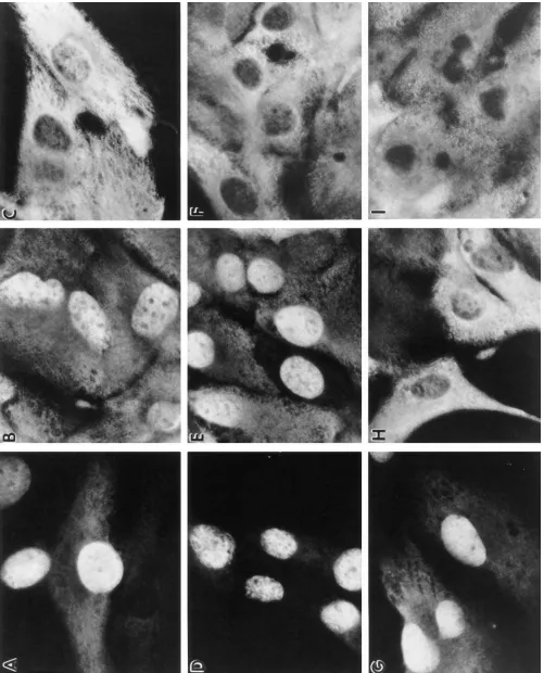
Figure 4. The effects of the protein-stabilizing agents on p53 protein folding are reversible. A1-5 cells growing on coverslips at 39.58C were incu-bated in media supplemented with either; D2O (A–C); TMAO (D–F); or glycerol (G–I) (concentrations as described in Fig. 3). After 2 d of
incu-bation at 39.58C, one coverslip from each group of cells was taken and processed for p53 indirect immunofluorescence. To the remaining cover-slips, the media was removed, and the cells were washed with (and further incubated in) fresh medium supplemented only with 10% serum. Following a further 1 or 2 d incubation at 39.58C, the cells were examined for the distribution of p53. A–C; cells after 0, 1, and 2 d, respectively, following removal of D2O; D–F, cells after 0, 1, and 2 d, respectively, following removal of TMAO containing media; G–I, cells after 0, 1, and 2 d,
Figure 5. The presence of glycerol corrects temperature-sensitive (ts) pp60src activities at the nonpermissive temperature. Cells expressing a ts
form of pp60src were incubated at 32.58C, or at 39.58C in the presence or absence of 1 M glycerol. After 3 d of treatment, the cells were examined
by phase-contrast microscopy. (A) Cells incubated at 32.58C for 3 d. (B) Cells incubated at 39.58C for 3 d. (C) Cells incubated at 39.58C for 3 d in the presence of 1 M glycerol.
Figure 6. Effects of pro-tein stabilizers on the morphology of ts 20 cells expressing a temperature-sensitive enzyme involved in protein ubiquitination. Equal numbers of cells expressing a temperature-sensitive mutant of the E1 enzyme were incubated at 32.58C (permissive temp.), or at 39.58C (non-permissive temp.) in ei-ther the presence or ab-sence of the different protein stabilizing agents. After 2 d of incu-bation the cells were ex-amined by phase-con-trast microscopy. (A) Cells incubated at 32.58C. (B) Cells incubated at 39.58C. (C) Cells incu-bated at 39.58C in me-dium prepared with 100% D2O. (D) Cells incubated
[image:7.612.56.546.402.740.2]1438 Brown et al.
different protein-stabilizing agents. For example, cells incu-bated at 39.58C in culture medium prepared with 100% D2O exhibited an apparent growth rate only slightly higher than that observed for the untreated cells maintained at the nonper-missive temperature. In addition, the morphology of the cells appeared to reflect an intermediate phenotype, with both spin-dle-shaped and well-rounded cells being observed (Fig. 6 C). Treatment of the ts20 cells with either TMAO (Fig. 6 D) or glycerol (Fig. 6 E) resulted in an apparent restoration of the wild-type phenotype. Like the control cells maintained at 32.58C, those cells incubated at 39.58C in the presence of TMAO or glycerol exhibited normal growth rates and a well-rounded morphology.
A more quantitative comparison of the growth rates of the cells incubated at 39.58C in the presence of the three protein-stabilizing compounds is shown in Fig. 7. Cells incubated at 32.58C in regular culture medium displayed an exponential rate of growth, while those maintained at 39.58C failed to grow. The actual reduction in cell number found for the cells maintained at 39.58C was observed over the course of many experiments, and likely represents cell death (although the mechanism, necrosis versus apoptosis, remains unclear). Simi-lar to our observations using phase-contrast microscopy, cells incubated at 39.58C in culture medium prepared with D2O failed to exhibit any significant growth. Cells incubated with either glycerol or TMAO at 39.58C did exhibit a recovery of cell proliferation, albeit at rates less than that observed for the control cells incubated at 32.58C.
Growth inhibition of the ts20 cells at the nonpermissive temperature has been suggested to be, at least in part, because of the failure of the cells to ubiquitinate and therefore degrade the normally short-lived p53 tumor suppressor protein (23). Accordingly, when the cells were maintained at the permissive temperature (at 32.58C where E1 is functional), p53 levels were undetectable as determined by indirect immunofluores-cence analysis (Fig. 8 B). In contrast, p53 easily was observed within the nucleus of those cells maintained at the nonpermis-sive temperature of 39.58C (Fig. 8 D). When the cells were in-cubated at 39.58C in the presence of either TMAO (Fig. 8 F) or glycerol, however (Fig. 8 H), the levels of p53 nuclear staining was significantly reduced, approaching that observed for the cells maintained at the permissive temperature (Fig. 8 B).
[image:8.612.57.297.58.237.2]Similar results were obtained when the levels of p53 in the ts20 cells were determined by Western blotting (Fig. 9). Cells incubated at 32.58C showed relatively low levels of p53, while those incubated at 39.58C had much higher levels of the pro-tein. When the cells were incubated at 39.58C in the presence of either TMAO (T) or glycerol (G) (Fig. 9 A), the levels of p53 were significantly reduced, now appearing similar to those observed for the cells maintained at 32.58C. Again, these ob-servations are consistent with the idea that the different pro-tein-stabilizing agents are effective in restoring a functional
Figure 7.ts20 cells incubated in the presence of the protein stabilizers now proliferate at the nonpermissive temperature. ts20 cells were plated on 60 mm dishes at low confluence and incubated for 24 hr at 32.58C. Control cells (no added chemicals), were then placed at either 32.58C (permissive temp.) or 39.58C (nonpermissive temp.). In paral-lel, some of the cells were incubated in the presence of the three dif-ferent compounds, and then incubated at 39.58C. After 1, 2, or 3 d of incubation, the cells were collected and cell number was determined as described in Methods. Cell number as a function of the days (D) of incubation at either 32.58C or 39.58C are presented.
type pp60src activity at the nonpermissive temperature (data not shown).
Correcting the folding defect of the E1 enzyme.The final tem-perature-sensitive mutant examined is an enzyme involved in the pathway of ubiquitin-dependent protein degradation. ts20 cells express a temperature-sensitive E1 enzyme that is ren-dered inactive at the nonpermissive temperature (39.58C). As a consequence, these cells are unable to carry out ubiquitin-dependent protein degradation events when maintained at 39.58C (22, 23). ts20 cells were plated at subconfluency and then incubated at 32.58C, or 39.58C in either the absence or presence of glycerol, TMAO, or D2O. 2 d later the cells were examined by phase-contrast microscopy (Fig. 6). The untreated control cells maintained at 32.58C (where the E1 enzyme is ac-tive) had grown to near confluence, and exhibited a well-spread morphology (Fig. 6 A). In contrast, when incubated at the nonpermissive temperature (i.e., 39.58C where E1 is inac-tive) the cells did not grow at all (Fig. 6 B). Note that the few cells that did survive at 39.58C exhibited a spindle-shaped mor-phology, markedly different than that observed for the cells grown at the permissive temperature. Interestingly, different morphological phenotypes and apparent growth rates were observed when the cells were incubated in the presence of the
1440 Brown et al.
the protein-stabilizing agents, especially considering the rela-tively long half-life ( z20 h) of this protein (24). Instead, we
observed that the addition of the protein synthesis inhibitor blocked the reversion of the cells back to the wild-type pheno-type. For example, in each case, in the control cells returned to 32.58C (Fig. 11 B), or the cells left at 39.58C in the presence of either TMAO (Fig. 11 C) or glycerol (Fig. 11 D), strong nu-clear staining of p53 was still observed. This is in marked con-trast to the results shown in Fig. 10, where the nuclear staining of p53 no longer was observed upon temperature shift-down, or upon the addition of the different chemicals to the cells maintained at 39.58C. Therefore, we conclude that in the case of the E1 enzyme, the different protein stabilizing agents ap-parently do not correct the already misfolded, mature protein. Instead, we suspect that the chemical chaperones are only ef-fective in influencing the folding pathway of the newly synthe-sized E1 protein. Presumably, when synthesynthe-sized at 39.58C in the presence of the chemicals, at least a portion of the newly synthesized E1 enzyme now adopts its properly folded and bi-ologically active conformation.
Discussion
Abnormalities in protein folding constitute the molecular basis for a number of diseases (15, 16). Oftentimes single point or deletion mutations give rise to subtle folding defects that result in either a loss of protein function, or a failure of the protein to be correctly localized. Over the past year we have been explor-ing new ways to affect the protein foldexplor-ing environment inside the cell, and thereby correct the folding of mutant proteins which fail to achieve their biologically active conformation. As we have shown here, a variety of low molecular weight com-pounds were effective in correcting the folding pathway of dif-ferent proteins which manifest temperature-sensitive protein folding defects. These different compounds—glycerol, tri-methylamine N-oxide, and deuterated water—all have been shown previously to be effective in stabilizing proteins against thermal or chemically-induced denaturation and aggregation in vitro (for review see reference 25). Based on their ability to stabilize proteins in vitro, as well as influence the pathway of protein folding in vivo, we now often refer to these different reagents as chemical chaperones. Like members of the protein family of molecular chaperones, the different chemical chaper-ones do not provide any direct information for the folding pro-cess, nor are they part of the final folded structure. Instead, they appear to influence the overall fidelity of protein folding, likely by reducing the probability of the nascent polypeptide entering into a nonproductive folding pathway.
Our previous results examining the effects of chemical chaperones on the folding of a mutant form of the cystic fibro-sis transmembrane conductance regulator (CFTR) protein was the impetus for the work presented here (8). This work, as well as the studies of Sato et al. (26), showed that the temperature-sensitive folding defect associated with the DF508 CFTR mu-tant could be corrected by incubation of the cells in culture medium supplemented with the same three protein-stabilizing agents used in the present study. In yet another study, we found that the different chemical chaperones also were effec-tive in correcting another medically important protein folding abnormality associated with neurological disease. For exam-ple, a neurological disorder referred to as scrapie or mad cow disease appears to be due to the accumulation of an
abnor-Figure 9. ts20 cells incubated at 39.58C in the presence of protein-sta-bilizing agents now exhibit low levels of the p53 protein as deter-mined by Western blotting. ts20 cells were plated on 35-mm dishes, and were incubated at 32.58C for 1 d. One plate of cells was main-tained at 32.58C, while the other plates were incubated at 39.58C in the absence or presence of either TMAO (T) or glycerol (G). 1 d later the cells were harvested, and the relative levels of p53 (A) or the cytosolic chaperones hsp73/72 (B) determined by Western blotting.
ubiquitin pathway (thereby resulting in normal p53 degrada-tion), even when the cells are maintained at the nonpermissive temperature. It should be noted that the chemical treatments did not have any obvious effects on general cellular metabo-lism, with the overall rates of protein synthesis, for example, being similar for each treatment (data not shown). Along these lines, we were also curious to know whether incubation of the ts20 cells with any of the different protein-stabilizing agents might have an effect on the overall levels of the cytosolic hsp70 chaperones. As shown in Fig. 9 B, the relative levels of hsp73 and hsp72 appeared identical in the cells regardless of the incu-bation temperature, or the inclusion of either glycerol or TMAO into the culture medium.
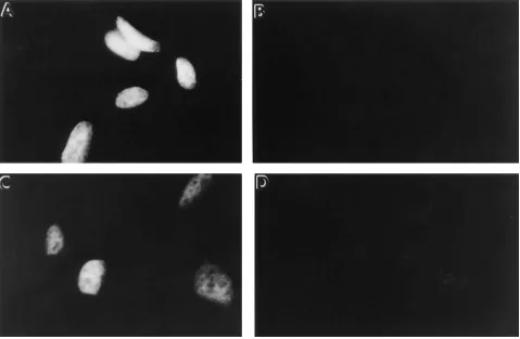
We next examined whether the mutant phenotype was re-versible in cells expressing the ts E1 protein. For these experi-ments, we again used the appearance of p53 staining (or lack thereof) as an assay for E1 enzyme activity. As shown in Fig. 10 A, cells maintained at 39.58C for 2 d in the absence of pro-tein stabilizers exhibited strong nuclear p53 staining, indicative that the E1 enzyme (and therefore the ubiquitin pathway) was inactive. Switching the 39.58C cells back to 32.58C for 1 d re-sulted in a disappearance of nuclear p53 staining (Fig. 10 B), indicative of a restoration of the ubiquitin-dependent protein degradation pathway. In those cells incubated at 39.58C for 1 d in normal culture medium, and then transferred into culture medium containing either TMAO or glycerol for an additional day at 39.58C, mixed results were obtained. Specifically, TMAO treatment resulted in a diminishment, but not com-plete abolition, of nuclear p53 staining (Fig. 10 C). In the case of glycerol treatment, however, p53 staining no longer was ob-served (Fig. 10 D).
Proper folding of the newly synthesized (but not the mature) form of the E1 protein is corrected by the chemical treatments.
[image:10.612.57.299.57.134.2]mally folded protein referred to as the prion protein. We have found that many of the same chemical chaperones used in the present study are effective in interfering with the formation of the pathogenic prion protein in a neuronal derived cell line (27). It is important to point out, however, that the conversion of wild-type prion into its pathogenic isoform does not appear to be a temperature-dependent process. Thus, in addition to their effectiveness for correcting temperature-sensitive protein fold-ing defects, chemical chaperones also may prove to have effi-cacy in correcting other types of protein folding abnormalities. The primary objective of the study presented here was to determine whether temperature-sensitive protein folding mu-tants in general could be corrected via the use of chemical chaperones. Hence, our choice of the different temperature-sensitive mutants being examined was based primarily on the ease of screening for the appearance of a wild-type–like phe-notype. Owing to the results presented here, we suggest that a number of temperature-sensitive protein folding mutants can be corrected following treatment with various protein-stabiliz-ing chemicals. Interestprotein-stabiliz-ingly, the overall extent of correction appeared to vary somewhat as a function of the particular compound used. For example, glycerol has proven to be the
most effective chemical chaperone for correcting all of the dif-ferent temperature-sensitive mutants we have examined to date. High concentrations of the polyol (between 0.5-1.0 M), however, are always required to correct the particular protein folding abnormality. While the methylamine TMAO perhaps works a little less effectively than glycerol, it does so at concen-trations of 100 mM or less. Finally, deuterated water has provided us with very mixed results. Although it appears to be effective in correcting some temperature-sensitive protein folding de-fects, oftentimes the compound can have obvious deleterious effects on the viability of the cells.
[image:11.612.59.538.60.373.2]The mechanisms by which these various compounds affect protein folding or stability have been examined in vitro using defined test proteins (28, 29). Most of the different low molec-ular weight compounds enhance the stability of proteins to thermal or chemically induced denaturation and aggregation. In the case of glycerol (and likely other polyols), it has been suggested that the polyol tends to be excluded from the imme-diate vicinity of a polypeptide. Therefore, at high concentra-tions, glycerol might be expected to increase the relative hy-dration around the protein, and thereby result in the polypeptide decreasing its relative surface area. This tighter
1442 Brown et al.
packing of the protein, driven in a sense by an increased hy-drophobic effect, would serve to enhance the stability of the protein in response to thermal or chemical treatments. Obvi-ously, replacement of water by D2O also would be expected to influence protein–solvent interactions, and thereby possibly affect protein folding pathways. We refer the reader to some excellent review articles which discuss in more detail the ef-fects of these different compounds on protein folding and sta-bility (25, 30, 31).
Two types of temperature-sensitive protein folding defects have been described in the literature: Class 1; correctly folded (mature) proteins which are destabilized upon a temperature shift, thereby resulting in their loss of activity (32); or Class 2; newly synthesized proteins which are able to fold properly at the permissive temperature, but which fold incorrectly at the nonpermissive temperature (33). All of our data indicate that it is this latter class of temperature-sensitive mutants which are being affected in our studies presented here. For example, for each of the ts mutants examined, the time needed to correct the mutant phenotype via exposure of the cells to the protein stabilizing agents was relatively long. Furthermore (at least for
[image:12.612.59.540.61.377.2]the E1 enzyme), in the absence of ongoing protein synthesis, in-clusion of the different chemicals into the culture medium did not result in an apparent restoration of mature E1 enzyme func-tion (albeit as determined indirectly by monitoring p53 levels). This observation, along with a number of our other unpub-lished findings, is consistent with the idea that it is primarily newly synthesized temperature-sensitive protein folding mu-tants which are amenable to correction via the chemical chap-erones. We emphasize, however, that proteins do exist that manifest destabilizing temperature-sensitive mutations (e.g., Class 1 mutants), and that some of these may in fact be amena-ble to correction via the strategies used here (currently under study). In the case of the Class 2 mutants, we suspect that the chemical chaperones affect a critical step in the folding path-way of the newly synthesized, temperature-sensitive protein folding mutant. For example, at the nonpermissive tempera-ture, the chemical chaperones may help in lowering a critical energy barrier which is rate-limiting in the folding pathway. In addition, the chemical chaperones might act to reduce the pro-pensity of the newly synthesized mutant protein to go off path-way and end up in a denatured or aggregated state. Whatever
the mechanism, all of our data indicate that once the particular temperature-sensitive protein folding mutant successfully has acquired its properly folded and biologically active state, it re-mains correctly folded at the nonpermissive temperature, even when the chemical chaperone was subsequently removed.
The concept of using protein stabilizing agents in vivo to correct protein folding defects has been in the scientific litera-ture for 30 yr or more. In both yeast and bacteria it was recog-nized that different mutations (usually missense mutations) could be corrected by either reducing the growth temperature or by increasing the osmotic strength of the growth medium. Indeed, Hawthorne and Friis in a 1964 report demonstrated that a variety of mutations in Saccharomyces cerivisiae could be corrected by the inclusion of compounds such as glucose, potassium chloride, glycerol, or diethylene glycol into the growth medium (7). Based on their success, they referred to their particular experimental protocol as osmotic remediation. Furthermore, in recent years it has been shown that cells from different organisms routinely employ osmotic remediation to deal with adverse changes in their environment which might lead to protein folding errors (34). For example, animal cells chronically exposed to hyperosmotic stress (e.g., cells in the kidney) respond by accumulating low molecular weight com-pounds in an effort to maintain their osmotic balance. These compounds, referred to as cellular osmolytes, are comprised primarily of three classes of organic compounds: carbohy-drates, free amino acids (and amino acid derivatives), and methylamines. Members of the first two groups also are re-ferred to as compatible osmolytes because of their ability to accumulate within the cell to high concentrations without sig-nificantly perturbing protein function. Alternatively, counter-acting osmolytes, represented by the methylamines, are usually produced to offset the protein denaturing effects of urea. For example, in many saltwater organisms as well as in the mam-malian kidney where urea can reach dangerously high levels, methylamines are found to accumulate as a way to offset the protein denaturing effects of urea (35, 36). Interestingly, at a 1:2 ratio of methylamine/urea, the potential protein denaturing ef-fects of urea are greatly reduced.
Thus, while the concept of correcting protein folding ab-normalities by changing the protein folding environment is not new, we suggest that this type of approach may represent a novel strategy to interfere with those protein folding abnor-malities which constitute the molecular basis of different dis-eases. Based on our observations with the DF508 CFTR pro-tein as well as the alanine to valine p53 tumor suppressor mutant (both of which are associated with disease in humans), we wonder how many other genetically inherited diseases re-sult in the production of mutant proteins which exhibit tem-perature-sensitive folding defects. Considering the results pre-sented here, we predict that most temperature sensitive protein folding mutants (at least of the Class 2 variety) will be amenable to correction in vivo by one or more of the chemical chaperones. Furthermore, we hope that our observations will encourage others to search for additional low molecular weight compounds which might be effective in correcting pro-tein folding abnormalities. Identifying small molecules that are: (a) able to passively move into cells, (b) effective in influ-encing the protein folding environment, and (c) nontoxic to the cell tissue and organism may prove to be both faster and more efficacious for treating disease than are current ap-proaches using gene therapy.
Acknowledgments
We thank members of the Welch lab for critical comments.
This work was supported by the National Cystic Fibrosis Founda-tion, Cystic Fibrosis Research, Inc., and the National Institutes of Health (GM33551).
References
1. Germsla, S.Y., and E.R. Stuur. 1972. The effects of combining urea and
an alcohol on the heat-induced reversible denaturation of the ribonuclease. Int.
J. Pept. Protein Res. 4:372–378.
2. Back, J.F., D. Oakenfull, and M.B. Smith. 1979. Increased thermal
stabil-ity of protein in the presence of sugars and polyols. Biochemistry. 18:5191–5199.
3. Gekko, K., and S. Koga. 1983. Increased thermal stability of collagen in
the presence of sugars and polyols. J. Biochem. (Tokyo). 94:199–208.
4. Lin, P.S., L. Kwock, and K. Hefter. 1981. Protection of heat induced
cyto-toxicity by glycerol. J. Cell. Physiol. 108:439–448.
5. Henle, K.J., J.W. Peck, and R. Higashikubo. 1983. Protection against
heat induced cell killing by polyols in vitro. Cancer Res. 43:1624–1633.
6. Edington, B.V., S.A. Whelan, and L.E. Hightower. 1989. Inhibition of heat shock (stress) protein induction by deuterium oxide and glycerol:
Addi-tional support for the abnormal protein hypothesis of induction. J. Cell. Physiol.
139:219–228.
7. Hawthorne, D.C., and J. Friss. 1964. Osmotic remedial mutants. A new
classification for nutritional mutants in yeast. Genetics. 50:829–839.
8. Brown, C.R., L.Q. Hong-Brown, J. Biwersi, A.S. Verkman, and W.J.
Welch. 1996. Chemical chaperones correct the mutant phenotype of the DF508
cystic fibrosis transmembrane conductance regulator protein. Cell Stress &
Chap. 1:117–125.
9. Cheng, S.H., R.J. Gregory, J. Marshall, S. Paul, D.W. Souza, G.A. White, C.R. O’Riordan, and A.E. Smith. 1990. Defective intracellular transport and
processing of CFTR is the molecular basis of most cystic fibrosis. Cell. 63:827–
834.
10. Yang, Y., S. Janich, J.A. Cohn, and J.M. Wilson. 1993. The common variant of cystic fibrosis transmembrane conductance regulator is recognized by
hsp 70 and degraded in a pre-golgi non-lysosomal compartment. Proc. Natl.
Acad. Sci. USA. 90:9480–9484.
11. Pind, S., J.R. Riordan, and D.B. Williams. 1994. Participation of the en-doplasmic reticulum chaperone calnexin (p88, IP90) in the biogenesis of the
cystic fibrosis transmembrane conductance regulator. J. Biol. Chem. 269:12784–
12788.
12. Li, C., M. Ramjeesingh, E. Reyea, T. Jensen, X. Chang, J.M. Rommens,
and C.E. Bear. 1993. The cystic fibrosis mutation (DF508) does not influence
the chloride channel activity of CFTR. Nature Genet. 3:311–316.
13. Drumm, M.L., D.S. Wilkinson, L.S. Smit, R.T. Worrell, and T.V.
Strong. 1991. Chloride conductance expressed by DF508 and other mutant
CFTRs in Xenopus oocytes. Science (Wash. DC). 254:1797–1799.
14. Denning, G.M., M.P. Anderson, J.F. Amara, J. Marshallo, A.E. Smith, and M.J. Welsh. 1992. Processing of mutant cystic fibrosis transmembrane
con-ductance regulator is temperature sensitive. Nature (Lond.). 358:761–764.
15. Thomas, P.J., B.H. Qu, and P.L. Pedersen. 1995. Defective protein
fold-ing as a basis of human disease. TIBS (Trends Biochem. Sci.). 20:456–459.
16. Welch, W.J., and C.R. Brown. 1996. Influence of molecular and
chemi-cal chaperones on protein folding. Cell Stress & Chap. 1:109–115.
17. Finlay, C.A., P.W. Hinds, T.H. Tan, D. Eliyahu, M. Oren, and A.J. Le-vine. 1988. Activating mutations for transformation by p53 produce a gene
product that forms an hsc70-p53 complex with an altered half-life. Mol. Cell.
Biol. 8:531–539.
18. Michalovitz, D., O. Halvey, and M. Oren. 1990. Conditional inhibition of transformation and of cell proliferation by a temperature-sensitive mutant of
p53. Cell. 62:671–680.
19. Martinez, J., I. Georgoff, J. Martinez, and A.J. Levine. 1991. Cellular lo-calization and cell cycle regulation by a temperature-sensitive p53 protein.
Genes & Dev. 5:151–159.
20. Gannon, J.V., and D.P. Lane. 1991. Protein synthesis required to anchor
a mutant p53 protein which is temperature sensitive. Nature (Lond.). 349:802–
806.
21. Maroney, A.C., S.A. Qureshi, D.A. Foster, and J.S. Brugge. 1992. Clon-ing and characterization of a thermolabile v-src gene for use in reversible
trans-formation of mammalian cells. Oncogene. 7:1207–1214.
22. Kulka, R.G., B. Raboy, R. Schuster, H.A. Parag, G. Diamond, A. Ciechanover, and M. Marcus. 1988. A Chinese hamster cell cycle mutant ar-rested at G2 phase has a temperature-sensitive ubiquitin-activating enzyme, E1.
J. Biol. Chem. 263:15726–15731.
23. Chowdary, D.R., J.J. Dermody, K.K. Jha, and H.L. Ozer. 1994.
Accu-mulation of p53 in a mutant cell line defective in the ubiquitin pathway. Mol.
Cell. Biol. 14:1997–2003.
1444 Brown et al.
the nucleus in a cell cycle-dependent manner. J. Biol. Chem. 271:15608–15614.
25. Schein, C.H. 1990. Solubility as a function of protein structure and
sol-vent components. Bio-Technology. 8:308–316.
26. Sato, S., C.L. Ward, M.E. Krouse, J.J. Wine, and R.R. Kopito. 1996. Glycerol reverses the misfolding phenotype of the most common cystic fibrosis
mutation. J. Biol. Chem. 271:635–638.
27. Tatzelt, J., S. Pruisner, and W.J. Welch. 1996. Chemical chaperones
in-terfere with the formation of scrapie prion proteins. EMBO (Eur. Mol. Biol.
Organ.) J. 15:6363–6373.
28. Gekko, K., and S.N. Timasheff. 1981. Mechanisms of protein
stabiliza-tion by glycerol: Preferential hydrastabiliza-tion in glycerol-water mixtures.
Biochemis-try. 20:4667–4676.
29. Gekko, K. and S.N. Timasheff. 1981b. Thermodynamic and kinetic
ex-amination of protein stabilization by glycerol. Biochemistry. 20:4677–4686.
30. Santoro, M.M., Y. Liu, S.M.A. Khan, L.X. Hou, and D.W. Bolen. 1992. Increased thermal stability of proteins in the presence of naturally occurring
os-molytes. Biochemistry. 31:5278–5283.
31. Lin, T.Y., and S.N. Timasheff. 1994. Why do some organisms use a urea-methylamine mixture as osmolyte? Thermodynamic compensation of urea and
trimethylanine N-oxide interactions with protein. Biochemistry. 33:12695–
12701.
32. Hawkes, R., M.G. Grutter, and J. Schellman. 1984. Thermodynamic
sta-bility and point mutations of bacteriophage T4 lysozyme. J. Mol. Biol. 175:195–
212.
33. Sturtevant, J.M., M. Yu, C. Haase-Pettingell, and J. King. 1989. Ther-mostability of temperature-sensitive folding mutants of the P22 tailspike
pro-tein. J. Biol. Chem. 264:10693–10698.
34. Burg, M.B. 1995. Molecular basis of osmotic regulation. Am. J. Physiol.
268:983–996.
35. Yancey, P.H., M.E. Clark, S.C. Hand, R.D. Bowlus, and G.N. Somero.
1982. Living with water stress; evolultion of osmolyte systems. Science (Wash.
DC). 217:1214–1222.
36. Somero, G. 1986. Protons, osmolytes and fitness of internal milieu for