STAT4 and STAT6 regulate systemic
inflammation and protect against lethal
endotoxemia
Alex B. Lentsch, … , Celia Chao, Michael J. Edwards
J Clin Invest.
2001;
108(10)
:1475-1482.
https://doi.org/10.1172/JCI13763
.
Members of the signal transducer and activator of transcription (STAT) family are
transcription factors that mediate many of the effects of pro- and anti-inflammatory cytokines.
The progressive systemic inflammatory response induced by endotoxin is mediated by
overzealous cytokine production. Here we identify STAT4 and STAT6 as critical regulators
of the systemic inflammatory response to endotoxin. Mice deficient for STAT4 or STAT6
were highly susceptible to lethal endotoxemia. In
STAT4
–/–mice, antibody blockade of
IL-12 prevented mortality, suggesting that STAT4 confers protection, while another signaling
pathway mediates the detrimental effects of IL-12. In
STAT6
–/–mice we observed
dysregulated activation of the transcription factor NF-
k
B, resulting in augmented production
of proinflammatory cytokines and chemokines. Furthermore,
STAT6
–/–mice displayed
increased organ accumulation of leukocytes and significant hepatocellular injury. These
findings demonstrate that STAT4 and STAT6 confer protection against endotoxin-induced
death and that for STAT6 these protective effects occur through the regulation of NF-
k
B
activation and subsequent production of proinflammatory cytokines and chemokines.
Article
Find the latest version:
Introduction
The septic state is associated with a generalized activa-tion of inflammatory pathways following infecactiva-tion. In Gram-negative infections endotoxin is a primary stim-ulus of the inflammatory response. Endotoxin binds to a variety of cell types, including macrophages, mono-cytes, and endothelial cells, triggering activation of the transcription factor NF-κB (1). Activation of NF-κB, which consists of nuclear translocation and DNA bind-ing, results in the expression of proinflammatory cytokines, chemokines, and VCAMs (2, 3). During sep-sis or endotoxemia, the dysregulated production of these mediators may result in an overwhelming sys-temic inflammatory response that contributes to dis-turbed cardiopulmonary function and hemodynamic instability, sometimes progressing to multiorgan failure and death (4−6). While NF-κB appears to be the primary endotoxin-induced transcriptional activator of proin-flammatory mediators, little is known about the tran-scriptional control mechanisms that attempt to regu-late the inflammatory response during endotoxemia. The signal transducer and activator of transcription (STAT) family of transcription factors includes seven members that are activated in response to different cytokine-receptor interactions (7). Of this family, STAT4 and STAT6 are highly relevant to the inflam-matory response to bacteria or endotoxin. STAT4 is the primary transcription factor utilized by IL-12 (8, 9). IL-12 is critical for bacterial containment and clearance
in septic peritonitis and for promotion of the inflam-matory response during endotoxemia (10−12). STAT6 is activated by IL-4 and IL-13 (13−15). No role for IL-4 has been established in either sepsis or endotoxemia, but IL-13 appears to be a major regulator of the inflammatory response of both models by preventing organ dysfunction and death (16, 17). In the cecal liga-tion and puncture model of septic peritonitis, mice lack-ing either STAT4 or STAT6 had less organ injury and mortality, suggesting that STAT4 and STAT6 are inti-mately involved in the host response to infection (18). The objective of the current study was to investigate the roles of STAT4 and STAT6 in the systemic inflam-matory response induced by endotoxin. Since this model is devoid of any infectious burden, it provides information strictly related to the systemic inflamma-tory response induced by this component of Gram-neg-ative bacteria. Our results demonstrate that mice lack-ing either STAT4 or STAT6 are much more susceptible to lethal endotoxemia than their wild-type counter-parts. Furthermore, blockade of endogenous IL-12 in STAT4–/–mice prevented the lethal effects of
endotox-in, suggesting that the deleterious effects of IL-12 are mediated by another signaling pathway. In STAT6–/–
mice, there was unregulated activation of NF-κB and increased chemokine expression in liver and lung. These findings suggest that STAT4 and STAT6 regu-late the systemic inflammatory response to endotoxin by distinct mechanisms.
STAT4 and STAT6 regulate systemic inflammation
and protect against lethal endotoxemia
Alex B. Lentsch, Atsushi Kato, Brian Davis, Warner Wang, Celia Chao,
and Michael J. Edwards
Department of Surgery, University of Louisville School of Medicine, Louisville, Kentucky, USA
Address correspondence to: Alex B. Lentsch, Department of Surgery, University of Louisville, James Graham Brown Cancer Center, Room 426, Louisville, Kentucky 40202, USA.
Phone: (502) 852-7698; Fax: (502) 852-2975; E-mail: [email protected].
Received for publication July 16, 2001, and accepted in revised form September 25, 2001.
Members of the signal transducer and activator of transcription (STAT) family are transcription fac-tors that mediate many of the effects of pro- and anti-inflammatory cytokines. The progressive sys-temic inflammatory response induced by endotoxin is mediated by overzealous cytokine production. Here we identify STAT4 and STAT6 as critical regulators of the systemic inflammatory response to endotoxin. Mice deficient for STAT4 or STAT6 were highly susceptible to lethal endotoxemia. In
STAT4–/–mice, antibody blockade of IL-12 prevented mortality, suggesting that STAT4 confers
pro-tection, while another signaling pathway mediates the detrimental effects of IL-12. In STAT6–/–mice
we observed dysregulated activation of the transcription factor NF-κB, resulting in augmented pro-duction of proinflammatory cytokines and chemokines. Furthermore, STAT6–/–mice displayed
increased organ accumulation of leukocytes and significant hepatocellular injury. These findings demonstrate that STAT4 and STAT6 confer protection against endotoxin-induced death and that for STAT6 these protective effects occur through the regulation of NF-κB activation and subsequent production of proinflammatory cytokines and chemokines.
Methods
Murine model of endotoxemia. STAT4–/–and STAT6–/–mice
(backcrossed ten generations to BALB/c) were purchased from The Jackson Laboratory (Bar Harbor, Maine, USA) and bred and maintained at the University of Louisville. Age- and sex-matched BALB/c mice were purchased from The Jackson Laboratory and used as wild-type con-trols. For induction of endotoxemia, mice received 3 mg/kg endotoxin (LPS from Escherichia coli, serotype 0111:B4; Sigma Chemical Co., St. Louis, Missouri, USA) in 0.75 ml PBS by intraperitoneal injection. Control mice received 0.75 ml PBS. For IL-12 blockade, 300 µg of affinity-purified polyclonal rabbit anti-mouse IL-12 IgG was administered via the tail vein just prior to endotox-in endotox-injection. We have previously shown that this dose of anti−IL-12 effectively neutralizes the biological activity of IL-12 in a murine model of hepatic ischemia/reperfu-sion (19). Controls received 300 µg of preimmune rabbit IgG. This project was approved by the University of Louisville Animal Care and Use Committee and was in compliance with NIH guidelines.
Immunoprecipitation and Western blot. Liver and lung tis-sues were homogenized in cold PBS supplemented with 1% NP-40, 0.5% deoxycholate, 0.1% SDS, 1 µg/ml leu-peptin, 1 µg/ml aprotonin, 10 µg/ml soybean trypsin inhibitor, 1 µg/ml pepstatin, and 100 µg/ml of PMSF. Protein concentration was determined by bicinchoninic acid protein assay (Pierce Chemical Co., Rockford, Illi-nois, USA). One milligram of total protein was used for immunoprecipitation. The protein samples were pre-cleared with 200 µl of protein G (Life Technologies Inc., Rockville, Maryland, USA) per sample for 1 hour at 4°C. Antibodies to STAT4 and STAT6 (Santa Cruz Biotech-nology Inc., Santa Cruz, California, USA) were prebound to protein G and 2 µg antibody/protein G conjugate was added to each sample and incubated for 2 hours at 4°C. The beads were washed three times with cold supple-mented PBS, resuspended in SDS sample buffer, and boiled for 3 minutes. The immunoprecipitated samples were separated by 10% SDS-PAGE and transferred to a PVDF membrane. The membranes were blocked in 5% BSA in Tris-buffered saline overnight at 4°C and reblocked in 5% milk in Tris-buffered saline plus 0.2% Tween-20 (TTBS) for 30 minutes at room temperature. Mouse anti-phosphotyrosine antibody (Santa Cruz Biotechnology Inc.) was diluted at 1:1000 with 5% milk in TTBS and incubated with the membranes for 1 hour at room temperature. The membranes were washed three times with TTBS and incubated with goat anti-mouse IgG horseradish peroxidase secondary antibody (Santa Cruz Biotechnology Inc.), diluted at 1:5000 with 5% milk in TTBS, for 30 minutes at room temperature. The mem-branes were washed as above and immunoreactive pro-teins were visualized using enhanced chemiluminescence. Electrophoretic mobility shift assay. Nuclear extracts of liver tissue were prepared by the method of Deryckere and Gannon (20) and analyzed by electrophoretic mobility shift assay (EMSA). Briefly, double-stranded NF-κB con-sensus oligonucleotide (Promega Corp., Madison,
Wis-consin, USA) was end-labeled with γ[32P] ATP (3,000 Ci/mmol at 10 mCi/ml; Amersham Life Sciences Inc., Arlington Heights, Illinois, USA). Binding reactions con-taining equal amounts of nuclear protein extract (20 µg) and 35 fmol (∼50,000 cpm, Cherenkov counting) of oligonucleotide were incubated at room temperature for 30 minutes. Reaction volumes were held constant to 15
µl. Reaction products were separated in a native 4% poly-acrylamide gel and analyzed by autoradiography.
Ribonuclease protection assay. Total RNA from lung and liver was extracted with TRIzol Reagent (Life Technolo-gies Inc.) Ribonuclease protection assays were per-formed using RiboQuant kits purchased from Pharmingen (San Diego, California, USA) as described by the manufacturer. A customized DNA template was used to analyze the expression of the chemokines lym-photactin (Ltn), RANTES, eotaxin, macrophage inflam-matory protein–1α (MIP-1α), MIP-1β, MIP-2, IFN-γ inducible protein–10 (IP-10), and monocyte chemoat-tractant protein-1 (MCP-1), as well as the housekeeping gene GAPDH. The probes were hybridized with 10 µg of total RNA for 16 hours at 56°C, after which free probe and other single-stranded RNA was digested with RNas-es. Double-stranded (protected) RNA was purified and resolved using the QuickPoint Gel system (Novex, San Diego, California, USA).
[image:3.576.318.484.575.657.2]Serum and tissue cytokine/chemokine assays. ELISA reagents for TNF-α, MIP-2, MCP-1, IFN-γ, IL-6, and GM-CSF were from PeproTech Inc. (Rocky Hill, New Jersey, USA). ELISA reagents for IL-10 and IL-13 were purchased from R&D Systems Inc. (Minneapolis, Min-nesota, USA), and reagents for IL-12 were from Pharmingen. Extraction of proteins from tissue samples for ELISA analyses was performed as described else-where (21). Briefly, tissues were weighed and immedi-ately placed in 10 volumes (wt/vol) of a protease inhibitor cocktail containing 10 mM EDTA, 2 mM PMSF, 0.1 mg/ml soybean trypsin inhibitor, 1.0 mg/ml BSA, 1% penicillin-streptomycin, and 0.02% sodium azide in isotonic PBS, pH 7.0. Tissues were disrupted with a tissue homogenizer and lysates were incubated at 4°C for 2 hours. Samples were clarified by two rounds of centrifugation at 12,500 gfor 10 minutes at 4°C. Pro-tein concentration was determined by bicinchoninic
Figure 1
acid assay with trichloroacetic acid precipitation using BSA as a reference standard (Pierce Chemical Co.).
Histology and clinical chemistry. Tissues were fixed in 10% buffered formalin, embedded in paraffin, sec-tioned, and stained with hematoxylin and eosin. Twen-ty high-power fields per tissue section were observed for each animal, and the number of granulocytes and mononuclear cells was recorded. Data are expressed as the number of cells per high-power field. Serum was analyzed for alanine aminotransferase (ALT) as an index of liver injury. Measurements of serum ALT were made using a diagnostic kit from Sigma Chemical Co. Myeloperoxidase assay. Liver myeloperoxidase (MPO) content was assessed by methods similar to those of Schierwagen et al. (22). Liver tissue (100 mg) was homogenized in 2 ml of buffer A (3.4 mM KH2HPO4, 16 mM Na2HPO4, pH 7.4). After centrifugation for 20 minutes at 10,000 g, the pellet was resuspended in 10 volumes of buffer B (43.2 mM KH2HPO4, 6.5 mM Na2HPO4, 10 mM EDTA, 0.5% hexadecyltrimethylam-monium, pH 6.0) and sonicated for 10 seconds. After heating for 2 hours at 60°C, the supernatant was react-ed with 3,3′, 3,5′-tetramethylbenzidine (Sigma Chemi-cal Co.) and optiChemi-cal density determined at 655 nm.
Statistical analysis. All data are expressed as mean ± SEM. Survival data were analyzed with Fisher’s exact test. All other data were analyzed with a one-way ANOVA with subsequent Student-Newman-Keuls test. Differences were considered significant at P < 0.05.
Results
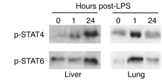
STAT4 and STAT6 are activated in liver and lung during endo-toxemia. Both IL-12 and IL-13 have been shown to be crit-ically important to the systemic inflammatory response to endotoxin (10, 16), suggesting potential roles for STAT4 and STAT6. In order to assess whether endotoxin resulted in activation of STAT4 and STAT6 in major
organs, we assessed the level of activated (phosphorylat-ed) STAT4 and STAT6 in liver and lung. Figure 1 shows the results of immunoprecipitation/Western blot analy-sis of phosphorylated STAT4 and STAT6 in liver and lung. In liver, little activated STAT4 was observed at time 0. STAT6 appeared to have a basal level of constitutive activation. One hour after endotoxin, levels of phospho-rylated STAT4, but not STAT6, were increased. However, 24 hours after endotoxin there was a dramatic increase in the amount of phosphorylated STAT4 and STAT6 in liver extracts. In lung, phosphorylated forms of both STAT4 and STAT6 were increased 1 hour after endotox-in. After 24 hours, the amount of phosphorylated STAT4 resembled that at time 0, while levels of phosphorylated STAT6 remained above that of time 0. Thus, it appears that activation of STAT4 and STAT6 occurs in distinct organ-specific, time-dependent fashions.
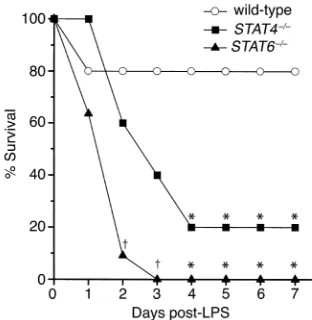
STAT4 and STAT6 are required for protection against lethal endotoxemia. To determine whether activation of STAT4 and STAT6 are important regulatory events in the response to endotoxin, we compared the survival rates of wild-type, STAT4–/–, and STAT6–/–mice after
admin-istration of 3 mg/kg endotoxin. Twenty percent of wild-type mice died within 24 hours, but none died there-after, yielding a survival rate of 80% (Figure 2). Forty percent of STAT4–/–mice died within 2 days, 60% died
after 3 days, and 80% were dead after 4 days (P= 0.023). No STAT4–/–mice died after 4 days, resulting in a 20%
survival rate. STAT6–/– mice died precipitously after
endotoxin, with 36% dead after 24 hours, 91% dead after 2 days (P= 0.002), and 100% dead by 3 days (P< 0.001). For all mice (wild-type, STAT4–/–, and STAT6–/–), those
surviving after 7 days were alive after 15 days. Mortality was significantly greater in STAT6–/– mice than in
STAT4–/–mice 2 days (P= 0.024) and 3 days (P= 0.035)
[image:4.576.74.230.54.214.2]after endotoxin (Figure 2). These data demonstrate that
Figure 2
[image:4.576.337.492.485.654.2]STAT4–/–and STAT6–/–mice are more susceptible to lethal endotox-emia than wild-type mice. Following intraperitoneal administration of 3 mg/kg endotoxin, wild-type, STAT4–/–, and STAT6–/–mice were mon-itored for survival. Those mice alive at day 7 were alive at day 15 (data not shown). For each group, n= 10−11. *P < 0.05 compared with wild-type mice; †P < 0.05 compared with wild-type and STAT4–/–mice.
Figure 3
STAT4- and STAT6-deficient mice are far more suscep-tible to lethal endotoxemia than wild-type mice.
Because STAT4 mediates many of the biological effects of IL-12 (8, 9), and blockade of IL-12 has been shown to be protective in a similar murine model of endotoxemia (10), we sought to determine whether, in this model, IL-12 has effects mediated by other signal transduction mechanisms. For these experiments, STAT4–/–mice were
treated with either 300 µg preimmune rabbit IgG or anti−IL-12 just prior to endotoxin administration. As shown in Figure 3, none of the STAT4–/–mice receiving
anti−IL-12 died, while 67% of those receiving preim-mune IgG died within 4 days (P = 0.005). These data sug-gest that IL-12 contributes to the lethal effects of endo-toxin by a STAT4-independent mechanism.
Augmented cytokine and chemokine expression in STAT6–/–
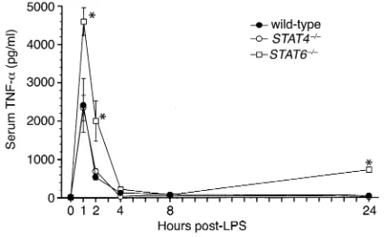
mice. Endotoxemia is characterized by a rapid increase in the expression of the proinflammatory cytokine TNF-α, as well as the subsequent multiorgan expression of CXC and CC chemokines (23−26). To investigate the potential mechanisms of the increased mortality observed in STAT4–/–and STAT6–/–mice, we measured serum levels of
TNF-αand expression of chemokine mRNA and protein in liver and lung. Wild-type mice showed the expected rapid increase in serum TNF-α, with peak levels occurring 1 hour after endotoxin (Figure 4). STAT4–/–mice had a
nearly identical pattern of TNF-αproduction. In STAT6–/–
mice, however, serum TNF-αwas greatly increased at 1 hour, 2 hours, and 24 hours, compared with both wild-type and STAT4–/–mice (Figure 4), indicating
dysregulat-ed production of this inflammatory cytokine.
A number of other cytokines have been implicated in the positive and negative regulation of systemic inflam-mation induced by endotoxemia. In order to ascertain whether the susceptibility of STAT4–/–and STAT6–/–may
be a result of altered tissue expression of pro- and/or anti-inflammatory cytokines, we measured the expression of the proinflammatory cytokines IL-12, IFN-γ, and GM-CSF and the anti-inflammatory cytokines, IL-6, IL-10, and IL-13 in the liver and lung by ELISA 1 hour and 24 hours after endotoxin administration. With the
excep-tion of IL-6, which was increased in liver and lungs of STAT6–/–mice after 24 hours (Table 1), we found no
dif-ferences in the protein expression of these mediators in wild-type, STAT4–/–, or STAT6–/–mice at either time point.
Because the multi-organ leukocyte recruitment observed during endotoxemia is, in part, dependent upon increased organ expression of chemokines, we assessed the expression of chemokine mRNAs in liver and lung tis-sues. STAT4–/–mice differed from wild-type mice, with
increased IP-10 and MCP-1 mRNA expression in liver 1 hour after endotoxin (Figure 5). Similarly, liver expression of IP-10 and MCP-1 mRNA was increased in livers of STAT6–/– mice after 1 hour. Twenty-four hours after
endotoxin, chemokine mRNA expression in STAT4–/–
mice appeared identical to that of wild-type mice. How-ever, STAT6–/–mice displayed greatly increased expression
of mRNA for MIP-1α, MIP-2, IP-10, and MCP-1 in both liver and lung 24 hours after endotoxin (Figure 5). Mea-surement of MIP-1α, MIP-2, and MCP-1 proteins in liver and lung 24 hours after endotoxin showed no differences between wild-type mice and STAT4–/–mice (Figure 6).
STAT6–/–mice showed increases in MIP-1α, MIP-2, and
MCP-1 proteins in liver and MIP-1αand MCP-1 in lung. Thus, STAT6, but not STAT4, appears to be required for the regulation of cytokine and chemokine production during endotoxemia.
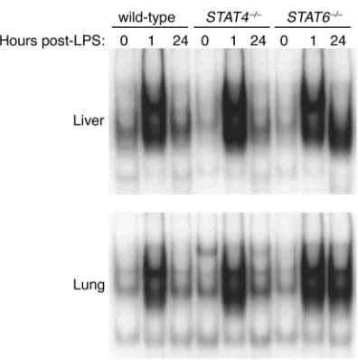
Activation of NF-κB is dysregulated in STAT6–/–mice. The
transcription factor NF-κB has been implicated in the development of the systemic inflammatory response in sepsis and endotoxemia (27, 28). NF-κB is a major tran-scriptional regulator of numerous proinflammatory mediators, including TNF-α, MIP-1α, MIP-2, and MCP-1 (3). In order to assess whether the function of NF-κB was altered in STAT6–/–mice as a potential
mech-anism for the unregulated production of cytokines and chemokines, we measured the nuclear translocation and DNA binding of NF-κB in liver and lung extracts. In wild-type and STAT4–/–mice, there was little NF-κB in
nuclear extracts of liver and lung at time 0 (Figure 7). One hour after endotoxin, NF-κB activation was dra-matically increased, returning to baseline levels after 24 hours. In contrast, NF-κB activation in liver and lungs of STAT6–/–mice showed the rapid activation of NF-κB
[image:5.576.65.280.52.183.2]after 1 hour, but this high level of activation was also present 24 hours after endotoxin (Figure 7), suggesting that STAT6 is critical for the regulation of NF-κB acti-vation and subsequent production of cytokines and chemokines during endotoxemia.
Figure 4
[image:5.576.304.540.652.719.2]Serum levels of TNF-αduring endotoxemia. TNF-αcontent in serum samples was measured by ELISA in wild-type, STAT4–/–, and STAT6–/– mice after intraperitoneal injection of 3 mg/kg endotoxin. Values are mean ± SEM with n= 5−8 mice per group. *P < 0.05 compared with wild-type and STAT4–/–mice.
Table 1
IL-6 expression 24 hours after endotoxin administration
IL-6 (pg/mg protein)
Liver Lung
Wild-type 59.6 ± 3.2 42.3 ± 13.9
STAT4–/– 57.5 ± 3.0 37.6 ± 5.5
STAT6–/– 114.8 ± 16.3A 251.0 ± 49.8A
Increased hepatocellular damage and organ leukocyte accu-mulation in STAT6–/–mice. Endotoxin causes the
accumu-lation of leukocytes in multiple organs. Therefore, we con-ducted experiments to determine whether the augmented production of cytokines and chemokines in STAT6–/–mice
was associated with increased leukocyte trafficking and organ dysfunction. Sections of liver and lung were evalu-ated for histopathological changes 24 hours after endo-toxin. No differences were observed in lung sections from wild-type, STAT4–/–, and STAT6–/–mice; all showed similar
thickening of the alveolar membranes (data not shown). In liver sections, tissue architecture appeared relatively normal in wild-type and STAT4–/–mice (Figure 8, a and b,
respectively). However, livers from STAT6–/–mice showed
abundant areas of hepatocyte necrosis (Figure 8c). These histopathological changes in the livers of STAT6–/–mice
were associated with a significant increase in serum ALT, a marker of hepatocellular injury (Figure 8d). There were no changes in serum ALT levels in either wild-type or STAT4–/–mice. Thus, the greater susceptibility of STAT6–/–
mice to the lethal effects of endotoxin may be related to the hepatocellular injury observed in these mice.
In both liver and lung sections, we observed large numbers of granulocytic and monocytic leukocytes. In lungs, these cells appeared to be adherent to the vascu-lar endothelium, and there was no evidence of cells in the alveolar spaces. In liver, these cells were localized primarily along the vascular walls of central veins and within sinusoids. To determine whether there were dif-ferences in the extent of leukocyte accumulation in liver and lung between wild-type, STAT4–/–, and
STAT6–/–mice, we performed MPO assays (for
quanti-tation of neutrophils) and morphometric analyses of granulocytes and mononuclear cells within these tis-sues. The results of our MPO assays are shown in Fig-ure 9. In liver, no significant changes in MPO activity were observed 2, 4, or 8 hours after endotoxin (Figure 9a). After 24 hours, liver MPO levels in STAT6–/–mice
were up to threefold higher than the levels in wild-type and STAT4–/–mice, which were themselves increased
above baseline. In lung, MPO activity in STAT4–/–mice
was significantly greater than in wild-type mice 1 and 2 hours after endotoxin, remained elevated after 8 hours, and decreased by 24 hours (Figure 9b). MPO
lev-Figure 5
[image:6.576.66.405.51.282.2]Chemokine mRNA expression in liver and lung tissues during endotoxemia. mRNA for the chemokines Ltn, RANTES, eotaxin, MIP-1α, MIP-1β, MIP-2, IP-10, and MCP-1 were evalu-ated by ribonuclease protection assay. GAPDH was used to confirm the equality of in vitro transcription efficiency and sample loading. The first lane contains unprotected probes and the corresponding lines indicate the location of the protected mRNA species.
Figure 6
[image:6.576.74.523.565.700.2]els in lungs from STAT6–/–mice were indistinguishable
from those in wild-type mice for up to 8 hours, but 24 hours after endotoxin they were nearly threefold high-er than in wild-type or STAT4–/–mice. Based on these
findings it appears that STAT4–/–mice have
augment-ed neutrophil accumulation in lungs shortly after endotoxin but, by 24 hours, have similar amounts of neutrophils to those of wild-type mice. Conversely, STAT6–/–mice appear to have a delayed but grossly
increased accumulation of neutrophils in liver and lung following endotoxin.
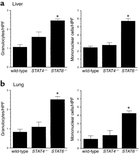
In addition to tissue MPO assays, we performed mor-phometric analyses to quantitate the number of gran-ulocytes and mononuclear cells in liver and lung tissues 24 hours after endotoxin. As shown in Figure 6, livers from STAT4–/–mice had similar amounts of
granulo-cytes and mononuclear cells when compared with wild-type mice (Figure 10a). STAT6–/–mice, however, had
sig-nificantly higher numbers of granulocytes and mononuclear cells in liver. Similarly, lungs from wild-type mice and STAT4–/–mice had equivalent numbers
of granulocytes and mononuclear cells, while STAT6–/–
mice had significantly higher numbers of these cells (Figure 10b). These observations support our MPO data and suggest that in addition to increased neu-trophil accumulation, STAT6–/–mice have augmented
recruitment of mononuclear cells (most likely lym-phocytes) to liver and lung 24 hours after endotoxin.
Discussion
STAT4 and STAT6 were originally defined as the criti-cal transcription factors mediating Th1 and Th2 cell differentiation, respectively (8, 9). However, cytokines that activate STAT4 and STAT6, IL-12 and IL-13, have prominent roles in a wide variety of inflammatory
responses, suggesting that STAT4 and STAT6 may have novel functions in these reactions. In the context of the systemic inflammatory response induced by endotoxin or sepsis, IL-12 and IL-13 appear to play con-trasting roles. IL-12 has been shown to propagate the inflammatory response to endotoxin by augmenting proinflammatory cytokine production and contribut-ing to endotoxin-induced death (10). Similarly, we have shown that IL-12 is a primary mediator of the hepatic inflammatory response to ischemia/reperfusion (19). However, IL-12 is also important for resistance to infec-tion, with a primary role in the clearance of bacteria (10, 12). Conversely, IL-13 plays similar roles in both endotoxemia and sepsis by downregulating the pro-duction of inflammatory cytokines and chemokines and reducing organ neutrophil recruitment (16, 17). Others have demonstrated that neutralization of IL-12 prevents the lethal effects of endotoxin (10). Until now STAT4 has been thought to mediate all of the known effects of IL-12; accordingly, we expected STAT4–/– mice to be protected against
endotoxin-induced death. However, we found that STAT4–/–mice
were not protected from endotoxin-induced death but were much more susceptible to death than wild-type mice. We observed significantly more neutrophils (meas-ured by MPO) in the lungs of STAT4–/–mice shortly after
endotoxin (1−4 hours), but these changes were not accompanied by any noticeable differences in lung histopathology after 24 hours. These data are particu-larly interesting since they suggest that STAT4 activation by IL-12 has a beneficial effect. Furthermore, we found that blockade of IL-12 in STAT4–/–mice completely
[image:7.576.59.257.50.249.2]abro-gated the lethal effects of endotoxin. This suggests that
Figure 7
[image:7.576.308.530.453.625.2]Activation of NF-κB during endotoxemia in wild-type, STAT4–/–, and STAT6–/–mice. Liver and lung nuclear extracts obtained at time 0 or 1 or 24 hours after administration of 3 mg/kg endotoxin were analyzed by EMSA.
Figure 8
during endotoxemia IL-12 has divergent functions involving different signaling pathways. Supportive of this concept is the recent report that bacterial clearance after cecal ligation and puncture is similar in wild-type and STAT4–/–mice (18). Blockade of IL-12 in the same
model resulted in decreased bacterial clearance (12), sug-gesting that STAT4 does not mediate the antibacterial effects of IL-12. Together with our current findings, it appears that activation of STAT4 confers protection from endotoxin lethality while an as-yet undefined sig-naling pathway mediates other effects of IL-12.
In contrast to our current findings, Matsukawa et al. showed that STAT4–/–mice were less susceptible to lethal
septic peritonitis than their wild-type counterparts (18). This effect appeared to be due to increased organ expres-sion of anti-inflammatory cytokines and reduced organ expression of CXC chemokines in STAT4–/–mice.
Subse-quently, STAT4–/–mice had less neutrophil
accumula-tion and organ injury (18). Here, in a model of endotox-emia, we found no differences in organ expression of cytokines or chemokines in wild-type and STAT4–/–mice.
These findings highlight the discrepancies between infectious (i.e., cecal ligation and puncture) and nonin-fectious (endotoxin) models of sepsis, which differ sub-stantially in their complexity as well as in their inflam-matory makeup (29). Histological assessment of all major organs (liver, lung, kidney, and heart) provided no further clues, as tissues from wild-type and STAT4–/–
mice were indistinguishable (data not shown). Thus, while it is clear that STAT4–/–mice are more vulnerable
to the lethal effects of endotoxin, the precise cause of this vulnerability remains an important question.
In wild-type mice, endotoxin induced an acute increase in STAT6 activation in lung, and more chronic activa-tion was noted in both liver and lung. STAT6–/–mice
were highly susceptible to the lethal effects of endotox-in. The mechanism of this increased mortality appears to be related to augmented expression of TNF-αand the chemokines MIP-2, IP-10, MIP-1α, and MCP-1 in liver and lung tissues. The increased cytokine and chemokine production in STAT6–/– mice was associated with
increased accumulation of neu-trophils and mononuclear cells in liver and lung. In parallel, recent work from our laborato-ry has demonstrated that the activation of STAT6 reduces inflammatory liver injury induced by ischemia/reperfu-sion by suppressing the gene transcription of proinflamma-tory cytokines (30). Subse-quently, others have shown that STAT6 suppresses NF-κB transcriptional activation by sequestering transcriptional coactivators (31). In the current studies, STAT6–/– mice had
greatly enhanced activation of NF-κB at 24 hours, suggesting that STAT6 plays a cru-cial role in the regulation of NF-κB and subsequent pro-duction of proinflammatory mediators. These effects were manifested most notably in the liver. STAT6–/–mice
displayed a marked increase in the accumulation of both neutrophils and mononuclear cells in association with histological evidence of hepatocyte necrosis. Increased serum levels of ALT confirmed our histological observa-tions. Together with our earlier findings in a model of liver ischemia/reperfusion (30, 32), these data provide strong evidence that STAT6 regulates systemic
inflam-Figure 9
[image:8.576.62.369.54.194.2]MPO activity in liver (a) and lung (b) tissues in wild-type, STAT4–/–, and STAT6–/–mice after intraperitoneal injection of 3 mg/kg endotoxin. Values are mean ± SEM with n= 8−10 mice per group. *P < 0.05 compared with wild-type controls.
Figure 10
[image:8.576.306.539.407.663.2]matory response to endotoxin by suppressing proin-flammatory gene expression in multiple tissues.
The role of STAT6 in sepsis is more complicated. In a model of cecal ligation and puncture-induced peritoni-tis, STAT6–/–mice have increased peritoneal production
of TNF-αand CC chemokines and increased neutrophil accumulation (18). These effects are quite similar to what we observed in liver and lung of STAT6–/–mice after
endo-toxin administration. Following cecal ligation and punc-ture, the initial response of the host is to wall off the infection, and thus, front-line defenses are directed toward the peritoneum. In this respect, both the local inflammatory response to bacterial infection and the generalized systemic inflammation induced by endotox-in appear to be similarly augmented endotox-in the absence of STAT6. However, STAT6–/–mice have reduced chemokine
production and less neutrophil accumulation in distal organs (18). Furthermore, STAT6–/–mice showed reduced
mortality to the septic peritonitis. A possible explanation for these opposing effects in models of sepsis and endo-toxemia may be attributable to the infectious burden. It appears that during septic peritonitis there is a differen-tial cytokine response such that there is a local (peri-toneal) Th1 response while a Th2 response is found in distal organs. As a result, alteration of the immune response by deletion of STAT6 may be beneficial to host survival during infection, by allowing for a more effective antibacterial defense. Conversely, enhancement of the inflammatory response observed in STAT6–/–during
ster-ile endotoxemia may be detrimental to host survival. Our studies provide new insights regarding the roles of STAT4 and STAT6 in a noninfectious model of systemic inflammation. Both STAT4 and STAT6 were found to function in a protective manner, albeit via divergent mech-anisms. Perhaps the most significant finding of our study is that IL-12 appears to utilize multiple signaling path-ways. STAT4, which previously was thought to be the pri-mary transcription factor induced by IL-12, confers pro-tective effects while an unknown pathway mediates other actions of IL-12, which may include induction of proin-flammatory cascades and antibacterial defenses. STAT6, on the other hand, appears to regulate proinflammatory cytokine and chemokine expression by preventing activa-tion of NF-κB. These studies suggest that manipulation of the function of these STAT factors may provide sub-stantial therapeutic benefit in a number of systemic and compartmentalized inflammatory disorders.
Acknowledgments
This work was supported in part by NIH grant DK-56029.
1. Guha, M., and Mackman, N. 2001. LPS induction of gene expression in human monocytes. Cell. Signal. 13:85–94.
2. Ghosh, S., May, M.J., and Kopp, E.B. 1998. NF-κB and Rel proteins: evo-lutionarily conserved mediators of immune responses. Annu. Rev. Immunol. 16:225–260.
3. Pahl, H.L. 1999. Activators and target genes of Rel/NF-κB transcription factors. Oncogene.18:6853–6866.
4. Hack, C.E., Aarden, L.A., and Thijs, L.G. 1997. Role of cytokines in sep-sis. Adv. Immunol. 66:101–195.
5. Martich, G.D., Boujoukos, A.J., and Suffredini, A.F. 1993. Response of man to endotoxin. Immunobiology.187:403–416.
6. Christman, J.W., Lancaster, L.H., and Blackwell, T.S. 1998. Nuclear fac-tor κB: a pivotal role in the systemic inflammatory response syndrome and new target for therapy. Intensive Care Med.24:1129–1130. 7. Takeda, K., and Akira, S. 2000. STAT family of transcription factors in
cytokine-mediated biological responses. Cytokine Growth Factor Rev.
11:199–207.
8. Kaplan, M.H., Sun, Y.-L., Hoey, T., and Grusby, M.J. 1996. Impaired IL-12 responses and enhanced development of Th2 cells in STAT4-deficient mice. Nature.382:174–177.
9. Thierfelder, W.E., et al. 1996. Requirement for Stat4 in interleukin-12-mediated responses of natural killer and T cells. Nature.382:171–174. 10. Zisman, D.A., et al. 1997. Anti-interleukin-12 therapy protects mice in lethal endotoxemia but impairs bacterial clearance in murine Escherichia coliperitoneal sepsis. Shock.8:349–356.
11. Wysocka, M., et al. 1995. Interleukin-12 is required for interferon-gamma production and lethality in lipopolysaccharide-induced shock in mice. Eur. J. Immunol. 25:672–676.
12. Steinhauser, M.L., et al. 1999. Multiple roles for IL-12 in a model of acute septic peritonitis. J. Immunol. 162:5437–5443.
13. Kaplan, M.H., Schindler, U., Smiley, S.T., and Grusby, M.J. 1996. Stat6 is required for mediating responses to IL-4 and for development of Th2 cells. Immunity.4:313–319.
14. Takeda, K., et al. 1996. Essential role of STAT6 in IL-4 signaling. Nature.
380:627–630.
15. Takeda, K., Kamanaka, M., Tanaka, T., Kishimoto, T., and Akira, S. 1996. Impaired IL-13-mediated functions of macrophages in STAT6-deficient mice. J. Immunol. 157:3220–3222.
16. Muchamuel, T., Menon, S., Pisacane, P., Howard, M.C., and Cockayne, D.A. 1997. IL-13 protects mice from lipopolysaccharide-induced lethal endotoxemia: correlation with down-modulation of TNF-alpha, IFN-gamma, and IL-12 production. J. Immunol. 158:2898–2903.
17. Matsukawa, A., et al. 2000. Expression and contribution of endogenous IL-13 in an experimental model of sepsis. J. Immunol. 164:2738–2744. 18. Matsukawa, A., Kaplan, M.H., Hogaboam, C.M., Lukacs, N.W., and Kunkel, S.L. 2001. Pivotal role of signal transducer and activator of tran-scription (Stat)4 and Stat6 in the innate immune response during sep-sis. J. Exp. Med. 193:679–688.
19. Lentsch, A.B., et al. 1999. Requirement for interleukin-12 in the patho-genesis of warm hepatic ischemia/reperfusion injury in mice. Hepatology.
30:1448–1453.
20. Deryckere, F., and Gannon, F. 1994. A one-hour minipreparation tech-nique for extraction of DNA-binding proteins from animal tissues.
Biotechniques.16:405.
21. Villavedra, M., Carol, H., Hjulstrom, M., Holmgren, J., and Czerkinsky, C. 1997. “PERFEXT”: a direct method for quantitative assessment of cytokine production in vivo at the local level. Res. Immunol. 148:257–266. 22. Schierwagen, C., Bylund-Fellenius, A.C., and Lundberg, C. 1990. Improved method for quantification of tissue PMN accumulation meas-ured by myeloperoxidase activity. J. Pharmacol. Methods. 23:179–186. 23. Standiford, T.J., et al. 1995. Macrophage inflammatory protein-1 alpha
mediates lung leukocyte recruitment, lung capillary leak, and early mor-tality in murine endotoxemia. J. Immunol.155:1515–1524.
24. Zisman, D.A., et al. 1997. MCP-1 protects mice in lethal endotoxemia. J. Clin. Invest. 99:2832–2836.
25. Rovai, L.E., Herschman, H.R., and Smith, J.B. 1998. The murine neu-trophil-chemoattractant chemokines LIX, KC, and MIP-2 have distinct induction kinetics, tissue distributions, and tissue-specific sensitivities to glucocorticoid regulation in endotoxemia. J. Leukoc. Biol. 64:494–502. 26. Frangogiannis, N.G., Mendoza, L.H., Smith, C.W., Michael, L.H., and Entman, M.L. 2000. Induction of the synthesis of the C-X-C chemokine interferon-gamma-inducible protein-10 in experimental canine endo-toxemia. Cell Tissue Res. 302:365–376.
27. Bohrer, H., et al. 1997. Role of NFκB in the mortality of sepsis. J. Clin. Invest. 100:972–985.
28. Ruetten, H., and Thiemermann, C. 1997. Effect of calpain inhibitor I, an inhibitor of the proteolysis of I kappa B, on the circulatory failure and multiple organ dysfunction caused by endotoxin in the rat. Br. J. Phar-macol. 121:695–704.
29. Remick, D.G., Newcomb, D.E., Bolgos, G.L., and Call, D.R. 2000. Com-parison of the mortality and inflammatory response of two models of sepsis: lipopolysaccharide vs. cecal ligation and puncture. Shock.
13:110–116.
30. Kato, A., Yoshidome, H., Edwards, M.J., and Lentsch, A.B. 2000. Regula-tion of liver inflammatory injury by signal transducer and activator of transcription-6. Am. J. Pathol. 157:297–302.
31. Ohmori, Y., and Hamilton, T.A. 2000. Interleukin-4/STAT6 represses STAT1 and NF-κB-dependent transcription through distinct mecha-nisms. J. Biol. Chem. 275:38095–38103.