Examples of in vivo isotype class switching in
IgM+ chronic lymphocytic leukemia B cells.
F Fais, … , M Ferrarini, N Chiorazzi
J Clin Invest.
1996;
98(7)
:1659-1666.
https://doi.org/10.1172/JCI118961
.
Chronic lymphocytic leukemia (CLL) usually involves the expansion of a clone of CD5+ B
cells synthesizing IgM antibodies. These B cells appear to be blocked at the antigen
receptor-expressing stage of B cell differentiation and are thought not to undergo an isotype
class switch to IgG or IgA production. In vivo and in vitro studies suggest, however, that in
some instances terminal differentiation and isotype switching can occur. To test the
hypothesis that in vivo isotype class switching occurs in IgM+ B-type CLL cells, we
analyzed the PBMC of 19 CLL patients for the presence of transcripts encoding the
rearranged CLL V(H)DJ(H) associated with either gamma or alpha H chains. The molecular
data indicate that approximately 50% of B-CLL patients have amplifications of IgM+ B cells
that undergo an isotype class switch. Switching to IgA appears to occur more often than to
IgG; also, switching can involve different IgG subclasses in individual patients. In many
instances, these CLL-related gamma and alpha transcripts are much more plentiful than
those of normal B cells that produce the same isotype. These switched transcripts do not
reveal evidence for the accumulation of significant numbers of new V(H) gene mutations.
The cellular data indicate that B cells with lesser amounts of surface membrane IgD and
higher IgM/IgD ratios are more likely to undergo this switching […]
Research Article
Find the latest version:
1
J. Clin. Invest.
© The American Society for Clinical Investigation, Inc. 0021-9738/96/10/1659/08 $2.00
Volume 98, Number 7, October 1996, 1659–1666
Examples of In Vivo Isotype Class Switching In IgM
1Chronic Lymphocytic
Leukemia B Cells
Franco Fais,* Brian Sellars,* Fabio Ghiotto,* Xiao-Jie Yan,* Mariella Dono,§ Steven L. Allen,* Daniel Budman,* Klaus Dittmar,*
Jonathan Kolitz,* Stuart M. Lichtman,* Philip Schulman,* Michael Schuster,* Vincent P. Vinciguerra,* Kanti Rai,i
Freda K. Stevenson,‡ Peter K. Gregersen,* Manlio Ferrarini,§ and Nicholas Chiorazzi*
*Department of Medicine, North Shore University Hospital and Cornell University Medical College, Manhasset, New York 11030;
‡Tenovus Laboratory, Southampton University Hospitals, Southampton, United Kingdom; §Laboratory of Clinical Immunology, Istituto
Nazionale per la Ricerca sul Cancro, Genova, Italy; and iLong Island Jewish Hospital, Hillside Medical Center, New Hyde Park, New York 11040
Abstract
Chronic lymphocytic leukemia (CLL) usually involves the expansion of a clone of CD51 B cells synthesizing IgM anti-bodies. These B cells appear to be blocked at the antigen re-ceptor–expressing stage of B cell differentiation and are thought not to undergo an isotype class switch to IgG or IgA production. In vivo and in vitro studies suggest, how-ever, that in some instances terminal differentiation and iso-type switching can occur.
To test the hypothesis that in vivo isotype class switching occurs in IgM1 B-type CLL cells, we analyzed the PBMC of 19 CLL patients for the presence of transcripts encoding the re-arranged CLL VHDJH associated with either g or a H chains.
The molecular data indicate that z 50% of B-CLL patients
have amplifications of IgM1 B cells that undergo an isotype class switch. Switching to IgA appears to occur more often than to IgG; also, switching can involve different IgG sub-classes in individual patients. In many instances, these CLL-related g and a transcripts are much more plentiful than those of normal B cells that produce the same isotype. These switched transcripts do not reveal evidence for the accumu-lation of significant numbers of new VH gene mutations.
The cellular data indicate that B cells with lesser amounts of surface membrane IgD and higher IgM/IgD ratios are more likely to undergo this switching process. Furthermore, B cells expressing IgG and IgA of the same idiotype or VH
family and the same CDR3 length as those of the CLL IgM1 clone can be identified in the blood of patients studied using multiparameter immunofluorescence analyses.
Collectively, these data suggest that not all members of a B-CLL clone are frozen at the surface membrane Ig-expres-sing stage of B cell maturation, and that some members can switch to the production of non-IgM isotypes. The occur-rence of switching without the accumulation of V gene
mu-tations indicates that the processes of differentiation and diversification are not linked. (J. Clin. Invest. 1996. 98: 1659–1666.) Key words: surface immunoglobulins • immu-noglobulin variable region •point mutation •autoantibodies
•autoimmunehemolytic anemia
Introduction
Most patients with B-type chronic lymphocytic leukemia (B-CLL)1 have clonal amplifications of IgM1 B lymphocytes that
are blocked at the surface membrane Ig (smIg)-expressing stage of B cell maturation (1). However, in vitro studies have suggested that B-CLL cells are not frozen permanently at this stage of differentiation, since appropriate stimulation can give rise to terminal differentiation (2, 3) and to isotype class switching (4, 5). Studies of patient samples also support the no-tion that these processes can occur in vivo. These include the findings of circulating plasma cells that produce Ig of the same isotype and idiotype as the CLL cell (6) and of non-IgM mono-clonal serum proteins in the serum of certain IgM1 B-CLL
pa-tients (7–9). However, the relationship of these monoclonal Igs to the smIg of the CLL clones is unclear. Finally, the existence of isotype-switched clonal members in other lymphoid malig-nancies has been documented (10–21).
Recently, our laboratory reported evidence for IgM-express-ing progenitors of IgG1 B-CLL cells (22). These progenitors
gave rise not only to the IgG1 CLL B cell, but also to
IgA-expressing progeny. Both the IgM1 progenitors and their IgA1
progeny were able to accumulate Ig VH gene mutations, de-spite evidence suggesting that they might be involved in the leukemogenic process. Since these data suggested that isotype switching could occur in vivo in “preleukemic” B-CLL clones, we investigated whether overt “leukemic” IgM1 B-CLL cells
also were able to undergo an Ig class switch.
Our new data indicate that isotype class switching can be detected in z 50% of IgM1 B-CLL patients. The B-CLL cells
of these patients express lower levels of smIgD. Cellular exam-ples of this switching process can be identified in the blood as smIgG1 and smIgA1 B cells that express the same V
H gene or gene family as those of the CLL B cell. Since significant num-bers of new VH gene mutations are not frequent in these iso-type-switched variants, these data support the hypothesis that somatic mutation is downregulated in overt B-CLL cells and their progeny.
This work was presented in part at the 9th International Congress of Immunology, San Francisco, CA 23–29 July 1995, and at the Euro-pean Science Foundation Meeting on “B Lymphocytes in Normal and Disease States,” Lunteren, The Netherlands 14–18 October 1995. Address correspondence to Dr. Nicholas Chiorazzi, North Shore University Hospital, 350 Community Drive, Manhasset, NY 11030. Phone: 516-562-1085; FAX: 516-562-1683.
Received for publication 27 December 1995 and accepted in re-vised form 23 July 1996.
Methods
Patients. 19 patients with B-CLL, 14 males and 5 females, were stud-ied. These patients were selected randomly from a cohort of 158 B-CLL patients seen over the past 8 yr by the members of the Don Monti Di-vision of Medical Oncology of North Shore University Hospital. Monoclonal serum proteins of the IgG or IgA isotypes were not de-tected in these patients through standard clinical serologic analyses. PBMC obtained from heparinized venous blood by density gradient centrifugation (Ficoll-Paque; Pharmacia LKB Biotechnology, Piscat-away, NJ) were used either immediately or after thawing samples that had been cryopreserved with a programmable cell-freezing machine.
Immunofluorescence analyses. The sm phenotypes of the CLL B cells were determined by direct immunofluorescence using a FAC-Scan® (Becton Dickinson & Co., Mountain View, CA). Polyclonal and monoclonal antibodies reactive with the following antigens were used: CD5, CD19, CD23, CD38, CD54, CD58, CD11a, CD18, CD3, CD8, CD16, CD56, Igk, Igl (Becton Dickinson & Co.), CD39 (PharMingen, San Diego, CA), IgM, IgD, IgG, IgA, (Southern Biotech-nology Associates, Birmingham, AL), and CD10 (Coulter ImmuBiotech-nology, Hialeah, FL). As controls, FITC- or PE-conjugated irrelevant anti-bodies were used (Becton Dickinson & Co.). 4 3 105 cells were incu-bated with the different antibodies for 30 min at 48C and washed in cold PBS. Cells then were fixed with 2% formaldehyde for 1 h and analyzed. In some instances, double and triple immunofluorescence analyses were performed using the above antibodies and biotinylated conjugates of modified Staphylococcal protein A (mSpA; gift of Dr. Gregg Silver-man, University of California at San Diego) or of the rat mAb 9G4. mSpA reacts with most VH3 gene products (23) and 9G4 reacts with a private idiotypic determinant of the VH4-34 (VH4.21) gene (24).
Preparation of RNA and cDNA synthesis. Total RNA was iso-lated from either fresh or cryopreserved PBMC using Ultraspec RNA (Biotecx Laboratories, Houston, TX) according to the manu-facturer’s instructions. 2 mg of RNA were reverse transcribed to cDNA using M-MLV reverse transcriptase (GIBCO BRL, Gaithers-burg, MD) and either a m, g, or a H chain–specific 12-mer primer (Table I). These reactions were carried out in 25 ml using 10 pmol of the appropriate primer at 428C for 1 h.
Conditions for PCR and cDNA sequencing. To determine the VH gene nucleic acid sequence of the CLL B cells, 3 ml of the m cDNA were amplified using a sense VH leader family–specific primer in con-junction with an antisense 19-mer Cm primer (M5; Table I). The reac-tion was carried out in 50 ml using 20 pmol of each primer and cycled
with a 9600 apparatus (Perkin-Elmer Cetus, Norwalk, CT) as follows: denaturation at 948C for 30 s, annealing at 558C for 30 s, and exten-sion at 728C for 1 min. After 35 cycles, extension was continued at 728C for an additional 10 min. The VH PCR product was either se-quenced directly after purification with Wizard PCR Preps (Promega, Madison, WI), or cloned into TA vector (Invitrogen, San Diego, CA) and then sequenced using an automated sequenator (Applied Biosys-tems, Inc., Foster City, CA). In some instances, the products of such PCR were reamplified using a seminested approach in which the H chain isotype–specific antisense primer was maintained and used in conjunc-tion with a VH FR1 family–specific sense primer. The conditions were the same as for the first round of PCR, except that the reaction was cy-cled 30 times. These products were cloned in TA vectors. Random col-onies were selected and processed using Wizard minipreps (Promega) and then sequenced using M13 forward and reverse primers.
Ig VH gene fingerprinting analyses. The lengths of the rearranged
H chain CDR3 were used as presumptive indicators of transcripts clonally related to the CLL B cell but expressing different H chain isotypes. To analyze CDR3 lengths, the following strategy was used: the m, g, and a cDNA were amplified using an appropriate VH leader–specific sense primer and an isotype-specific H chain anti-sense primer (Table I). These first PCR products were then reampli-fied using two nested consensus primers, a sense FR3 and an end-labeled (with g32P; Promega) antisense J
H primer. Since the same two primers were used in each reaction, the lengths of the radiolabeled products for an individual VH CDR3 were identical, regardless of their H chain isotype. The technical details for these reactions have been reported elsewhere (22).
Results
Identification of B-CLL patient-specific, isotype-switched mRNA transcripts. The relevant clinical features and laboratory data for the 19 patients studied are listed in Table II. The VH gene sequences used by the leukemic cells of these patients were determined in separate analyses (Fais et al., manuscript in preparation). These data (summarized in Table II) allowed us to use the Ig fingerprinting approach (27) to search the blood of these patients for isotype-switched progeny of the CLL B cell clone. This technique takes advantage of the fact that B cells are heterogeneous and relatively unique for their
rear-Table I. Oligonucleotide Primers
VH leader primers VH FR1 primers
VH1: 59 ATGGACTGGACCTGGAGGGTCTTCT VH1: 59 GTGAAGGT(TC)TCCTGCAAGGCT [18–24] VH2: 59 ATGGACATACTCTGTTCCACGCTTCC VH4: 59 CTGTCCCTCACCTGC(AG)CTG [18–24] VH3: 59 ATGGAGTTTGGGCTGAGCTGGGTTT
VH4: 59 ATGAAACACCTGTGGTTCTTCCTCC VH5: 59 ATGGGGTCAACCGCCATCCTCGCCC
VH6: 59 ATGTCTGTCTCCTTCCTCATCTTCTT FR3 consensus primer
VH7: 59 TTCTTGTTGCAGCAGCCACAGG FR3: 59 ACACGGC(CT)(AG)TGTATTACTGTGC [94–101} CH primers
M3: 59 TGGAAGAGGCAC [223c–223] IgM5: 59 CCAAGCTTAAGGAAGTCCTGTGCGAG [149–143]
G3: 59 CCTTGGTGTTGC [218–215] IGG: 59 GTAGGACAGC(C/T)GGGAAGGTGTGCAC [172–164]
A3: 59 CATCCTGGCTGG [221–216] IgA: 59 GAGGCTCAGCGGGAAGACCTT [126–120]
JH consensus primer
JH: 59 CTGAGGAGACGGTGACC [108–113]
The numbers in brackets represent codon positions according to Kabat et al. (Sequences of Proteins of Immunological Interest. U.S. Department of
1 ranged VHDJH CDR3 lengths, and therefore, the identification of
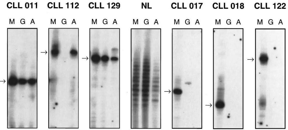
cDNA with identical CDR3 lengths suggests clonal relatedness. Total mRNA from the PBMC of the 19 patients were ana-lyzed with this assay using a VH leader primer corresponding to that of the CLL B cell and CH primers specific for m, g, and a. In z 50% of the patients studied, dominant cDNA bands of
the g and/or a isotypes were found that corresponded in size to those of the m CLL cDNA. In three cases, these bands were solely of the IgA isotype (Nos. 008, 079, and 112), and in one patient, they were only of the IgG isotype (No. 059). In six in-stances (Nos. 002, 011, 129, 130, 141, and 153), cDNA for both isotypes were found. These data suggest strongly that certain subclones of these CLL B cells have switched isotype produc-tion in vivo. Fig. 1 illustrates results for six representative CLL samples, three in which switching has been detected (left side) and three in which it has not (right side), and a tonsil sample as a polyclonal B cell control.
DNA sequence analyses of the g and a H chain cDNA.
To assure that the products observed in these fingerprinting assays were clonally related to the IgM1 CLL B cells, the
ini-tial PCR products (VH leader → CH) of four samples (Nos. 002, 011, 112, and 129) were reamplified using a seminested strategy that yielded products spanning the rearranged V gene from FR1 → CH1. These products then were cloned and se-quenced. For brevity, these sequences are not presented;
how-ever, they are available from EMBL/GenBank/DDBJ under accession numbers U71103–U711106.
The data can be summarized by the following points. First, the sequences of the g and a cDNA were virtually identical to those of the B-CLL m cDNA, with only rare differences that were consistent with Taq error. In only three instances two nu-cleotide discrepancies from the m CLL sequence were found, and these were distributed randomly throughout the rear-ranged VH genes. These data confirm the clonal relatedness of the various cDNA. In addition, the high level of sequence sim-ilarity between the IgM1 CLL clone and the clonally related
g- and a-expressing progeny indicates that significant
intra-clonal diversification had not occurred. Next, since the cDNA that were sequenced spanned the junction between the VHDJH and the CH, these data confirm that the g and a H chain gene segments were physically linked to the VHDJH in the initial mRNA transcripts and, therefore, were unlikely the result of PCR priming artefacts. Sequences illustrating this union for the IgG and IgA clones from patient No. 129 are shown in Fig. 2. Finally, these data demonstrate semiquantitative differ-ences in the numbers and types of H chain switch variants de-tected in individual CLL patients. Every IgG1 cDNA clone
[image:4.612.55.560.74.370.2]was found to be related to the corresponding CLL B cell (Ta-ble III). Among the IgA clones, however, heterogeneity was observed. In only one patient (No. 112), all the a cDNA were
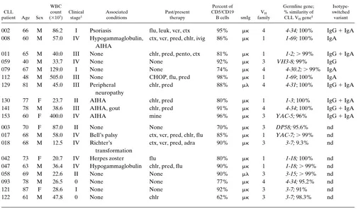
Table II. Clinical and Laboratory Characteristics of the CLL Patients Studied
CLL
patient Age Sex WBC count (3103)
Clinical stage‡
Associated conditions
Past/present therapy
Percent of CD5/CD19
B cells smIg VH
family
Germline gene; % similarity of CLL VH gene§
Isotype-switched variant
002 66 M 86.2 I Psoriasis flu, leuk, vcr, ctx 95% mk 4 4-34; 100% IgG 1 IgA
008 60 M 57.0 IV Hypogammaglobulin, AIHA
ctx, vcr, pred, chlr, ivig 86% mk 1 1-69; 100% IgA
011 65 M 40.0 III None chlr, pred, pento, ctx 81% mk 1 1-2; . 99% IgG 1 IgA
059 40 M 33.7 IV None None 92% mk 3 VH3-8; 99% IgG
079 67 M 129.0 I None None 74% mk 4 4-30.2; . 99% IgA
112 48 M 505.0 III None CHOP, flu, pred 98% mk 1 1-69; 100% IgA
129 81 M 45.0 III Peripheral neuropathy
chlr, pred 88% ml 4 4-31; 100% IgG 1 IgA
130 77 F 23.7 II AIHA chlr, pred 80% mk 1 1-3; 100% IgG 1 IgA
141 78 M 38.6 III AIHA, gout chlr, pred 91% mk 4 4-34; 100% IgG 1 IgA
153 60 F 400.0 IV AIHA mine 96% mk 3 YAC-5; 96% IgG 1 IgA
003 70 F 87.0 II None None 70% mk 3 DP58; 95.6% nd
017 68 M 58.0 IV Bell’s palsy ctx, vcr, pred, chlr, flu 85% mk 1 YAC-7; . 99% nd 018 68 M 12.5 IV Richter’s
transformation
ctx, vcr, pred, adra 90% mk 3 3-7; 9.3% nd
042 73 F 20.7 IV Herpes zoster flu 80% mk 1 1-18; 100% nd
047 63 M 36.4 IV Hypogammaglobulin chlr, pred, flu 90% mk 1 1-18; . 99% nd
058 69 M 22.6 II None None 90% ml 3 3-15; .99% nd
093 78 M 26.5 0 None None 77% mk 4 4-34; 95.2% nd
121 87 F 28.6 I None None 92% mk 3 3-7; 91% nd
122 61 M 47.8 0 None chlr 62% mk 3 3-7; 98.3% nd
PBMC from 19 patients with B-CLL were studied. Institutional Review Board approval was obtained for these studies. ‡Based on the classification of
Rai et al. (25). §When possible, genes are identified as suggested by Shin et al. (26), initial descriptions are used otherwise. adria, adriamycin; chlr,
chlorambucil; ctx, cyclophosphamide; flu, fludarabine; ivig, intravenous gammaglobulin; mine, mesna, ifosfamide, mitoxantrone, etoposide; pred, prednisone; vcr, vincristine; wbc, white blood cell.
of CLL origin; in patients 002 and 011, 40% of the IgA clones were not related to the CLL. In addition, the data indicate dif-ferences in the relative frequencies of the individual IgG sub-classes detected within and among patients. Thus, for the two IgG-producing patients studied (Nos. 011 and 129), the major-ity of the clones expressed IgG1 and/or IgG3 (No. 011, 90% IgG1; No. 129, 63% IgG1 and 30% IgG3); a minority ex-pressed IgG2 and none exex-pressed IgG4. (Table III).
Surface membrane phenotypic analyses. The preceding data indicate that a subpopulation of IgM-expressing B-CLL cells undergoes an isotype class switching event in vivo. Since this phe-nomenon was observed in only z 50% of the patients studied,
however, surface phenotyping analyses were performed in an attempt to distinguish those B-CLL clones capable of this process. Although a total of 16 different markers were analyzed, sig-nificant differences among the two groups were found only for
the intensity of smIgM and smIgD expression (Table IV). Thus, among the CLL patients in whom switching was not de-tected, mean channel fluorescence intensity for IgM was 63.50 (SD 5 38.007) and for IgD it was 40.75 (SD 5 26.926) with an IgM/IgD ratio of 2.00. In contrast, among the CLL patients that did switch, mean channel fluorescence intensity for IgM was 52.714 (SD 5 15.524) and for IgD was 22.45 (SD 5 19.715); IgM/IgD 5 4.60. Statistical comparisons (nested ANOVA) of each of these values between the two groups are significantly different, although the most significant differences are in the levels of smIgD expression (P , 0.0001) and the IgM/IgD ratio (P , 0.0001).
Detection of circulating isotype class–switched CLL B cells.
To confirm that the CLL-related, non-IgM mRNAs were trans-lated into Ig molecules, triple-color immunofluorescence was used to detect smIgG1 and smIgA1 B cells expressing the
ap-Figure 1. Ig VH gene fingerprinting analyses. 7.5% acrylamide gel showing the relative lengths of the VH CDR3 cDNA generated from PBMC RNA of six patients with CLL. The CLL m bands are indicated by arrows. Note that RNA from patients 011, 112, and 129 yield bands in the g
[image:5.612.60.553.58.281.2]and/or a lanes with lengths identical to those of the leukemic cells, suggesting clonal relatedness, whereas RNA from patients 017, 018, and 122 yield only the CLL m band. The center panel represents PCR products generated from tonsillar B cell RNA using a VH4 leader primer. These il-lustrate the typical ladder-like appearance of normal polyclonal B cells.
Figure 2. Comparisons of the cDNA sequences derived from the PBMC RNA of the IgM1 CLL patient No. 129 with representative IgG
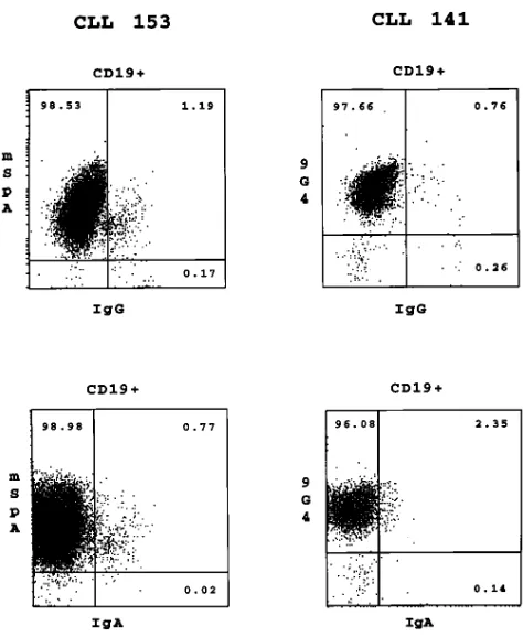
[image:5.612.54.546.562.705.2]1 propriate VH gene or gene family of the CLL B cell. To
ad-dress this, we used the 9G4 mAb that reacts with the VH4-34 (VH4.21) gene product (24) and mSpA that reacts with VH3 gene products (23). Preliminary immunofluorescence data us-ing B-CLL cells expressus-ing these two VH gene families con-firmed these selected reactivities (data not shown). Table V and Fig. 3 provide the results obtained when three patients whose CLL B cells use the VH3 gene YAC-5 (No. 153) and the VH4-34 gene (Nos. 002 and 141) were studied in this manner.
As would be expected in IgM1 CLL patients, in each case
the majority of the circulating CD191 cells do not express IgG
or IgA (Table V). In all instances, however, there exist a small number of CD191 B cells that express either smIgG1 (CLL
No. 141, 0.37%; No. 153, 1.15%; No. 002, 0.60%) or smIgA1
(No. 141, 1.89%; No. 153, 0.80%; No. 002, 0.32%). Triple-color immunofluoresecence studies indicate that the vast majority of these CD191 IgG1 and CD191 IgA1 B cells also express the
VH gene products of the corresponding CLL B cell (Fig. 3). When sorted for IgG/IgA expression by FACS®, the V
H gene of these cells had a CDR3 length identical to that of the IgM1
CLL clone, confirming their leukemic origin (data not shown). Note, however, that in patient No. 141 there is a relatively
large number of IgG-bearing cells (6.52%; Table III) that ex-press the VH4-34–specific marker (6.39%) but do not express CD19. These IgG1, 9G41 (non-B) cells coexpress CD16 and
[image:6.612.315.555.81.253.2]CD56, but lack CD3/CD8 coexpression, indicating that they are most likely natural killer cells. Thus, these data suggest that these IgG1 CLL B cells have secreted sufficient amounts
Table III. Summary of the cDNA Sequences of the Isotype-switched CLL Variants
CLL patient
Total number of cDNA clones
sequenced
Percentage of
g or a cDNA clones related to the leukemic clone
IgG subclasses of the g cDNA clones
IgG IgA IgG IgA g1 g2 g3 g4
002 — 17 0% 58% — — — —
011 10 12 100% 58% 90% 10% — —
112 — 10 0% 100% — — — —
129 10 10 100% 70% 63% 7% 30% —
[image:6.612.57.297.83.199.2]The nucleotide sequences of these CLL cells are available form EMBL/ GenBank/DDBJ under accession numbers U71103–U71106.
Table IV. Comparison of smIgM and smIgD
Immunofluorescence Intensities of CLL Patients in Whom Switching Was Detected vs Those in Whom it Was Not Detected
Group
smIgM smIgD IgM/IgD
MCFI SD MCFI SD MCFI SD
Switching
detected‡ 52.71 15.524 22.45 19.715 4.6 4.0 Switching not
detected§ 63.50 38.007 40.75 26.926 2.0 1.1 Statistical
significance P5 0.017 P50.0001 P5 0.0001
*Values represent mean channel fluorescence intensities determined
using direct immunofluoresence and a FACScan® flow cytometer, and
were calculated using a nested ANOVA. ‡Mean of 14 determinations of
eight samples. §Mean of 16 determinations of seven samples. iStatistical
[image:6.612.316.553.395.684.2]significance as calculated by the Student’s t test. Significant differences in expression were not seen for the following markers: CD5, CD19, CD23, CD38, CD39, CD10, CD54, CD58, CD11a, CD18, CD3, CD8, CD16, and CD56.
Table V. Immunofluorescent Analyses of Circulating IgG1 and
IgA1 Cells from Three Patients with IgM1-CLL CLL patient number
141 153 002
IgG1/CD191 0.37% 1.15% 0.60%
IgG1/CD192 6.52% 0.95% 0.02%
IgG1/V
H1 6.39% 1.52% 0.60%
IgG1/V
H2 0.45% 0.91% 0.02%
IgA1/CD191 1.89% 0.80% 0.32%
IgA1/CD192 1.02% 0.08% 0.00%
IgA1/V
H1 1.10% 0.81% 0.34%
IgA1/V
H2 0.06% 0.02% 0.01%
IgG1/CD161 9.25% 0.64% 0.11%
IgG1/CD561 5.62% 0.43% 0.11%
PBMC from three patients were stained with the marker combinations listed and then analyzed by flow cytometry. Results of sm expression of
CD19, IgG or IgA, and VH for patients Nos. 153 and 141, as determined
by triple immunofluorescence, are shown in Fig. 3. For patient 141, VH
was determined by the VH4-34–specific mAb 9G4; for patients 153 and
002, VH was determined by the VH3 family–specific reagent mSpA.
Figure 3. Three-color immunofluorescent analyses of smIgG1 and
smIgA1 PBMC from two patients with IgM1 B-CLL. PBMC were
[image:6.612.56.299.538.654.2]of Ig to permit FcgR-mediated loading of the large granular lymphocytes (LGL) with the VH4-34 IgG. A similar, albeit less prominent example of this phenomenon is seen in patient No. 153 (Table V).
Discussion
The preceding data indicate that isotype class switching can be detected in z 50% of IgM1 CLL patients (Table I; Fig. 1). This
switching appears to occur more frequently to IgA than to IgG, inasmuch as 9 of the 10 patients in whom switching was detected involved this isotype. Although switching to IgG was found in five patients, in four of these cases switching to IgA occurred also. In one patient, switching was restricted to IgG (Table I). In three instances, serial samples spanning several years were analyzed. In two cases (Nos. 002 and 003), the iden-tical isotype expression pattern was found, whereas in the third patient (No. 112), a new IgG band appeared. Thus, these find-ings suggest that this switching is an ongoing, nonrandom event that may be influenced by B cell, accessory cell, and/or antigenic signals.
The notion that switching is not random is supported by the differences in the expression of CLL-related transcripts that are associated with the various IgG subclasses. Consistent with our previous studies of 10 IgG1 B-CLL cases (28), the
CLL-related clones were enriched in IgG1 and IgG3 expression (Nos. 011 and 129; Table III). It is possible that our inability to detect transcripts for every isotype in each patient may be a function of the sensitivity of the PCR assay and the primers used. Nevertheless, our data suggest a differential level of mRNA expression for the various isotypes within an individ-ual patient. Supportive of this contention is our recent finding that the PBMC of only three (Nos. 11, 79, and 141) of six pa-tients who were found to switch to IgA and/or IgG (Nos. 11, 79, 141, 129, 130, and 153) were found to also express CLL-spe-cific transcripts associated with the e H chain (data not shown).
Our Ig VH fingerprinting and sequencing data indicate two points about the numbers of the clonally related CLL vs. nor-mal B cells in the blood of these patients. First, in many in-stances, the CLL-related a and g clones were expressed in sig-nificant excess over the normal B cell clones. Indeed, in most cases, radiolabeled bands representative of normal B cells were barely visible (Fig. 1) unless the radiographs were ex-posed for longer periods of time (data not shown). These data suggest an in vivo amplification of the isotype-switched CLL clones. Indeed, this impression is supported by the immunofluo-rescent analyses that document that the majority of the IgG1
and IgA1 B cells in the blood of these patients express the
CLL VH gene (Fig. 3) and have a CDR3 length identical to that of the IgM1 CLL clone. The frequency of B cells
express-ing these switched Ig molecules in relation to those expressexpress-ing IgM, however, appears to be very small, approximating a 98:1:1 IgM/IgG/IgA ratio (Fig. 3).
Second, there appear to be more residual normal B cells producing IgA than IgG in the blood of these patients (Table III and data not shown). Similar phenomena were seen in our previous study of IgG1 B-CLL cells (22). This may be a
reflec-tion either of an increased number of IgA precursors in the blood of CLL patients and normal subjects or of specific types of antigenic stimuli in these patients. In this regard, previous studies have suggested that IgA is the dominant isotype pro-duced in vivo and in vitro by normal B cells (29, 30).
Decreased expression of smIgM and smIgD characterized those CLL B cells in which switching was detected (Table IV). Since the diminution in smIgD far exceeded that of smIgM, these cells had higher IgM/IgD ratios. Analyses of 14 surface markers, including adhesion molecules, did not reveal other significant differences between these two groups. Since stimu-lation and maturation of normal B cells results in decreased expression of smIgD (31), these B-CLL cells may have re-ceived or continue to receive antigenic and/or accessory cell signals driving them to mature and undergo isotype class switching. Although the antigens driving these cells are un-known, they may be autoantigens, based on the known pro-pensity for B-CLL cells to produce Ig molecules with autore-activity (32–34, reviewed in 35).
Not infrequently, antigenic stimulation of normal B cells results in the accumulation of mutations in their Ig VH and VL genes. Therefore, those B-CLL cells with diminished smIgD and evidence of isotype switching might be expected to have a greater likelihood of having accumulated somatic mutations in these genes. Such a correlation was not found in these cases, since the VH genes of these IgM1 B-CLL cells and their switched progeny were virtually identical to those of the corre-sponding germline gene (Table II). It is unlikely that these switched transcripts were generated from a preleukemic pro-genitor cell that gave rise to the IgM1 B-CLL cell and other
normal cells, since our previous data suggest that such progen-itors can and do develop somatic mutations that can distin-guish them from the B-CLL cell (22).
The finding that isotype class switching was not accompa-nied by the accumulation of new V gene somatic variants is consistent with a large body of data suggesting that the level of somatic mutation detected in IgM1 B-CLL cells is minimal
(36–38, reviewed in 39). Somatic mutations, however, are more common in B-CLL cells of switched isotype (40–42), even though in several instances similar VH genes were ex-pressed in these two sets of CLL B cells. We recently demon-strated IgM1 progenitor cells for IgG1 CLL B cells that
exhib-ited certain features of “premalignant” B-CLL cells (e.g., in vivo expansion) but retained the ability to undergo an isotype class switch and to accumulate somatic mutations (22). This suggested that these two processes (switching and mutation) were active in the CLL precursors and their clonal relatives, but were downregulated in the overt B-CLL cell. Our present findings further refine this notion by indicating that the B-CLL cell retains the ability to switch isotype classes while downreg-ulating the accumulation of significant numbers of new VH mu-tations.
1 to IgG and/or IgA production (Nos. 008, 130, 141, and 153).
Since studies of patients with AIHA have identified warm-reac-tive antierythrocyte antibodies of all three major isotypes (43), one must consider the possibility that in certain patients the CLL B cell may be the source of such antibodies. Several pre-vious studies support such a hypothesis. For example, antibod-ies eluted from erythrocytes in some AIHA patients may be of restricted heterogeneity, as defined by serologic markers (44), Ig L chain type (45, 46), and electrophoretic mobility (47). In addition, murine CD51 B cells frequently react with red blood
cell antigens (48) and can cause hemolysis in vivo (49). There-fore, it is possible that the causes of AIHA and other autoim-mune phenomena in CLL are heterogeneous, and the poten-tial role of CLL-derived autoantibodies in these conditions requires further study.
Acknowledgments
These studies were supported in part by U.S. Public Health Service grant AI 10811 from the National Institutes of Health National Insti-tute of Allergy and Infectious Diseases, by the Joseph Eletto Leuke-mia Research Fund, by the Richard and Nancy Leeds Fund of the Department of Medicine of North Shore University Hospital, by the Sass Foundation for Medical Research, and by Consiglio Nazionale delle Ricerche–Applicazioni Cliniche della Ricerca Oncologica.
References
1. Dighiero, G., P. Travade, S. Chevret, P. Fenaux, C. Chastang, and J.L. Bi-net. 1991. B-cell chronic lymphocytic leukemia: present status and future direc-tions. Blood. 78:1901–1914.
2. Fu, S.M., N. Chiorazzi, H.G. Kunkel, J.P. Halper, and S. Harris. 1978. In-duction of in vitro differentiation and immunoglobulin synthesis of human leu-kemic B lymphocytes. J. Exp. Med. 148:1570–1578.
3. Totterman, T.H., N. Nilsson, and C. Sundstrom. 1980. Phorbol ester-induced differentiation of chronic lymphocytic leukemia cells. Nature (Lond.).
288:176–178.
4. Julliusson, G., K.-H. Rober, L. Hammarstrom, C.I.E. Smith, G. Biber-feld, and G. Gahrton. 1983. Mitogen-induced switching of immunoglobulin heavy-chain class secretion in chronic B-lymphocytic leukemia and immunocy-toma cell populations. Scand. J. Immunol. 17:51–59.
5. Sarfati, M., H. Luo, and G. Delespesse. 1989. IgE synthesis by chronic lymphocytic leukemia cells. J. Exp. Med. 170:1755–1780.
6. Fu, S.M., R.J. Winchester, T. Feizi, P.D. Walzer, and H.G. Kunkel. 1974. Idiotypic specificity of surface immunoglobulin and the maturation of leukemic bone-marrow derived lymphocytes. Proc. Natl. Acad. Sci. USA. 71:4487–4490.
7. Deegan, M.J., J.P. Abraham, M. Sawdyk, and E.J. Van Slyck. 1984. High incidence of monoclonal proteins in the serum and urine of chronic lymphocytic leukemia patients. Blood. 64:1207–1211.
8. Miller, R.H., M.S. Linet, M.L. Van Natta, L.D. McCaffery, and R.L. Humphrey. 1987. Serum protein electrophoresis patterns in chronic lympho-cytic leukemia. Arch. Intern. Med. 147:1614–1617.
9. Pangalis, G.A., N.M. Papadopoulos, and H.A. Moutsopoulos. 1987. Para-proteins in the serum of B-chronic lymphocytic leukemia. In Chronic Lympho-cytic Leukemia: Recent Progress and Future Direction. R.P. Gale and K. Rai, editors. Alan R. Liss, Inc., New York. 245–252.
10. Kubagawa, H., L.B. Vogler, J.D. Capra, M.E. Conrad, A.R. Lawton, and M.D. Cooper. 1979. Studies on the clonal origin of multiple myeloma: Use of individually specific (idiotype) antibodies to trace the oncogenic event to its earliest point of expression in B cell differentiation. J. Exp. Med. 150:792–807.
11. Grogan, T.M., B.G. Durie, C. Lomen, C. Spier, D.P. Wirtz, R. Nagle, G.S. Wilson, L. Richter, L. Vela, V. Maxey, K. McDaniel, and C. Rangel. 1987. Delineation of a novel pre-B cell compartment in plasma cell myeloma: immu-nochemical, immunophenotypic, genotypic, cytologic, cell culture and kinetic features. Blood. 70:932–942.
12. Jensen, G.S., M.J. Mant, and L.M. Pilarski. 1992. Sequential maturation stages of monoclonal B lineage cells from blood, spleen, lymph node and bone marrow from a terminal myeloma patient. Am. J. Hematol. 41:199–208.
13. Billadeau, D., G. Ahmann, P. Greipp, and B. Van Ness. 1993. The bone marrow of multiple myeloma patients contains B cell populations at different stages of differentiation that are clonally related to the malignant plasma cell. J. Exp. Med. 178:1023–1031.
14. Corradini, P., M. Boccadoro, C. Voena, and A. Pileri. 1993. Evidence for a bone marrow B cell transcribing malignant plasma cell VDJ joined to Cm
sequence in immunoglobulin (Ig)G- and IgA-secreting multiple myelomas. J. Exp. Med. 178:1091–1096.
15. Bertoli, L.F., H. Kubagawa, G.V. Borzillo, P.D. Burrows, M.T. Schreeder, A.J. Carrol, and M.D. Cooper. 1988. Bone marrow origin of a B-cell lymphoma. Blood. 72:94–101.
16. Jain, R., S. Roncella, S. Hashimoto, A. Carbone, P. Francia di Celle, R. Foa, M. Ferrarini, and N. Chiorazzi. 1994. A potential role for antigen selection in the clonal evolution of Burkitt’s lymphoma. J. Immunol. 153:45–52.
17. Richter, M.N. 1982. Generalized reticular cell sarcoma of lymph nodes associated with lymphatic leukemia. Am. J. Pathol. 4:285–292.
18. Harousseau, J.L., G. Flandrin, G. Tricot, J.C. Brouet, M. Seligmann, and J. Bernard. 1981. Malignant lymphoma supervening in chronic lymphocytic leukemia and related disorders. Richter’s syndrome: a study of 21 cases. Can-cer. 48:1302–1308.
19. Sun, T., M. Susin, M. Desner, R. Pergolizzi, J. Cuomo, and P. Koduru. 1990. The clonal origin of two cell populations in Richter’s syndrome. Hum. Pathol. 21:722–728.
20. Mayumi, M., H. Kubagawa, G.A. Omura, W.E. Gathings, J.F. Kearney, and M.D. Cooper. 1982. Studies on the clonal origin of human B cell leukemia using monoclonal anti-idiotype antibodies. J. Immunol. 129:904–910.
21. Vogler, L.B., J.L. Preud’homme, M. Seligmann, W.E. Gathing, W.M. Crist, M.D. Cooper, and F.J. Bollum. 1981. Diversity of immunoglobulin ex-pression in leukemic cells resembling B lymphocyte precursors. Nature (Lond.).
290:339–341.
22. Dono, M., S. Hashimoto, F. Fais, V. Trejo, S.L. Allen, S.M. Lichtman, P. Schulman, V.P. Vinciguerra, B. Sellars, P.K. Gregersen, M. Ferrarini, and N. Chiorazzi. 1996. Evidence for progenitors of B-CLL cells that undergo intra-clonal differentiation and diversification. Blood. 87:1586–1594.
23. Silverman, G.J., M. Sasano, and S.B. Wormsley. 1993. Age-associated changes in binding of human B lymphocytes to a VH3 restricted unconven-tional bacterial antigen. J. Immunol. 151:5840–5855.
24. Stevenson, F.K., M. Wrightham, M.J. Glennie, D.B. Jones, A.R. Cattan, T. Feizi, T.J. Hamblin, and G.T. Stevenson. 1986. Antibodies to shared idio-types as agents for analysis and therapy for human B cell tumors. Blood. 68: 430–436.
25. Rai, K.R., A. Sawitsky, E.P. Cronkite, A.D. Chanana, R.N. Levy, and B.S. Pasternack. 1975. Clinical staging of chronic lymphocytic leukemia. Blood.
46:219–234.
26. Shin, E.K., F. Matsuda, H. Nagaoka, Y. Fukita, T. Imai, K. Yokoyama, E. Soeda, and T. Honjo. 1991. Physical map of the 39 region of the human im-munoglobulin heavy chain locus: clustering of autoantibody-related variable segments in one haplotype. EMBO (Eur. Mol. Biol. Organ.) J. 10:3641–3645.
27. Deane, M., and J.D. Norton. 1991. Immunoglobulin gene “fingerprint-ing”: an approach to analysis of B lymphocyte clonality in lymphoproliferative disorders. Br. J. Haematol. 77:274–281.
28. Wakai, M., S. Hashimoto, M. Omata, Z.M. Sthoeger, S.L. Allen, S.M. Lichtman, P. Schulman, V.P. Vinciguerra, B. Diamond, M. Dono, M. Ferrarini, and N. Chiorazzi. 1994. IgG1, CD51 human chronic lymphocytic leukemia B
cells. Production of IgG antibodies that exhibit diminished autoreactivity and IgG subclass skewing. Autoimmunity. 19:39–48.
29. Mestecky, J., R.J. Winchester, T. Hoffman, and H.G. Kunkel. 1977. Par-allel synthesis of immunoglobulins and J chain in pokeweed mitogen–stimu-lated normal cells and in lymphoblastoid cell lines. J. Exp. Med. 145:760–765.
30. Mestecky, J., and J.R. McGhee. 1987. Immunoglobulin A (IgA): molec-ular and cellmolec-ular interactions involved in IgA biosynthesis and immune re-sponse. Adv. Immunol. 40:153–245.
31. MacLennan, I.C., Y.J. Liu, S. Oldfield, J. Zhang, and P.J. Lane. 1990. The evolution of B-cell clones. Curr. Top. Microbiol. Immunol. 159:37–63.
32. Broker, B.M., A. Klajman, P. Youinou, J. Jouquan, C.P. Worman, J. Murphy, L. Mackenzie, R. Quartey-Papafio, M. Blaschek, P. Collins, S. Lai, and P.M. Lydyard. 1988. Chronic lymphocytic leukemia cells secrete multispe-cific antibodies. J. Autoimmunity. 1:469–481.
33. Sthoeger, Z.M., M. Wakai, D.B. Tse, V.P. Vinciguerra, S.L. Allen, D.R. Budman, S.M. Lichtman, P. Schulman, L.R. Weiselberg, and N. Chiorazzi. 1989. Production of autoantibodies by CD5-expressing B lymphocytes from pa-tients with chronic lymphocytic leukemia. J. Exp. Med. 169:255–268.
34. Borche, L., A. Lim, J.-L. Binet, and G. Dighiero. 1990. Evidence that chronic lymphocytic leukemia B lymphocytes are frequently committed to pro-duction of natural autoantibodies. Blood. 76:562–569.
35. Kipps, T.J., and D.A. Carson. 1993. Autoantibodies in chronic lympho-cytic leukemia and related systemic autoimmune diseases. Blood. 81:2475–2487. 36. Kipps, T.J., E. Tomhave, P.P. Chen, and D.A. Carson. 1988. Autoanti-body associated k light chain variable region gene expressed in chronic lympho-cytic leukemia with little or no somatic mutation. Implications for etiology and immunotherapy. J. Exp. Med. 167:840–852.
37. Meeker, T.C., J.C. Grimaldi, R. O’Rourke, J. Loeb, G. Juliusson, and S. Einhorn. 1988. Lack of detectable somatic hypermutation in the V region of the Ig H chain gene of a human chronic B lymphocytic leukemia. J. Immunol. 141: 3994–3998.
39. Kipps, T. 1993. Immunoglobulin genes in chronic lymphocytic leukemia.
Blood. Cells. 19:615–625.
40. Friedman, D.F., J.S. Moore, J. Erikson, J. Manz, J. Goldman, P.C. Now-ell, and L.E. Silberstein. 1992. Variable region gene analysis of an isotype-switched (IgA) variant of chronic lymphocytic leukemia. Blood. 80:2287–2297.
41. Ebeling, S.B., M.E.M. Schutte, and T. Logtenburg. 1993. Molecular analysis of VH and VL regions expressed in IgG-bearing chronic lymphocytic leukemia (CLL): further evidence that CLL is a heterogenous group of tumors.
Blood. 82:1623–1626.
42. Hashimoto, S., M. Dono, M. Wakai, S.L. Allen, S.M. Lichtman, P. Schulman, V.P. Vinciguerra, M. Ferrarini, J. Silver, and N. Chiorazzi. 1995. So-matic diversification and selection of immunoglobulin heavy and light chain variable region genes in IgG1 CD51 chronic lymphocytic leukemia B cells. J. Exp. Med. 181:1507–1517.
43. Engelfreit, C.P., M.A.M. Overbeeke, and A.E.G.Kr. von dem Borne. 1992. Autoimmune hemolytic anemia. Semin. Hematol. 29:3–12.
44. Litwin, S.D., S. Balaban, and M.E. Eyster. 1973. Gm allotype preference in erythrocyte IgG antibodies of patients with autoimmune hemolytic anemia.
Blood. 42:241–246.
45. Leddy, J.P., and R.F. Bakemeier. 1965. Structural aspects of human erythrocyte autoantibodies. I. L chain types and electrophoretic dispersion. J. Exp. Med. 121:1–17.
46. Sthoeger, Z.M., D. Sthoeger, M. Shtairid, E. Sigler, D. Geltner, and A. Berrebi. 1993. Mechanism of autoimmune hemolytic anemia in chronic lym-phocytic leukemia. Am. J. Hematol. 43:259–264.
47. Andrzejewski, C., Jr., P.J. Young, D.B. Cines, and L.E. Silberstein. 1990. Heterogeneity of human red cell autoantibodies assessed by isoelectric focusing. Transfusion. 31:236–244.
48. Mercolino, T.J., L.W. Arnold, L.A. Hawkins, and G. Haughton. 1988. Normal mouse peritoneum contains a large population of Ly-11 (CD5) B cells
that recognize phosphatidyl choline: relationship to cells that secrete hemolytic antibody specific for autologous erythrocytes. J. Exp. Med. 168:687–698.