Thesis submitted to the University of London for the degree of Doctor of Medicine (MD)
by
Dr. Brian P. Saunders MB BS MRCP Endoscopy Department
All rights reserved
INFORMATION TO ALL USERS
The quality of this reproduction is dependent upon the quality of the copy submitted.
In the unlikely event that the author did not send a complete manuscript
and there are missing pages, th ese will be noted. Also, if material had to be removed, a note will indicate the deletion.
uest.
ProQuest 10106781
Published by ProQuest LLC(2016). Copyright of the Dissertation is held by the Author.
All rights reserved.
This work is protected against unauthorized copying under Title 17, United States Code. Microform Edition © ProQuest LLC.
ProQuest LLC
789 East Eisenhower Parkway P.O. Box 1346
Title page Abstract
Table of contents List of tables List of figures Statement Dedication 1 2 3 9 11 14 16
Chapter 1 : Introduction 1.1 : The challenge
1.2 : Historical perspective
1.3 : Colonoscopy vs barium enema
1.4 : Large bowel endoscopy today: indications/present facilities 1.5 : Colonic anatomy
1.5.1 : Embryology of the colon 1 .5.2 : Anatomical variations
1.5.3 : Relevance for colonoscopy
1 .5 .4 : Racial variations in colonic anatomy 1.6 : Defining the procedure of colonoscopy
1.6.1 : Patient selection
1 .6 .2 : Psychological preparation
1.6.3 : Bowel preparation
1 .6.3.1 : Purge + enema regimes
1 .6 .3 .2 : Mannitol
1 .6 .3 .3 : Normal Saline
1 .6 .3 .4 : Picolax
1 .6 .3 .5 : Polyethylene glycol/BES
1 .6 .3 .6 : Sodium phosphate
1 .6 .4 : Sedation/analgesia for colonoscopy 1.6 .4 .1 : Colonoscopy without sedation 1 .6 .4 .2 : "Conscious sedation"
1.6 .4 .3 : Benzodiazepines
1.6.4.4 : Diazepam vs midazolam 38
1.6.4.5 : Benzodiazepine antagonists 40
1.6.4.6 : Analgesia for colonoscopy 41
1.6.4.7 : Safety with benzodiazepine/opiate sedation 41
1.6.4.8 : Other drugs used for sedation 42
1.6.4.9 : Epidural analgesia 43
1.6.4.10: Patient-controlled sedation 44
1.6.4.11 : Nitrous oxide/oxygen inhalation 45
1.6.5 : Anti-spasmodic premedication 45
1.6.6 : Colonoscope insertion 46
1.6.6.1 : Recurrent colonoscope looping 47
1.6.6.2 : Bowel adhesions 47
1.6.7 : Insertion technique 48
1.6.7.1 : Hand pressure 48
1.6.7.2 : Changes in patient position 49
1.6.7.3 : Different types of endoscope 49
1.6.7.4 : Stiffening devices 50
1.6.8 : Imaging colonoscopy 50
1.6.8.1 : Accuracy of tip location 51
1.6.8.2 : Fluoroscopy 52
1.6.8.2.1 : Advantages 52
1.6.8.2.2 : Disadvantages 53
1.6.8.3 : Alternative methods of tip location 54
1.7 : Aims of the thesis 55
Chapter 2 : Peroperative comparison of Western and Oriental colonic anatomy and mesenteric attachments
2.1 : Introduction 57
2.2 : Methodology 57
2.3 : Statistical Analysis 63
2.4 : Results 63
2.5 : Discussion 67
3.1 : Introduction
3.2 ; Method 71
3.3 : Statistical analysis 71
3.4 : Results 71
3.5 : Discussion 74
3.6 : Conclusions 75
Chapter 4 : Why are women more difficult to colonoscope than men ?
4.1 : Introduction 77
4.2 : Method 77
4.3 : Statistical analysis 82
4.4 : Results 82
4.5 : Discussion 84
4.6 : Conclusions 85
Chapter 5 : Can difficulty at colonoscopy be predicted from a previous barium enema ?
5.1 : Introduction 86
5.2 : Method 86
5.3 : Statistical analysis 87
5.4 : Result 89
5.5 : Discussion 95
6.1 : Introduction 97
6.2 : Risk factors for poor preparation. 97
6.3 : Experience at St.Mark's 97
6.4 : Taste trial with mannitol/Picolax mixture 100
6.5 : Pilot study assessing mannitol/Picolax mixture
6.5.1 : Method 102
6.5.2 : Results 102
6.5.3 : Discussion 103
6.6 : Two dose PEG-BES vs a mannitol/Picolax mixture as bowel preparation for day-case colonoscopy
6.6.1 : Patients and Methods 103
6.6.2 : Statistical analysis 105
6.6.3 : Results 108
6.6.4 : Discussion 110
6.7 : Conclusions 111
Chapter 7 : Sedation/analgesia for colonoscopy
7.1 : Introduction 113
7.2: Method 114
7.3 : Statistical method and analysis 116
7.4 : Results 116
7.5 : Discussion 127
7.6 : Conclusions 130
Chapter 8 : Anti-spasmodic premedication for colonoscopy
8.1 : Introduction 131
8.2 : Method 131
8.3 : Statistical analysis 132
8.4 : Results 133
8.5 : Discussion 135
9.1 : Introduction 137 9.1.1 : Sedation/analgesia for flexible sigmoidoscopy 137 9.1.2 : Anti-spasmodic medication for flexible sigmoidoscopy 138
9.2 : Aim 138
9.3 : Method 139
9.4 ; Statistical analysis 141
9.5 : Results 141
9.6 : Discussion 145
9.7 ; Conclusions 147
Chapter 10 : Magnetic imaging of colonoscopy.
10.1 : Introduction 148
1 0 .2 : The principle of the method 148
10.3 : Accuracy of the system 150
1 0 .4 : Preliminary clinical evaluation
10.4.1 : Method 157
1 0 .4 .2 : Statistical analysis 162
10.4.3 : Results 162
10 .4 .3 .1 : Looping during intubation 163 1 0 .4 .3 .2 : Effect of hand pressure 163 1 0 .4 .3 .3 : Change of patient position 169 1 0 .4 .3 .4 : Accuracy without the imager view 169
1 0 .4 .4 : Discussion 171
1 0 .4 .4 .1 : The imaging system 171 1 0 .4 .4 .2 : Comparison with/without the imager view 172 1 0 .4 .4 .3 : Accuracy of tip location with the imager 174 1 0 .4 .4 .4 : Limitations of the imaging system 177
1 0 .4 .4 .5 : Safety 177
1 0 .4 .4 .6 : Electronic imaging and technique 178
Chapter 12 : The future of large bowel endoscopy.
12.1 ; Diagnostic and therapeutic endoscopy 187
12.2 : Screening colonoscopy/flexible sigmoidoscopy 188
12.3 : Tcaching colonoscopy 189
12.4 : Colonoscope design 191
Bibliography 193
Appendix 1 218
Magnetic imaging of colonoscopy: a selection of teaching material demonstrating the accurate straightening of loops encountered during colonoscope insertion.
Appendix 2 (see video) 228
Video footage demonstrating the magnetic imaging system.
Publications in support of the thesis 229
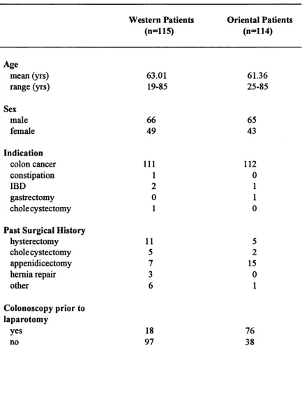
Table 1 : Details of Western and Oriental patients undergoing laparotomy.
Table 2 : Measurements of abdominal dimensions and sigmoid loop in Western and Oriental patients undergoing laparotomy.
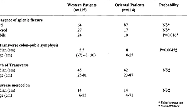
Table 3 : Measurements and appearance of the splenic flexure and transverse colon in Western and Oriental patients undergoing laparotomy.
Table 4 : Patient details in the difficult to colonoscope and not difficult groups.
Table 5 : A comparison of segmental colonic lengths and degree of mobility of the descending colon and transverse colon between the sexes.
Table 6 : Patient details of the difficult colonoscopy and control groups.
Table 7 : Measurement of colonic segmental length and mobility in difficult colonoscopy and control groups.
Table 8 : Association between indication for colonoscopy and poor preparation.
Table 9 : Association between age and poor preparation.
Table 10 : Volunteers' taste preferences for 4 different bowel preparations.
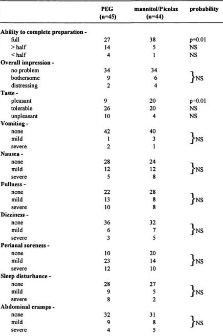
Table 11 : Patients' experience of bowel preparation.
Table 12 : Description of bowel preparation grading and comparison between PEG and mannitol/Picolax mixture.
Table 13 : Patient details in the PEG and mannitol/Picolax groups.
Table 15 : Side effects reported in the intravenous sedation, nitrous oxide and placebo groups.
Table 16 : Patient details and endoscopic findings in the hyoscine and placebo groups.
Table 17 : Subject details and endoscopic findings in hyoscine, nitrous oxide and placebo groups.
Table 18 : Details of patients examined with or without the imager view.
Table 19 : Incidence and type of loop that occurred in the two patient groups.
Table 20 ; Number of straightening attempts per procedure in the two study groups.
List of figures
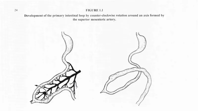
Figure 1.1: Development and rotation of the primary intestinal loop.
Figure 1.2 : Rotation of the primitive caecum and final fixations of the colon.
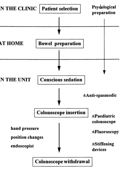
Figure 1.3 : Schematic representation of day-case colonoscopy.
Figure 2.1 : Sketches showing the method of measurement of the sigmoid loop and descending colon.
Figure 2.2 : Sketches of the different appearances of the splenic flexure.
Figure 2.3 : Sketches demonstrating the method of measurement of the transverse colon, ascending colon and caecum.
Figure 4.1 : Barium enema demonstrating the method of measurement of colonic segmental lengths.
Figure 4.2 : Prone enema film demonstrating the method for measuring transverse mobility.
Figure 4.3 : Barium enema film demonstrating "redundancy" of the transverse colon.
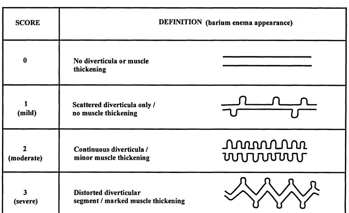
Figure 5.1 : Radiological classification of diverticular disease severity.
Figure 5.2 : Barium enema film of a patient who was technically difficult to intubate demonstrating severe sigmoid colon diverticular disease.
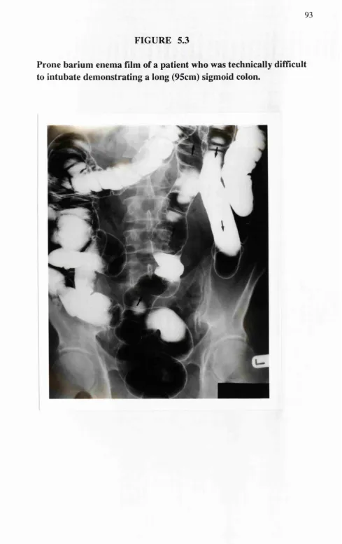
Figure 5.3 : Barium enema film of a patient who was technically difficult to intubate demonstrating a long sigmoid colon.
Figure 7.1 : Column charts demonstrating observed pain episodes and need for additional intravenous sedation in the three study groups.
Figure 7.2 : Box-whisker plot of patients perception of pain during colonoscopy.
Figure 7.3 : Scatter plot comparing the nurse observers estimation of patient pain to the patients' own perception of pain during colonoscopy.
Figure 7.4 : Histogram of difference between nurse and patient pain scores.
Figure 7.5 : Scatter plot of patient anxiety scores prior to colonoscopy compared to patients' perception of pain during the examination.
Figure 7.6 : Box-whisker plot showing patients perception of sedation during colonoscopy in the three patient groups.
Figure 7.7 : Box-whisker plot showing the duration of stay within the recovery area for patients in the three study groups.
Figure 9.1 : Data plot showing duration of procedure in the three study groups.
Figure 9.2 : Data plot of patients pain scores during flexible sigmoidoscopy.
Figure 10.1 : Schematic representation of the magnetic imaging system.
Figure 10.2 : Diagram demonstrating location of an individual point using triangulation.
Figure 10.3 : AP view of the colonoscope using magnetic imaging system.
Figure 10.4 : Magnetic imaging system view of catheter in a 2-D shape: actual vs imager measurements.
Figure 10.6 : Magnetic imaging system view of catheter in a 3-D shape: actual vs imager measurements.
Figure 10.7 : Magnetic imager view of the colonoscope during caecal intubation demonstrating the position of surface body markers.
Figure 10.8 : Lateral view of the colonoscope using magnetic imaging demonstrating accurate sigmoid abdominal pressure.
Figure 10.9 : AP imager views of the colonoscope demonstrating common loop configurations.
Figure 10.10 : AP imager views of the colonoscope demonstrating uncommon loop configurations.
Figure 10.11 : AP and lateral views of the colonoscope using magnetic imaging demonstrating caecal intubation with an alpha loop.
Figure 10.12 : Lateral view of the colonoscope within the sigmoid colon using magnetic imaging.
Figure 10.13 : AP view of the colonoscope using magnetic imaging demonstrating accurate placement of abdominal hand pressure to counter a deep transverse loop.
Figure 10.14 : Lateral views of the colonoscope using magnetic imaging demonstrating incorrect use of abdominal pressure for removal of an alpha loop.
Figure 10.15 : AP and lateral views of unsuccessful descending colon intubation.
Statement
While having prime responsibility for the implementation, co-ordianation and analysis of the studies comprising this thesis, I wish to acknowledge the immense support provided by my colleagues at St Mark's Hospital, particularly Dr. Christopher Williams my supervisor and mentor who was closely involved with the design of all the work described.
By the nature of clinical research much of the work presented has been a a team effort, involving collaboration with many clinicians and clinical staff. Advice was sought from my surgical colleagues Mr. Robin Phillips (StMark's Hospital) and Mr. Roger Leicester (St. George's Hospital) regarding the methodology of the anatomy studies described in Chapter 2 and I am indebted to the surgeons of the St.Mark's Association who carried out the colonic measurements, particularly Dr. Testsu Muto and Dr. Tadahiko Masaki (1st Department of Surgery, Tokyo University Hospital, Japan). Dr. Clive Bartram, Dr Steve Halligan and Dr. Craig Jobling of the Department of Radiology St. Mark's Hospital contributed to the design of the studies recorded in Chapters 4 and 5 and Drs Halligan and Jobling performed measurements of the barium enemas. Assistance in the running of the studies described in Chapter 4, 5, 6 and 7 was given by Dr. Manabu Fukumoto during a 1 year research attachment to St.Mark's Hospital from Toho University Hospital, Japan.
Most of the work described in Chapter 10 was carried out in the Ipswich General Hospital with the collaboration of Dr.Duncan Bell (Ipswich Hospital). Development of the imaging system would not have been possible without the expert technical assistance of Mr. John Bladen (Department of Engineering, Sheffield University) who also helped during the clinical assessment.
Dedication
Chapter 1 : Introduction
1.1 : The Challenge
1.2 : Historical perspective
limited tip angulation; guidance required fluoroscopy, as there was no means of viewing down the instrument. During the 1960's several attempts were described to develop the first forward viewing flexible sigmoidoscope, mostly with little or no success (Kanazawa & Tanaka, 1965; Niwa et al, 1969; Oshiba & Watanabe, 1965; Turell, 1963; Fox, 1969). Frustrated by the lack of progress in intubating from below, an Italian group attempted retrograde intubation of the colon (Provenzale & Revignas, 1969). Their patients were required to swallow a long, soft, polyvinyl tube with a mercury bag at its tip which was allowed to traverse the entire intestinal tract and emerge from the anus. This then served as a pulley to draw back a flexible sigmoidoscope tied to its tip. Though success in intubating the sigmoid colon was reported, the procedure was clearly impractical, taking several days to complete and involving large doses of screening radiation for the patient. It was not until the late 1960's that true flbreoptic sigmoidoscopy came into its own with several reports of success in intubating the sigmoid colon (Overholt & Pollard, 1967; Overholt, 1969). Fibreoptic technology was first invented by Hopkins (Hopkins & Kapanay, 1954) in 1952 and was further developed by Hirschowitz, whose team were the first to construct a flexible optical bundle for image transmission (Hirschowitz et al, 1957). Their work culminated in the development and clinical evaluation of the first fibreoptic gastroscope (Hirschowitz, 1961).
Following the introduction of the 108cm flexible colonoscope, Shinya and Wolf were the first to report routine intubation of the entire colon (Wolff & Shinya, 1971). Soon afterwards they showed that colonoscopic polypectomy could also be achieved, and so began the modem age of colonoscopy (Wolff & Shinya, 1973). During the 1970's fibreoptic colonoscopy became an established procedure at many centres throughout the world and began to rival barium enema as the investigation of choice for the diseased colon (Wolff et al, 1975; Loose & Williams, 1974). The next major advance came in the mid 1980's with the introduction of video technology. The charge coupled device or CCD chip, positioned at the tip of the colonoscope and containing between 30-100,000 individual photo cells or pixels (picture elements), relays images direct to a TV monitor, so removing the need for the endoscopist to hold the colonoscope close to the face (Sivak & Fleischer, 1984). This made colonoscopy not only more hygienic, avoiding splash contamination, but also more comfortable to perform.
1.3 : Colonoscopy vs barium enema
and then, if examination of the entire colon is mandatory to detect synchronous cancers or other coexistent lesions (Ekelund & Pihl, 1974; Peabody & Smithwick, 1961; Gelfand et al, 1979), supplementary barium enema is indicated. When colonoscopy is technically difficult for anatomical reasons, barium enema in combination with flexible sigmoidoscopy may be a more acceptable method of cancer surveillance (Williams et al, 1982). Barium enema is superior to colonoscopy in demonstrating gross colonic anatomy, diverticular disease, colonic fistulae and in localising a lesion prior to surgery (Frager et al, 1987). It also has the advantage that it provides a permanent record of the colon, which can be subsequently reviewed. Patient preference for colonoscopy or barium enema is unclear. Van Ness et al. questioned 107 patients who had recently undergone both colonoscopy (with light sedation) and barium enema (Van Ness et al, 1987). They found that patients reported less procedure discomfort during colonoscopy and required less time off work afterwards. However a recent report in the radiological literature suggests that despite sedation, patients find colonoscopy more painful than double contrast barium enema (Steine, 1994).
1.4 : Large bowel endoscopy today: indications and present facilities
endoscopists or hospital gastroenterologists (Johnson, 1982; DiSaro & Sanowski, 1993; Traid et al, 1983). Current opinion also suggests that it could be a cost- effective method of screening for colorectal cancer (Atkin et al, 1993).
Colonoscopy in the UK is performed at teaching centres and most district general hospitals and is the investigation of choice for rectal bleeding (Irvine et al, 1988), diarrhoea (Durdey et al, 1987), detection of colorectal neoplasia (Aldridge & Sim, 1986) and assessment of inflammatory bowel disease (Lindsay et al, 1988). It is also considered to be the procedure of choice in the surveillance of patients at increased risk of colorectal cancer i.e. those with previous colonic neoplasia (Waye & Braunfeld, 1982), a strong family history of cancer (Hodgson et al, 1993) or long-standing ulcerative colitis (Yardley et al, 1979).
Demand for flexible sigmoidoscopy and colonoscopy is increasing and a recent report by the Endoscopy Section Committee of the British Society of Gastroenterology suggests that present availability in the UK both of equipment and skilled endoscopists falls well below the desired level (Report by the Endoscopy Section Committee British Society of Gastroenterology, 1987). There is therefore a perceived need to increase the numbers of patients who can be examined without compromising accuracy, patient safety or satisfaction and to improve, and if possible accelerate, the training of skilled endoscopists.
1.5. : Colonic anatomy
many of the causes of difficulty during colonoscopy and will be addressed in detail in this thesis.
1.5.1 : Embryology of the colon
remain suspended in the abdominal cavity on their respective mesocolons. The sigmoid mesocolon is V-shaped and attached to the pelvic wall between the point where the external iliac artery bifurcates and the third sacral vertebrae in the mid line (Williams & Warwick, 1980). The transverse mesocolon is a broad V shape and is attached across the posterior wall of the upper abdomen crossing the duodenum and the head of the pancreas.
1.5.2 : Anatomical variations
inches (2.5-5cm) in length, and the descending mesocolon 1-3 inches (2.5- 7.5cm). The presence of a large persisting descending mesocolon has also been described by Morgenstem who comments that this variation may be more common than was previously thought (Morgenstem, 1960). Mobility of the right colon due to a persisting ascending mesocolon, associated with caecal volvulus, has been described by Wolfer et al. (Wolfer et al, 1942). Variations in the degree of mobility of the transverse colon have also been noted: in a barium enema study. Moody assessed the variation in the position of the transverse colon in the supine and erect positions, finding that the transverse colon dipped as low as the tme pelvis in 17 % of individuals (Moody, 1927).
1.5.3 : Relevance for colonoscopy
increased frictional resistance to insertion of the instrument tip, and so result in secondary looping in the sigmoid colon as well, frustrating attempts at inward passage of the colonoscope.
1.5.4 : Racial variations in colonic anatomy
Racial variation in the size of the sigmoid colon has been noted by Lisowski who found a high incidence of long suprapelvic sigmoid loops (in association with a high incidence of sigmoid volvulus) in Ethiopians (Lisowski, 1955). No study has compared colonic anatomy between Caucasian and Oriental patients but anecdotal reports from visiting endoscopists (CB Williams, 1994 personal communication) have suggested that colonoscopy is easier in Orientals (see Chapter 2), possibly due to differences in colonic anatomy.
1.6 : Defining the procedure of colonoscopy
Colonic anatomy defines the anatomical challenge faced by the colonoscopist but many other factors both before and during the procedure contribute towards the relative success or failure of the procedure. In attempting to improve colonoscopy all aspects of the procedure need to be examined (figure 1.3).
1.6.1 : Patient selection
Schematic representation of day-case colonoscopy
IN THE CLINIC
Patient selection
Psydological
preparation
AT HOME
Bowel preparation
IN THE UNIT
Conscious sedation
±Anti-spasm odic
Colonoscope insertion
hand pressure
position changes
endoscopist
iP a ed ia tric
colonoscope
±Fluoroscopy
^Stiffening
devices
be unstable, or when severe inflanmiatory disease weakens the bowel wall (Williams, 1990).
1.6.2 : Psychological preparation
1.6.3 : Bowel preparation
Bowel preparation is a fundamental preliminary for safe and accurate colonoscopy. An "ideal" preparation would be easy to administer, have a rapid onset of action, produce satisfactory cleansing in approaching 100% of all patients, be cheap, easily packaged for postal delivery and above all be safe and acceptable to the patient. At present no such ideal exists. Available preparations may be effective in cleansing the bowel but cause distress to some patients, or can be well tolerated but ineffective in cleansing. Recently the importance of good bowel preparation has been further stressed by a report suggesting that patients' tolerance of colonoscopy may be directly influenced by the success or failure of the preparation (Probert et al, 1993).
1.6.3.1 : Purge + enema regimes
Historically the earliest preparations for colonoscopy involved purge (castor oil, senna or bisacodyl) and enema regimes accompanied by dietary restrictions for several days (Teague & Manning, 1977). Though adequate preparation was usually achieved, such involved and time-consuming regimens were often unacceptable to patient and endoscopy staff alike. This has led to an on-going search for a rapidly cleansing, oral means of bowel preparation for colonoscopy.
1.6.3.2 : Mannitol
tolerate (Gilmore et al, 1981). The use of mannitol declined after a case report of an explosion during electrosurgery (Bigard et al, 1979) related to an increase in explosive gas formation with mannitol preparation; mannitol can be fermented by colonic bacteria under anaerobic conditions into hydrogen. However ignition of colonic gas is only possible in the presence of oxygen-rich air. Use of carbon dioxide as the insufflating agent during colonoscopy effectively eliminates the risk of combustion (Taylor et al, 1981).
1.6.3.3 : Normal Saline
Oral lavage with 3-4 litres of physiological saline produces rapid and effective cleansing of the bowel (Levy et al, 1976). However the salty taste of the solution makes it difficult for patients to tolerate. Increased absorbtion of salt and water during preparation with normal saline may occur and be dangerous for elderly patients or those with cardiac or renal problems.
1.6.3.4 : Picolaxi
Picolax is a combination of the osmotic laxative magnesium citrate and the stimulant laxative sodium picosulphate. It is cheap, easily packaged and has a lemon taste that is generally well tolerated by patients. It has been shown to be effective preparation for barium enema (Bartram, 1984) but few studies have assessed its use for bowel cleansing at colonoscopy. When compared to a standard 4 litre preparation of polyethylene glycol-balanced electrolyte solution (PEG-BES) as preparation for colonoscopy, 2 sachets of Picolax was shown to
be better tolerated but a worse cleansing agent than PEG-BES (Dakkak et al, 1992). In this study an unacceptable 41% of patients receiving Picolax were found to have a poor preparation.
1.6.3.5 : Polyethylene glycol-balanced electrolyte solution (PEG-BES)
consuming and involved for out-patients. In order to reduce fullness and vomiting, addition of prokinetic agents such as metoclopramide or cisapride to the 4 litre PEG-BES preparation has been suggested, but no convincing evidence of an advantage over a standard administration technique has been demonstrated (Brady et al, 1985; Haringsma et al, 1986). One of the more successful approaches is to reduce the volume of PEG-BES drunk by the patient at any one time. It has been shown that fractionating the administration of PEG-BES improves patient acceptability without compromising bowel preparation (Rosch
& Classen 1987).
In an attempt to improve the taste of PEG-BES, a new low sodium solution "Nulytely" was introduced in 1986. A comparative trial with "Golytely" appeared to show an improvement in palatability with the new solution (DiPalma & Marshall, 1990). However a subsequent trial assessing the taste of both solutions in healthy volunteers has not confirmed any advantage in palatability with "Nulytely" (Froelich et al, 1991). A recent paper has assessed the palatability of flavouring PEG-BES and concludes that the addition of lemon flavouring improves the taste and acceptability compared to other flavours or no flavouring (Matter et al. 1993). Despite problems with the taste and volume of PEG-BES preparation, and its high cost^, it has become the most widely used bowel cleansing agent and the gold standard with which to compare new products.
1.6.3.6 : Sodium phosphate
Recently much interest has been generated by the use of the highly osmotic cathartic, sodium phosphate as bowel preparation for colonoscopy. Sodium phosphate preparation has been compared to a standard 4 litre PEG-BES regime and was found to have similar cleansing ability but better patient acceptability (Vanner et al, 1990). It has the advantage that it is a small volume preparation; two 45ml quantities being drunk at 6-OOpm the evening before and 6-00 am the morning of colonoscopy. However a question mark remains regarding safety, due to the transient hyperphosphataemia that may occur following administration (Physicians Desk Reference, 1987). This can potentially lead to concomitant hypocalcaemia and cardiac arrhythmias (McConnell, 1971). Sodium phosphate is not currently licensed for use as bowel preparation in the UK.
1.6.4 : Sedation/analgesia for colonoscopy
Ideally during colonoscopy all patients would be relaxed and comfortable yet still able to respond appropriately to commands. In an attempt to achieve this aim, sedative and analgesic drugs are often administered to the patient before or during the examination.
UK and America routinely use conscious sedation with benzodiazepine and opiate drugs (Keeffe & O'Connor, 1990) but in Japan many endoscopists use no sedation at all (Ueno et al, 1991), whilst in France examinations are frequently performed with the patient under a general anaesthetic (Guem et al, 1993). Present methods tend to be endoscopist-controlled, relying on the endoscopist to assess the sedation needs of the patients. Endoscopy staff however may be poor judges of patient discomfort during endoscopy (Schultz et al, 1994).
1.6.4.1 : Colonoscopy without sedation: selective sedation
reported severe pain. No attempt was made in the two larger studies described earlier to ascertain patients' preferences with regard to sedation or to assess pain levels during the examinations.
1.6.4.2 : "Conscious sedation"
The term "conscious sedation" has been defined as "the administration of medications that allow the introduction and manipulation of endoscopes, yet provide a relaxed patient able to respond and maintain vital fimctions" (Benjamin & Lightdale, 1990). Conscious sedation for colonoscopy is usually achieved by the intravenous administration of a benzodiazepine drug such as diazepam or midazolam, in combination with an opiate drug such as pethidine (Keeffe & O'Connor, 1990). Many questions regarding the type of drugs used, the method of administration and optimal methods of maintaining patient safety have been posed over the last 15 years.
1.6.4.3 : Benzodiazepines
"Valium"^, (diazepam 5mg/ml in an aqueous vehicle) is a relatively viscous solution and causes pain at the injection site and a high incidence of thrombophlebitis (Kawar & Dundee, 1982). This led to the development of an emulsion preparation ("Diazemuls''^) which has been shown to be associated with fewer local complications (Jensen et al, 1981). When given intravenously diazepam has a rapid onset of action (1-2 minutes) but a relatively long elimination half life that varies from 24-52 hours (McCloy & Pearson, 1990). Diazepam is eliminated in the liver but produces active metabolites with some pharmacological activity. In addition it is excreted in the bile and undergoes enterohepatic circulation which may result in a second peak effect 4-6 hrs after the initial dosing (Bell, 1990).
1.6.4.4 : Diazepam vs midazolam
During the 1970's and early 1980's, intravenous diazepam became established as the standard method of inducing anxiolysis, sedation and amnesia during colonoscopy. In 1986 the synthetic benzodiazepine midazolam was introduced which offers potential advantages over diazepam. Compared to diazepam, midazolam is water soluble, more potent, has a quicker onset of action, shorter elimination half-life (1.5-5hrs) and causes greater amnesia (Carrougher et al, 1993; Al-Khudhari et al, 1982; Reves et al, 1985; Gilvarry et al, 1990). Because of these factors midazolam was adopted enthusiastically and early clinical experience showed it to be superior to diazepam for upper gastrointestinal endoscopy (Cole et al, 1983). However soon after the introduction of midazolam a series of major cardiorespiratory events, including some deaths, were reported
to the US Food and Drug Administration. These reports raised concerns that the use of midazolam for endoscopic conscious sedation might constitute a serious risk (FDA's regulation on the new drug Versed^, 1988). In 1989, in a double blind study, bolus administration of midazolam (0.07mg/kg) was compared to diazepam (0.15mg / kg) (Lewis et al, 1989). This study showed a significant increase in over-sedation with midazolam and raised further concerns regarding its use for colonoscopy. However midazolam is 2-5 times as potent as diazepam and in many cases was being administered in too large a dose, particularly in elderly patients (Whitwam et al, 1983). In addition although midazolam has a more rapid onset of action, peak drug effect occurs later for midazolam than diazepam (Buhrer et al, 1990). This may have resulted in inadequate intervals of observation between repeat dosing of midazolam, thereby contributing to over sedation. These factors led to a reduction in the recommended initial dose of midazolam to 0.03mg/kg and the recommendation that further doses be titrated with suitable periods (minimum of 2 minutes) of observation in between (Lewis & Benjamin, 1990). Following these changes there was a significant reduction in the frequency of reported adverse cardiopulmonary events (Arrowsmith et al, 1991). Ginsberg et al. in 1992 compared the new lower dose of midazolam to the current recommended dose of diazepam (O.lOmg/kg) as sedation for colonoscopy and found that midazolam was comparable to diazepam in efficacy but produced more amnesia (Ginsberg et al, 1992). Surprisingly 21% of their patients receiving diazepam were found to be over-sedated, which raises the possibility that diazepam doses may be unnecessarily high in a number of patients, particularly those over 65 years. Midazolam is now more commonly used as sedation for endoscopic procedures than diazepam; in the 1989 survey of sedation and
momtoring practices by the American Society of Gastrointestinal Endoscopy, midazolam rather than diazepam was used routinely by 73% of respondents. Despite early worries regarding the use of midazolam, the new lower doses recommended appear safe as sedation for colonoscopy, provided that individual factors which may reduce dosage requirements, such as age and medical history, are taken into consideration.
1.6.4.5 : Benzodiazepine antagonists
The effects of benzodiazepines can be rapidly reversed in 1-5 minutes by the competitive benzodiazepine antagonist, flumazenil (Haefely et al, 1988). Flumazenil has a shorter elimination half-life than midazolam (53mins vs 120mins) and therefore rebound sedation is a theoretical possibility (Lauven et al, 1985). However, when flumazenil has been used to reverse conscious sedation, rebound sedation has not been found (Bartelsman et al, 1990, Jensen et al, 1989, Zuurmond et al, 1989). In a recent study the effects of flumazenil reversal after midazolam premedication for colonoscopy has been evaluated (Saletin et al,
1991). Compared to placebo, patients receiving reversal with flumazenil recovered quicker and could be discharged earlier. This allowed information regarding the examination to be discussed soon after the procedure and, in many cases, effectively saved a further out-patient appointment. It remains to be seen however whether the savings in bed-space and nursing time afforded by the routine administration of flumazenil will out weigh its present high cost (£16.32, per 5ml, 500 microgram ampoule^)
1.6.4.6 : Analgesia for colonoscopy
Patients may experience pain during colonoscope insertion due to excessive gas insufflation or more commonly, stretching of the bowel mesentery (Williams, 1990). For this reason most colonoscopists choose to administer an analgesic as well as an anxiolytic for the examination. The opiate analgesic pethidine (meperidine) is the most commonly used drug for colonoscopy (Keeffe & O'Connor, 1990) but because of the risk of hypotension and respiratory depression associated with its use (Shafer & Varvel, 1991), other opiate analgesic drugs (sufentanil, alfentanyl and fentanyl), with shorter half lives than pethidine, have been introduced. Compared to pethidine however, these drugs are less effective when used for analgesia during colonoscopy (Chokhavatia et al, 1993). Ketorolac triomethamine, a new parenteral non-steroidal anti-inflammatory drug (NSAID) has been shown to be as effect as pethidine for the control of post operative (Yee et al, 1986) and dental pain (Fricke & Angelloci, 1987). However when used for colonoscopy discomfort it was found to be no more effective than placebo (Deal et al, 1993), probably because NSAIDs have little central analgesic activity and are less effective than opiate drugs against visceral pain, perceived via sympathetic neurones (Snyder et al, 1974).
1.6.4.7 : Safety with benzodiazepine/opiate sedation
(Branch et al, 1976; Vinik et al, 1982; Fee et al, 1984). Benzodiazepines and opiates given independently may cause cardiopulmonary depression (Reeves et al, 1985; Laurence & Bennett 1987); given together, clinically-significant synergism may occur (Loebel & Danzig 1970). Caution is therefore advised when both a benzodiazepine and an opiate are given in combination. It has been recommended that, if an opioid-benzodiazepine combination is used, the opioid should be given first, before titrating in the benzodiazepine, which should be administered in a reduced dosage (McCloy & Pearson, 1990). Electronic monitoring should be available, as well as access to opiate and benzodiazepine antagonists (Bell et al, 1991). In addition, at-risk patients such as the elderly and those with significant cardiovascular or pulmonary disease should receive supplemental oxygen (2-4 litre/min). This has been shown to reduce oxygen desaturation during colonoscopy (Gross & Long, 1990; Simon et al, 1991).
1.6.4.8 : Other drugs for sedation
Major tranquillisers such as the phenothiazines or butyrophenones, the hypnotic propofol and the hypnoanalgesic ketamine have all been suggested as alternative methods of producing sedation/analgesia for colonoscopy.
The short-acting hypnotic and mild analgesic drug propofol has also been used to induce sedation in patients during colonoscopy (Gepts et al, 1985). Because of the risk of complete loss of consciousness this approach, despite computerisation of the delivery system (Church et al, 1991), necessitates electronic monitoring, supplemental oxygen and the presence of a trained anaesthetist. The use of propofol for colonoscopy has been reported in a series of 500 French patients (Guem et al, 1993). Recently a single blinded study comparing propofol to midazolam as sedation for colonoscopy has suggested that recovery is faster but amnesia less with propofol (Munoz-Navas et al, 1994).
The hypno-analgesic agent ketamine produces minimal respiratory depression. Its use has been proposed as an adjunct to midazolam as sedation/analgesia for colonoscopy (Rosing et al, 1991). However the use of ketamine is limited because of its association with a marked dose-dependent increase in blood pressure so that it is contraindicated in patients with borderline hypertension, or ischaemic heart disease. It can also produce myoclonic twitching and nystagmus as well as unpleasant post-sedation emergence reactions, such as bad dreams (Lauven, 1990).
1.6.4.9 : Epidural analgesia
epidural analgesia to re-examine 29 patients who had previously proved difficult to intubate (Bleiberg et al, 1982). Although total colonoscopy was achieved in 27 of the 29 patients, 2 developed significant hypotension and one sustained a colonic perforation, an unacceptably high complication rate.
1.6.4.10 : Patient controlled analgesia/sedation : parenteral administration
In the last 4 years patient-controlled analgesia (PCA) has become an acceptable and desirable method of pain relief in a number of clinical settings (Rowbotham,
1.6.4.11 : Patient-controlled nitrous oxide/oxygen inhalation
Nitrous oxide/oxygen 50/50 gaseous mixture has analgesic, sedative, and amnesic qualities (Pinell & Linscott, 1987). It has been widely used for patient- controlled analgesia during childbirth, as well as for painful procedures in traumatology, dentistry and for the relief of the pain of myocardial infarction (Minnitt, 1934; Thai et al, 1979; Allen, 1976; Kerr et al, 1975). It is safe, fast- acting (analgesia noted approximately 60 seconds after inhalation) and has a short duration of action; most of the gas is eliminated unchanged via the lungs in 1-5 mins (Brandt & Bugg, 1984).
The pharmacological profile of nitrous oxide/oxygen mixture and the patient- controlled method of administration suggested that it could be a usefid alternative as conscious sedation/analgesia for colonoscopy. Encouraging results have been obtained in a previous uncontrolled study (Beimet et al, 1971), and recently Lindblom et al. compared the use of patient-controlled nitrous oxide/oxygen mixture to a midazolam/ketobemidone combination as analgesia for colonoscopy (Lindblom et al, 1994). They found that analgesia was comparable in both patient groups but that recovery times were less for patients receiving nitrous oxide/oxygen mixture.
1.6.5 : Anti-spasmodic premedication
intramuscular dicyclomine hydochloiide or intravenous atropine (Norfleet, 1974, Bond et al, 1974, Waxman et al, 1991). However a recent abstract suggests that intravenous glucagon given prior to colonoscopy reduces colonoscope insertion times compared to placebo (Jamal et al, 1992).
1.6.6 : Colonoscope insertion
1.6.6.1 : Recurrent looping of the colonoscope
As the flexible colonoscope is inserted there is a tendency for loops to form, particularly in the sigmoid and transverse colon where the colon hangs freely on the sigmoid and transverse mesocolons, (although occasionally other parts of the colon may be mobile, as described earlier, see Chapter 2). Once a loop has formed, pushing the colonoscope inwards tends only to make the loop larger, imparting force through the apex of the loop and stretching the colonic mesenteries, which risks perforation (Wu, 1978). In most cases the loop must be straightened before further progress can be made (Webb, 1991). However when the colon is mobile or redundant, a similar or different loop may occur once further inward force is used (Hunt, 1981).
1.6.6.2 : Bowel adhesions
1.6.7 : Insertion technique and aids to colonoscope insertion
A detailed explanation of basic insertion technique is beyond the scope of this thesis and already covered in standard texts on the subject (Williams, 1990; Sakai, 1987). Fundamental principles include minimising air insufflation, frequent withdrawal manoeuvres to straighten the instrument and shorten the colon and awareness of patient discomfort during the procedure. Ancillary and technical aids to colonoscope insertion are discussed below.
1.6.7.1 : Hand pressure
1.6.7.2 : Changes in patient position
For convenience, colonoscope insertion is usually commenced in the left lateral position (Williams, 1990). Several reports have suggested that changes in patient position from the left lateral position may be helpful in facilitating passage of the endoscope (Waye et al, 1991b; Herrera, 1991; Church, 1993; Williams, 1990). Williams points out that in the left lateral position the sigmoid/descending colon junction, descending colon and splenic flexure are dependent and therefore often fluid filled (Williams, 1990). A change to the right lateral position causes air to replace fluid in these areas, improving visualisation and hence facilitating the steering of the colonoscope tip around acute bends. Waye et al. found that a change to the supine position was successful in advancing the colonoscope 66% of the time, particularly when passing the transverse colon and that this also facilitated hand pressure application (Waye et al, 1991b). It has also been suggested that placing the patient prone aids passage of the colonoscope across the transverse colon, possibly by the patients own weight pressed against the endoscopy couch splinting the abdomen and acting in the same way as non specific abdominal pressure (Herrera, 1991). Church found that turning the patient to the right lateral position was successful in passing the colonoscope from the ascending colon into the caecum 77% of the time (Church, 1993).
1.6.7.3 : Different types of endoscope
required Kozarek et al. have recommended the use of a 9.8mm gastroscope (Olympus XQ-10), which also has acutely angling tip characteristics and proved successftil in passing 77% of fixed/strictured sigmoid colons when the adult 1.3cm colonoscope had failed (Kozarek et al, 1989). Bat and Williams suggest that the shaft characteristics of the floppy 10mm paediatric colonoscope (Olympus PCF floppy, PCF 10-floppy) are more suited to the negotiation of fixed sigmoid bends: in (92%) of cases where the sigmoid colon could not be passed with an adult colonoscope, a change to the more floppy paediatric colonoscope allowed successfiil intubation (Bat & Williams, 1989).
1.6.7.4 : Stiffening devices
Many of the technical problems of colonoscopy are due to looping of the colonoscope in the sigmoid colon. In the early 1970's rigid overtubes were developed to stiffen the colonoscope and prevent sigmoid looping (Dehyle, 1972). However initial devices were awkward to use as they had to be in place on the colonoscope before intubation and required fluoroscopy for safe insertion. Williams has described a split over-tube stiffening device which can be inserted at any time during the examination and does not require fluoroscopy (Williams, 1983). An internal stiffening device has also been developed which is passed down the biopsy channel of the colonoscope and can be set to produce varying degrees of stiffness (Sullivan, 1990).
1.6.8 : Imaging colonoscopy
colonoscope within the abdomen. Detailed images of the mucosal surface are given from the tip of the colonoscope but the endoscopist can only guess at the configuration of the colonoscope or the exact anatomical location of the colonoscope tip within the abdomen. An inability to visualise the colonoscope has many consequences for the procedure. Firstly, lesions encountered may be incorrectly located anatomically, with resultant conftision at surgery (Frager et al, 1987). Secondly, caecal intubation may be wrongly assessed (Cirocco & Rusin, 1991) resulting in failure to identify right sided lesions, though this is rare if clear visualisation of the ileocaecal valve and appendix orifice are insisted upon. Thirdly, loop formation during intubation may be incorrectly assessed and the manoeuvres to straighten the loop and complete the examination may be prolonged or missed altogether. Finally, learning to perform colonoscopy may be prolonged because the withdrawal and torqueing manoeuvres essential to good technique are difficult to appreciate when the shaft of the colonoscope cannot be visualised.
1.6.8.1 : Accuracy of tip location
incorrectly identified the location of the colonoscope tip in 34% of cases. The caecum had not been reached in 20% of cases when the endoscopist thought it had. In 33% the endoscopist incorrectly identified the farthest area reached and in 63.6% the clinical impression of the anatomical site of a small lesion was inaccurate. Inaccuracy in the location of the colonoscope tip has also been described (Cirocco & Rusin, 1991); 15% of fluoroscopic checks showed the colonoscope to be in a different position from that estimated by the endoscopist. Frager et al. have also highlighted the difficulty encountered by the endoscopist in accurately identifying the position of colonic neoplasia (Frager et al, 1987).
1.6.8.2 : Fluoroscopy
Fluoroscopy has been used as an aid to intubation since the inception of colonoscopy in 1969. Many early colonoscopic series relied on fluoroscopy to ensure safe passage of the colonoscope (Classen, 1971; Williams & Teague, 1973; Wolff & Shinya, 1971). However as endoscopists have gained more experience and improvements have occurred in colonoscope design, the use of fluoroscopy has become less frequent (Teague et al, 1973). In a recent survey in America, 75% of endoscopists routinely perform colonoscopy without fluoroscopy, despite the belief of the majority of non-users that access to fluoroscopy would improve colonoscopic practice (Rauh et al, 1989).
1.6.8.2.1 : Advantages of fluoroscopy
1972; Gaisford, 1973). Secondly it allows the endoscopist to check the proximal depth of insertion and confirm that caecal intubation has been achieved (Wolff, 1979; Hunt, 1981; Waye, 1989). Thirdly it allows accurate diagnosis of looping configurations and acts as an aid to their straightening (Geenan et al, 1974; Webb, 1991); fluoroscopy is considered mandatory for the placement of rigid sigmoid over-tubes (Hunt, 1981; Talbott & MacKeigan, 1978; Berci et al, 1973). Finally, fluoroscopy can be used as a teaching aid, particularly when the trainee is learning the manoeuvres necessary to straighten sigmoid loops (Hunt, 1981; Geenan et al, 1974; Webb, 1991).
1.6.8.2.2 : Disadvantages of fluoroscopy
1.6.8.5 : Alternative methods of locating the colonoscope
Transillumination of the abdominal wall has been used as an adjunctive method of detecting the position of the colonoscope tip. With the colonoscope tip in the caecum, transillumination was possible in 90% of cases with a fibreoptic colonoscope but in just 54% with the less brilliant illumination afforded by a video colonoscope (Waye et al, 1988). Taking into account the variable anatomy of the colon, transillumination, even when possible, remains an unreliable marker of anatomical location.
Leicester and Williams have described the use of a metal detector to assess the position of an endoscope during flexible sigmoidoscopy or limited colonoscopy (Leicester & Williams, 1981). Although effective in locating the tip of the endoscope at the splenic flexure, when the endoscope is straight, this device lacks accuracy at other locations, particularly when different parts of the metallic colonoscope are in close proximity.
1.7 : Aims of the thesis
The studies presented in this thesis will attempt to contribute to understanding of large bowel endoscopy, highlight areas of clinical difficulty and investigate possible improvements in clinical practice.
AIM 1 : to define, in vivo, variations in colonic anatomy and mesenteric attachments and gain further understanding of the anatomical basis for difficulty during colonoscopy (Chapter 1).
AIM 2 : to assess the incidence and causes of difficulty at colonoscopy (Chapter 2).
AIM 3 : to attempt to define who is "at risk" of a technically difficult colonoscopy (Chapter 3 and 4).
AIM 4 : to investigate whether assessment of a previous barium enema can predict difficulty at colonoscopy (Chapter 5).
AIM 5 : to study a potentially more pleasant and effective bowel preparation for colonoscopy (Chapter 6).
AIM 6 : to evaluate alternative strategies for sedation/analgesia during colonoscopy (Chapter 7).
AIM 8 : to evaluate the possible advantages of patient-controlled analgesia and anti-spasmodic premedication for screening flexible sigmoidoscopy in a primary health care setting (Chapter 9).
Chapter 2 : Peroperative comparison of Western and Oriental colonic anatomy and mesenteric attachments
2.1 : Introduction
During colonoscope insertion the endoscopist attempts to pass the colonoscope to the caecum. The ease with which this is possible will depend to a certain extent upon the colonic anatomy encountered. If the colon is long or mobile there is a tendency for the colonoscope to loop during insertion, which impedes forward progress and may prevent total colonoscopy. The degree of variation in colonic length and fixation of relevance to colonoscopy has not been clearly defined and no attempt has been made to compare anatomy in different racial groups. Anecdotal reports from visiting endoscopists have suggested that colonoscopy is easier in Oriental than in Western patients (Williams, 1994 personal communication). Compared to their Western counterparts Oriental physicians routinely achieve total colonoscopy, with short intubation times and without patient sedation (Parry & Goh, 1993; Ueno et al, 1991). If colonoscopy is easier in Oriental patients there are likely to be underlying anatomical reasons for this. In this Chapter an attempt is made to define and compare the variations in colonic anatomy and mesenteric attachments (with relevance to colonoscopy) in Western and Oriental patients at laparotomy.
2.2 : Methodology
previous colonic resection or who were found to have malignant or inflammatory disease that distorted the appearance of their colonic anatomy. Details of patients' age, sex, past surgical history, previous colonoscopy and indication for laparotomy were recorded (table 1).
Details of Western and Oriental patients undergoing laparotomy.
Western Patients Oriental Patients
(n=115) (n=114)
Age
mean (yrs) 63.01 61.36
range (yrs) 19-85 25-85
Sex
male 66 65
female 49 43
Indication
colon cancer 111 112
constipation 1 0
IBD 2 1
gastrectomy 0 1
cholecystectomy 1 0
Past Surgical History
hysterectomy 11 5
cholecystectomy 5 2
appenidicectomy 7 15
hernia repair 3 0
other 6 1
Colonoscopy prior to laparotomy
yes 18 76
FIXED
("normal" variant - phrenico-colic ligament
MOBILE
(lax^ong or absent phrenico-colic ligament) anchors flexure in the left upper quadrant
COVERED
h = mid-transverse colon - pubic symphysis, i + j = maximum length of the transverse colon, k = maximum length of
transverse mesocolon, 1 = maximum length of ascending mesocolon, m = maximum length of ascending colon + caecum.
mobility of the ascending colon was measured using a similar technique to that for the descending colon (figure 2.3).
2.3 : Statistical Analysis
Length measurements were compared using the Mann-Whitney test and categorical data by Fisher's exact test. A P value of < 0.05 was considered significant.
2.4 : Results
Twenty three UK-based and 13 South East Asian based surgeons (Japan, Hong Kong and Singapore) contributed measurements to the study. Full sets of data were obtained from 115 Western (Caucasian) and 114 Oriental patients. The two study populations were similar with regard to age, sex, indication for laparotomy and past surgical history, (table 1). More Oriental (87%) than Western (16%) patients had been examined by colonoscopy prior to laparotomy.
There was no overall difference in abdominal dimension between the two study groups as assessed by pubic symphysis to xiphisternal or superior anterior iliac spine to superior anterior iliac spine lengths, (table 2).
Measurements of abdominal dimensions and the sigmoid loop in Western and Oriental patients undergoing laparotomy. Western Patients (n=l 15) Oriental Patients (n=114) Probability
A.S. Iliac spine - A.S. Iliac spine median (cm)
range (cm)
Pubic symphysis - xiphistemum median (cm)
range (cm)
Length of rectum + sigmoid colon median (cm)
range (cm)
Length of sigmoid mesocolon median (cm)
range (cm)
mesocolon or distance from the pubic symphysis to the first retroperitoneal point of attachment of the descending colon (sigmoid/descending junction), (table 2). Medial mobility of the descending colon was similar in the Western (median = 5cm, range 0-17cm) compared to the Oriental patients (median = 4cm, range 0- 10cm), however a colonoscopically "significant" descending mesocolon of > 10cm was seen in 10 (8%) Western patients but only 1 (0.9%) Oriental patient, P^O.Ol. The descending colon was slightly shorter in the Western (median =
18cm, range 9-42cm) than the Oriental patients (median = 20 cm, range 6- 38cm), P=0.04.
A mobile splenic flexure was seen more often in Western (20%) compared to Oriental (9%) patients, P=0.016. When pulled towards the symphysis pubis the distance from the splenic flexure to the symphysis pubis was less in the Western (median = 24 cm, range 7-40 cm) than in the Oriental patients (median = 27cm, range 4-40cm), P=0.003.
Mobility of the transverse colon was greater in the Western patients: when the transverse colon was pulled maximally downwards, the distance from the symphysis pubis to the mid transverse colon was less in the Western compared to Oriental patients, (table 3). In 34 (30 %) of the Western patients the mid- transverse colon reached the symphysis pubis or lower when pulled downwards in contrast to 12 (11%) of the Oriental patients, P< 0.001. However there was no significant difference in either the length of the transverse mesocolon or the maximum length of the transverse colon, (table 3).
Measurements and appearance of the splenic flexure and transverse colon in Western patients undergoing
laparotomy
Western Patients (n=115)
Oriental Patients (n=114)
Probability
Appearence of splenic flexure
fixed 64 87 NS*
covered 27 17 NS*
mobile 24 10 P=0.016*
Mid-transverse colon-pubic symphysis
median (cm) 5.5 8 P=0.0045*
range (cm) (-7) - (+ 30) 0-25
Length of Transverse
median (cm) 45 42 NS*
range (cm) 25-81 23-87
Transverse mesocolon
median (cm) 14 14 NS*
range (cm) 6-35 4-71
was no difference between the two groups with regard to length of the ascending colon + caecum, (Western median length = 17cm, range 7-31cm; Oriental median length =16.5 cm, range 9-26 cm. An ascending and descending mesocolon of >
10cm was present in 6 (5%) Western patients but was not found in Orientals. After adding the segmental lengths of the colon together (excluding those patients with sigmoid adhesions), there was no significant difference in total colonic length between the Western (median 114 = cm, range 68-159 cm) and Oriental patients (median = 111cm, range 78-161cm).
2.5 : Discussion
This study represents the first in vivo attempt to define variations in colonic anatomy and mesenteric attachments. Compared to the findings of Treves (Treves, 1885) this study has shown a much larger variation in colonic mobility in Western patients. In 86% some degree of medial deviation of the descending colon was possible and 8 % possessed a colonoscopically "significant" (> 10cm) persisting descending mesocolon (likely to cause unusual looping such as the reversed splenic flexure loop during colonoscope insertion). In 81% of Western patients some degree of medial deviation of the ascending colon was also possible, and 9% of patients possessed an ascending mesocolon of > 10cm. There was a tendency for patients with a mobile descending colon to possess also a mobile ascending colon, a finding in keeping with the observations of Treves (Treves, 1885).
explaining the apparent difficulty of colonoscopy in Western patients. Ease of intubation is also related to other factors apart from colonic anatomy, such as the skill of the colonoscopist, the endoscopic equipment available and the patients' tolerance of the procedure. However the increased mobility of the Western colon would suggest that atypical colonoscope looping would be more likely in Western patients. This study has also shown a greater incidence of sigmoid adhesions in Western compared to Oriental patients. This suggests that difficulty during intubation, due to acute angulation of the colonoscope in the sigmoid colon, could occur more frequently in Western patients. The increased incidence of sigmoid adhesions in Western patients may be a reflection of the known differences in distribution and incidence of diverticular disease between East and West (Guo-zung et al, 1984).
The figures for total colonic length in Oriental patients (median =112 cm) are slightly less than those described by Sadahiro et al, who measured colonic length fiom double contrast barium enema examinations and found that after correction for magnification the total length of the colon was on average 129.54cm ± 15.95 cm (Sadahiro et al, 1992). This discrepancy may be explained by the fact that Sadahiro et al. took measurements from patients whose barium enema series had been facilitated by the use of anti-spasmodic medication, which may have reduced muscle tone and hence increased the length of the colon.
2.6 : Conclusions
Chapter 3 : What makes colonoscopy difficult ?
3.1 : Introduction
3.2 : Method
Consecutive patients attending for colonoscopy were prospectively studied. All examinations were performed by one experienced endoscopist (CBW). Patients with failed bowel preparation and those found to have a colonic stricture were excluded from the study. All patients were premedicated with hyoscine n-butyl bromide (20mg iv.) and then sedated with titrated doses of pethidine and diazepam. Though available, pulse oximetry was not routinely used. An independent observer timed the procedures and difficulty was said to occur during insertion when the tip of the colonoscope had not advanced for a 5 minute period. When difficulty occurred, fluoroscopy was used to assess the configuration of the colonoscope, to aid in the straightening of loops and to determine further intubation strategy. Patient details including age, sex, past surgical history, indication for colonoscopy, endoscopic findings, patient sedation, time taken to complete colonoscope insertion and depth of insertion were recorded.
3.3 : Statistical analysis
Continuous data were compared using un-paired t-tests and categorical data Fisher's exact or the chi-squared test.
3.4 : Results
the finding of diverticular disease there were no significant differences between the dijficult and not difficult groups. However, significantly more female (23%) than male (11%) patients were found to be difficult to colonoscope (table 4). There was no difference in the incidence of previous pelvic surgery comparing those female patients who were difficult to colonoscope (32%) and those who were not (25%).
Total colonoscopy was eventually achieved in 99% of patients. In all difficult cases the median time taken to complete intubation was 24 minutes (max. Ihr) and the median sedation used was pethidine 40mg iv., "Diazemuls" 4mg iv. (maximum sedation = pethidine lOOmg iv., "Diazemuls" lOmg iv.). There were no complications related to colonoscope insertion recorded in this study.
In 65 (80%) of the 80 difficult examinations recurrent looping of the colonoscope was found to be the cause of difficulty. Fluoroscopy showed that in 13 of these patients a reversed splenic flexure loop had occurred, in 12 an "alpha loop", in 10 "N"-shaped sigmoid loops, in 1 a transverse "gamma" loop, and in 16 there were multiple loops. In a further 13 patients recurrent looping was considered by the endoscopist to be the cause of difficulty though this could not be confirmed by fluoroscopy because of equipment failure. The split over-tube stiffening device (Gortex PTFE, Olympus Ltd) was used successfully to prevent recurrent sigmoid colon looping in 9 patients. Total colonoscopy was eventually achieved in all patients where recurrent looping of the colonoscope occurred.