ESMO–ESGO consensus conference
recommendations on ovarian cancer:
pathology and molecular biology, early and
advanced stages, borderline tumours and
recurrent disease
N Colombo,1 C Sessa,2 A du Bois,3 J Ledermann,4 WG McCluggage,5 I McNeish,6 P Morice,7
S Pignata,8 I Ray-Coquard,9 I Vergote,10,11 T Baert,3 I Belaroussi,7 A Dashora,12 S Olbrecht,10,11
F Planchamp,13 & D Querleu,14 on behalf of the ESMO–ESGO Ovarian Cancer Consensus
Conference Working Group
►Additional material is published online only. To view please visit the journal online (http:// dx. doi. org/ 10. 1136/ ijgc- 2019- 000308).
For numbered affiliations see end of article.
Correspondence to:
Prof Nicoletta Colombo, ESMO Guidelines Committee, ESMO Head Office, Via Ginevra 4, 6900 Lugano, Switzerland. E-mail: clinicalguidelines@ esmo.org
Prof Denis Querleu, ESGO Guidelines Committee, ESGO Office, YMCA Palace, Na Porici 12/1041, 110 00 Prague, Czech Republic. Email: [email protected] See Appendix for members of the ESMO-ESGO Ovarian Cancer Consensus Conference Working Group.
Received 25 January 2019 Accepted 25 February 2019 Published Online First 2 May 2019
To cite: Colombo N, Sessa C, du Bois A, et al. Int J Gynecol Cancer 2019;29:728–760. © The Authors 2019. No commercial re-use. See rights and permissions. Published by BMJ.
Highlights
• ESGO and ESMO organised a joint consensus conference on ovarian cancer to address clinically-relevant questions regarding pathology and molecular biology, early-stage and borderline tumours, advanced stage disease and recurrent disease.
• Results of this consensus conference, including questions, recommendations and a summary of evidence supporting each recommendation, are detailed in this article.
AbStrACt
The development of guidelines recommendations is one of the core activities of the European Society for Medical Oncology (ESMO) and European Society of Gynaecologial Oncology (ESGO), as part of the mission of both societies to improve the quality of care for patients with cancer across Europe. ESMO and ESGO jointly developed clinically relevant and evidence-based recommendations in several selected areas in order to improve the quality of care for women with ovarian cancer. The ESMO–ESGO consensus conference on ovarian cancer was held on April 12–14, 2018 in Milan, Italy, and comprised a multidisciplinary panel of 40 leading experts in the management of ovarian cancer. Before the conference, the expert panel worked on five clinically relevant questions regarding ovarian cancer relating to each of the following four areas: pathology and molecular biology, early-stage and borderline tumours, advanced stage disease and recurrent disease. Relevant scientific literature, as identified using a systematic search, was reviewed in advance. During the consensus conference, the panel developed recommendations for each specific question and a consensus was reached. The recommendations presented here are thus based on the best available evidence and expert agreement. This article presents the recommendations of this ESMO–ESGO consensus conference, together with a summary of evidence supporting each recommendation.
IntrOduCtIOn
The development of guidelines recommendations is one of the core activities of both the European Society for Medical Oncology (ESMO) and the Euro-pean Society of Gynaecologial Oncology (ESGO), as
part of their mission to improve the quality of care for patients with cancer across Europe. The objectives of these recommendations are to improve and to harmo-nise the management of patients with ovarian cancer. ESMO and ESGO decided to jointly hold a consensus conference aiming at updating current knowledge relevant to the management of ovarian cancer.
Ovarian cancer is the leading cause of death among all gynaecological cancers in developed coun-tries, with most patients presenting with advanced stage tumours, as defined by the spread of the disease outside the pelvis [International Federation of Obstetrics and Gynecology (FIGO) stage III and IV]. The estimated number of new ovarian cancer cases
in Europe in 2012 was 65 538 with 42 704 deaths.1
More than two-thirds of patients are diagnosed at an advanced stage. More than 90% of malignant ovarian tumours are of epithelial origin, designated epithelial ovarian cancer (EOC). The most common and most lethal EOC is high-grade serous carcinoma (HGSC). Recent evidence suggests that most ‘extrauterine’ HGSCs arise from the fallopian tube and recommen-dations are presented for designating the site of origin of these neoplasms based on our current knowledge of the site of origin and precursor lesions.
rESpOnSIbIlItIES
These recommendations are a statement of evidence and consensus of the authors regarding their views of currently accepted approaches to diagnosis and
on September 6, 2020 by guest. Protected by copyright.
Table 1 Levels of evidence and grades of recommendation (adapted from the Infectious Diseases Society of America-United States Public Health Service Grading System*)
Levels of evidence
I Evidence from at least one large
randomised, controlled trial of good methodological quality (low potential for bias) or meta-analyses of well-conducted randomised trials without heterogeneity
II Small randomised trials or large
randomised trials with a suspicion of bias (lower methodological quality) or meta-analyses of such trials or of trials with demonstrated heterogeneity
III Prospective cohort studies
IV Retrospective cohort studies or case–
control studies
V Studies without control group, case
reports, expert opinions
Grades of recommendation
A Strong evidence for efficacy with a
substantial clinical benefit, strongly recommended
B Strong or moderate evidence for
efficacy but with a limited clinical benefit, generally recommended C Insufficient evidence for efficacy or
benefit does not outweigh the risk or the disadvantages (adverse events, costs, etc), optional
D Moderate evidence against efficacy
or for adverse outcome, generally not recommended
E Strong evidence against efficacy
or for adverse outcome, never recommended
*By permission of the Infectious Diseases Society of America.2 treatment. They do not include any economic analysis of the
strat-egies. Any clinician applying or consulting these recommendations is expected to use independent medical judgement in the context of individual clinical circumstances to determine any patient’s care or treatment. These recommendations make no representations or warranties of any kind regarding their content, use or application, and the authors disclaim any responsibility for their application or use in any way.
MEtHOdS
Two consensus conference chairs (n. Colombo, d. Querleu) were
appointed. The consensus panel comprised 40 experts in the management of ovarian cancer and included representation from ESMO and ESGO (see Appendix). Each panel member was assigned to one of four working groups (WGs), with a WG chair and co-chair appointed for each group. Each WG was assigned a subject area as follows:
1. Pathology and molecular biology (Chair: W.G. McCluggage;
Co-Chair: I. Mcneish)
2. Early-stage and borderline tumours (Chair: p. Morice; Co-Chair:
I. ray-Coquard)
3. Advanced stage disease (Chair: S. pignata; Co-Chair: I. Vergote)
4. Recurrent disease (Chair: A. du bois; Co-Chair: J. ledermann)
The methodology and medical writing support was provided by F. Planchamp and each WG was assisted by a fellow (T. Baert, I. Belaroussi, A. Dashora, S. Olbrecht). These five individuals did not participate in the voting of consensus recommendations.
The consensus conference was held on April 12–14, 2018 in Milan, Italy. Before this consensus conference, the WG chairs were asked to identify five clinically relevant questions for each subject area/WG, giving a total of 20 clinically relevant questions.
To ensure that the recommendations were evidence-based, the literature was reviewed. A systematic literature review of the studies published between January 2007 and December 2017 was carried out using the Medline database (see Section
1 of supplementary data, IJGC, available online). The
litera-ture search was limited to publications in English. Priority was given to high-quality systematic reviews, meta-analyses and randomised controlled trials (RCTs), but lower levels of evidence were also evaluated. The reference list of each identified article was reviewed for other potentially relevant papers. Each WG was responsible for reviewing the relevant literature in order to draft preliminary recommendations relating to each of their assigned questions.
During the conference, in parallel sessions, the four WGs discussed and reached agreement on recommendations relating to each of their assigned questions. Recommendations from each group were then presented to the entire panel of experts, where they were discussed and modified as required. An adapted version of the ‘Infectious Diseases Society of America-United States Public Health
Service Grading System’2 was used (see Table 1) to define the level
of evidence (LoE) and grade of recommendation (GoR) for each of the recommendations proposed by the group. Finally, members were asked to vote on each recommendation; members were allowed to abstain from voting in cases where they either had insufficient exper-tise to agree/disagree with the recommendation, or if they had a
conflict of interest that could be considered as influencing their vote. The recommendations from this consensus conference, together with a summary of evidence supporting each recommendation, are detailed in this article. A summary of all recommendations is included in supplementary table S1, IJGC, available online.
rESultS
pathology and Molecular biology
1. How to determine the site of origin of extrauterine HGSC? Despite growing evidence in support of the fallopian tube origin of
a significant majority of extrauterine HGSCs,3–5 there continues to
be disagreement on primary site assignment. This has implications for cancer registration and epidemiological analyses, and results in
differences in the staging of low-stage disease.6 Continuing doubt
on origin perpetuates the belief that there is a true biological entity of ‘primary peritoneal HGSC’, currently defined in the 2014 World
on September 6, 2020 by guest. Protected by copyright.
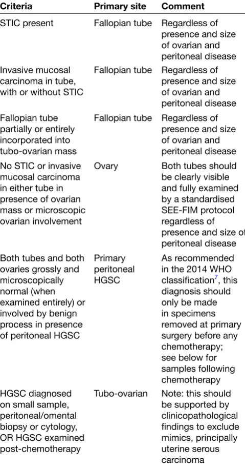
Table 2 Criteria for assignment of primary site in extrauterine HGSC
Criteria Primary site Comment
STIC present Fallopian tube Regardless of presence and size of ovarian and peritoneal disease Invasive mucosal
carcinoma in tube, with or without STIC
Fallopian tube Regardless of presence and size of ovarian and peritoneal disease Fallopian tube
partially or entirely incorporated into tubo-ovarian mass
Fallopian tube Regardless of presence and size of ovarian and peritoneal disease No STIC or invasive
mucosal carcinoma in either tube in presence of ovarian mass or microscopic ovarian involvement
Ovary Both tubes should
be clearly visible and fully examined by a standardised SEE-FIM protocol regardless of presence and size of peritoneal disease Both tubes and both
ovaries grossly and microscopically normal (when examined entirely) or involved by benign process in presence of peritoneal HGSC
Primary peritoneal HGSC
As recommended in the 2014 WHO classification7, this
diagnosis should only be made in specimens removed at primary surgery before any chemotherapy; see below for samples following chemotherapy HGSC diagnosed
on small sample, peritoneal/omental biopsy or cytology, OR HGSC examined post-chemotherapy
Tubo-ovarian Note: this should be supported by clinicopathological findings to exclude mimics, principally uterine serous carcinoma
HGSC, high-grade serous carcinoma; SEE-FIM, Sectioning and Extensively Examining the FIMbriated End; STIC, serous tubal intraepithelial carcinoma; WHO, World Health Organization.
Health Organization (WHO) classification7 as a disease of
exclu-sion, to be designated only in cases with no gross or microscopic evidence of mucosal disease in either the tubes or the ovaries. Most significantly, continuing skepticism regarding the tubal origin is an obstacle to studying the impact of ovary-conserving preventa-tive strategies that have potential to reduce HGSC incidence and mortality.
Studies on the origin of sporadic HGSC in the past have been hampered by its presentation with disseminated disease, technical challenges in performing molecular studies on formalin-fixed paraf-fin-embedded tissues and incomplete tubal examination; complete tubal sampling using detailed Sectioning and Extensively Examining the FIMbriated End (SEE-FIM) protocols is an essential prerequisite for identifying and sampling the microscopic precursor lesion of HGSC, serous tubal intraepithelial carcinoma (STIC). While STIC is reported to be present in 11–61% of HGSC cases, reports on low-stage and optimally examined cases clearly demonstrate that virtually all contain
STIC or small microscopic tubal HGSC.8–11 These studies also show
that examples of single-site disease are always tubal and never ovarian. Furthermore, while ovarian involvement in HGSC is typically bilateral, as is common in metastasis to a paired organ, tubal
involve-ment is unilateral in the majority of cases.12 These observations are
supported by detailed molecular analysis demonstrating shared TP53
mutation between STIC and HGSC, and that the majority of mutational and copy abnormalities seen in HGSC are also identified in
accom-panying STIC.13 Clonal evolution studies demonstrate the same
result14 15 but also show that, in advanced cases, intraepithelial tubal
metastasis can produce lesions indistinguishable from STIC, further demonstrating the futility of studying advanced HGSC to answer ques-tions about its origin. What these and other studies have demonstrated irrefutably is that, despite being widely disseminated at presentation in the majority of cases, HGSC arises from a single precursor clone,
and there is no molecular evidence of multifocal origin.16 17 A proposal
for primary site assignment in extrauterine HGSC is recommended
for reproducible categorisation (see Table 2), with its basis in
scien-tific evidence in favour of traditional beliefs7 18; this has been
recom-mended for use in international ovarian cancer pathology reporting
guidelines.19 This evidence also forms the basis for recommendations
on uniform staging of low-stage HGSC in cases that are left to the
pathologist’s and clinician’s discretion in the current FIGO system,20 21
resulting in potential for identical cases to be staged differently.6 It
should be emphasised that these criteria are only to be used for HGSC and not for other histological types of EOC.
recommendation 1.1
A large majority of extrauterine HGSCs arise in the fallopian tube from STIC. SEE-FIM sectioning of both fallopian tubes should be carried out in all cases of extrauterine HGSC where the tubes are grossly normal, and also in risk-reducing prophylactic surgery specimens.
Level of evidence: III
Strength of recommendation: A
Consensus: 100% (40) yes, 0% (0) no, 0% (0) abstain (40 voters)
recommendation 1.2
Extrauterine HGSC can only be assigned as ovarian in origin if both fallopian tubes are grossly normal, and histologically contain no mucosal disease following examination using a SEE-FIM protocol.
Level of evidence: III
Strength of recommendation: A
Consensus: 100% (40) yes, 0% (0) no, 0% (0) abstain (40 voters)
recommendation 1.3
Cases in which HGSC is present in the endometrium and the tube/ ovary are very likely to represent a primary at one site with metas-tasis to the other; these are very unlikely to represent synchronous independent neoplasms.
Level of evidence: V
Strength of recommendation: A
Consensus: 97.5% (39) yes, 2.5% (1) no, 0% (0) abstain (40 voters)
on September 6, 2020 by guest. Protected by copyright.
recommendation 1.4
The distinction between primary endometrial and primary tubal/ ovarian HGSC requires assessment of a constellation of patholog-ical features; negative wild-type 1 (WT1) staining favours an endo-metrial primary, but this is not always definitive.
Level of evidence: V
Strength of recommendation: A
Consensus: 92.5% (37) yes, 0% (0) no, 7.5% (3) abstain (40 voters)
recommendation 1.5
The use of uniform criteria is important in site assignment in extra-uterine HGSC for cancer registry and epidemiological reasons. The use of International Collaboration on Cancer Reporting (ICCR) and College of American Pathologists (CAP) guidelines is recommended.
Level of evidence: V
Strength of recommendation: A
Consensus: 100% (40) yes, 0% (0) no, 0% (0) abstain (40 voters)
recommendation 1.6
Correct and uniform use of site assignment criteria is particularly important for accurate staging of early HGSC.
Level of evidence: III
Strength of recommendation: A
Consensus: 100% (40) yes, 0% (0) no, 0% (0) abstain (40 voters)
recommendation 1.7
STIC should count as a disease site for staging purposes; for example, a case with a STIC and HGSC confined to the ovary should be staged as stage IIA fallopian tube HGSC.
Level of evidence: IV
Strength of recommendation: A
Consensus: 95% (38) yes, 0% (0) no, 5% (2) abstain (40 voters)
recommendation 1.8
True primary peritoneal HGSC is extremely rare. Level of evidence: IV
Strength of recommendation: A
Consensus: 100% (40) yes, 0% (0) no, 0% (0) abstain (40 voters)
recommendation 1.9
Multifocal origin of extrauterine HGSC is exceptionally rare and thus HGSC currently staged as IB should be considered as stage IIA.
Level of evidence: IV
Strength of recommendation: A
Consensus: 95% (38) yes, 5% (2) no, 0% (0) abstain (40 voters)
2. How to identify tumours that will respond to targeted therapies, including poly(adenosine diphosphate-ribose) polymerase inhibitors and immune checkpoint inhibitors? The targeted therapies that are under investigation include antian-giogenic agents, poly(adenosine diphosphate-ribose) polymerase (PARP) inhibitors, hormone receptor modulators and immune checkpoint inhibitors. Bevacizumab, an antivascular epithelial growth factor (anti-VEGF) monoclonal antibody has shown posi-tive results in first-line therapy with standard chemotherapy and also in both platinum-sensitive and platinum-resistant relapsed disease, with improved progression-free survival (PFS) in various
large RCTs.22–25 Improvements in overall survival (OS) have been
harder to demonstrate and are currently limited to a retrospective
analysis of high-risk patients within the ICON7 trial.22 Although
therapy targeting VEGF has become the standard of care in tubo-ovarian carcinomas as well as other solid malignancies, attempts to identify predictive molecular biomarkers for efficacy have failed to identify any that could help oncologists decide who should and, more importantly, who should not, receive VEGF-targeted therapies,
including bevacizumab.26
Angiogenic markers, such as CD31 expression, microvessel density and tumour VEGF-A levels, may provide prognostic infor-mation in recurrent/persistent EOC, and were identified in a retro-spective analysis of the Gynecologic Oncology Group (GOG) 218
study as potential predictive biomarkers,27 but further prospective
evaluation will be required. Another study showed a
discrimina-tory signature comprising mesothelin, FLT4, α-1 acid glycoprotein
(AGP) and cancer antigen 125 (CA125) as potentially identifying
those patients with EOC more likely to benefit from bevacizumab.28
A potential role of combined values of Ang1 and Tie2 as predic-tive biomarkers for improved PFS in bevacizumab-treated patients with EOC has also been suggested. However, these findings need
to be validated in larger trials.29 Currently, only clinical biomarkers
(including stage, debulking status and presence of ascites) appear to have predictive utility in selecting patients for first-line treatment with bevacizumab, and thus prospective studies evaluating predic-tive biomarkers of bevacizumab benefit are urgently required.
At the time of diagnosis, ∼50% of EOCs may exhibit defective
DNA repair via homologous recombination (HR) due to genetic and
epigenetic alterations of HR pathway genes.30 Defective HR is an
important therapeutic target in EOC as exemplified by the efficacy of platinum analogs in this disease, as well as the advent of PARP inhibitors that exhibit synthetic lethality when applied to HR-defi-cient cells. PARP inhibitors, such as olaparib, niraparib and ruca-parib, are being utilised in the clinic to manage recurrent EOCs that display defects in the HR repair pathway. However, PARP inhibitors also show significant clinical benefit in patients without demon-strable defects in known HR genes. Various studies validated this and extended the usefulness of PARP inhibitors in the treatment
setting beyond BRCA-mutated tumours.31 32
The strongest clinical evidence for the use of PARP inhibitors
comes from patients with germline or somatic mutations in BRCA1
or BRCA2, both as single-agent therapy and as maintenance
following response to platinum chemotherapy in the first-line33
and relapsed34–36 settings. Rucaparib also has robust activity as
single-agent therapy in relapsed BRCA-mutated HGSC,32 and the
ARIEL2 study32 demonstrated that tumours harbouring mutations
in RAD51C alterations are BRCA-like [high genomic loss of (LOH)]
and responded to rucaparib at very similar rates to BRCA
-mu-tated disease. However, attempts to identify robust predictive biomarkers of response to PARP inhibitors in HGSC beyond key
HR gene mutations have proven difficult. The ARIEL2 study32
utilised genome-wide LOH as a potential predictive biomarker,
and showed that BRCA WT/LOH high tumours did indeed have
higher response rates and improve PFS compared with BRCA
WT/LOH low, but lower than BRCA-mutated. However, attempts
to use LOH as a predictive marker in the maintenance setting
were less successful. The ARIEL3 study37 evaluated rucaparib
versus placebo as maintenance treatment in patients with recur-rent platinum-sensitive cancer and found rucaparib maintenance
on September 6, 2020 by guest. Protected by copyright.
treatment significantly improved PFS versus placebo in the
nested BRCA-mutated and HR deficiency (HRD) cohorts and in the
overall intention-to-treat (ITT) population. PFS was improved with rucaparib maintenance treatment versus placebo in patients with
BRCA WT EOC (LOH high and LOH low) as well. The NOVA study38
utilised a different algorithm to identify potential HRD tumours and again found that, in patients who had responded to platinum in the relapse setting, the median PFS was significantly longer among those receiving niraparib than among those receiving placebo, regardless of the presence or the absence of germline
BRCA mutations or HRD status. Thus, in the maintenance setting,
response to platinum chemotherapy remains the most robust predictive biomarker for PARP inhibitor benefit.
A major limitation of the current HR assays is that they are largely insensitive to reversion of HRD, which may occur on development of resistance to platinum and PARP inhibitors. True functional assays of HR function exist, but they require the cancer specimen to be exposed to some form of DNA damage, which precludes use of formalin-fixed, paraffin-embedded specimens, increases the technical complexity, and limits the reproducibility of these assays. Overall, there is currently no prospectively validated biomarker of HRD that has been incorporated into clinical practice, and this
remains an active area of investigation.39
Bowman et al40 demonstrated that higher levels of oestrogen
receptor (ER) expression in EOC resulted in disease stabilisation and CA125 response after treatment with the aromatase inhibitor letrozole, and suggested the presence of an endocrine-sensitive group that could be targeted in future studies. Similar results were later published by other groups, suggesting that ER/progesterone receptor (PR) expression status may be a predictive biomarker for
hormonal therapy.41 42 There are no positive prospective randomised
data for the use of hormone therapies as alternatives to chemo-therapy or as maintenance chemo-therapy in first-line or recurrent disease, even in low-grade serous carcinoma (LGSC). RCTs incorporating hormone therapy are required, especially in LGSC. Prospective vali-dation of ER score as a predictive biomarker is also required, as there is no validated or universally used ER score in EOCs.
recommendation 2.1
There are no validated predictive molecular biomarkers of bevaci-zumab benefit.
Level of evidence: IV
Strength of recommendation: A
Consensus: 100% (40) yes, 0% (0) no, 0% (0) abstain (40 voters)
recommendation 2.2
PARP inhibitors have greatest activity in patients with BRCA1/2
mutations.
Level of evidence: I
Strength of recommendation: A
Consensus: 100% (40) yes, 0% (0) no, 0% (0) abstain (40 voters)
recommendation 2.3
Testing for BRCA1/2 mutations is recommended for all patients
with non-mucinous ovarian cancer. Level of evidence: I
Strength of recommendation: A
Consensus: 95% (38) yes, 0% (0) no, 5% (2) abstain (40 voters)
recommendation 2.4
Testing for mutations in other HR genes, in particular RAD51C/D,
BRIP1 and PALB2, should be considered. Level of evidence: III
Strength of recommendation: A
Consensus: 100% (40) yes, 0% (0) no, 0% (0) abstain (40 voters)
recommendation 2.5
Current assays of HR function cannot be used to exclude patients from PARP inhibitor therapy.
Level of evidence: I
Strength of recommendation: A
Consensus: 100% (40) yes, 0% (0) no, 0% (0) abstain (40 voters)
recommendation 2.6
Moderate-strong ER staining may be a predictor of response to hormone therapy.
Level of evidence: III
Strength of recommendation: B
Consensus: 100% (40) yes, 0% (0) no, 0% (0) abstain (40 voters)
recommendation 2.7
There are currently no prospectively validated predictive biomarkers of response to immune checkpoint inhibitors that are specific to ovarian cancer.
Level of evidence: V
Strength of recommendation: A
Consensus: 100% (40) yes, 0% (0) no, 0% (0) abstain (40 voters)
3. How to identify patients with acquired/intrinsic resistance to chemotherapy?
Although most patients with HGSC initially respond to plati-num-based chemotherapy, the large majority of patients will relapse. Thus, resistance to platinum-based treatment is common, with roughly 20% of women experiencing disease progression ≤6 months after completing a platinum-based regimen (previously classified as ‘platinum-resistant’ relapse) or who fail to respond at all to first-line treatment or relapse within 4–6 weeks after last
platinum dose (previously classified as ‘platinum-refractory’).43
There have been many efforts over the years to develop accurate predictors of outcomes in patients treated with chemotherapy to
help inform treatment decisions.44
Elucidation of why platinum resistance occurs and how it can be reversed or prevented is essential for improving survival. However, the WG unanimously agreed that there are no validated predictive biomarkers that can be used in clinical practice for determining likelihood of primary platinum-refractory or platinum-resistant disease.
It is widely accepted that most HGSCs (60–80%) show a good response to conventional platinum-based chemotherapy. However, low-grade serous, mucinous, clear cell and endometrioid ovarian carcinomas are considered to be less chemoresponsive and to have a different prognosis, although in many cases they present at an early stage, in contrast to HGSCs, which usually present at an advanced stage. The large majority of patients enrolled in clinical trials have HGSC histology and thus the results from these studies cannot automatically be applied to all histological types, where numbers recruited to all-comer studies are low and where there
are generally very few specific studies.45
on September 6, 2020 by guest. Protected by copyright.
With better understanding of the molecular biology of EOCs, DNA damage repair through HR is known to play a vital role in contrib-uting to genomic stability and preventing malignant transformation.
Numerous studies have reported that mutation in BRCA1 or BRCA2
is a prognostic marker in EOC and concluded that patients with
BRCA mutation, especially BRCA2, have better survival outcomes,
which is likely to reflect increased response rates to platinum-based
chemotherapy.46–48
Germline or somatic mutations in HR genes are present in up to one-third of EOCs, including both serous and non-serous
histologies. In addition, Pennington et al49 looked at somatic and
germline mutations in 13 HR genes (BRCA1, BRCA2, ATM, BARD1,
BRIP1, CHEK1, CHEK2, FAM175A, MRE11A, NBN, PALB2, RAD51C, RAD51D). They concluded that somatic mutations in other HR genes have a similar positive impact on OS and platinum
respon-siveness as germline BRCA1/2 mutations. HR mutations were more
successful in predicting platinum sensitivity at primary treatment
than at relapse.49 Other potentially important mutations include
CDK12, loss of which may induce an HRD phenotype,50 although this needs further validation, as not all alterations will have the same effect on HR repair and sensitivity to platinum. Whole-genome studies in HGSC reveal that gene breakage commonly inactivates
the tumour suppressors RB1, NF1, RAD51B and PTEN and
contrib-utes to acquired chemotherapy resistance. CCNE1 amplification is
common in primary resistant and refractory disease, demonstrating the role of non-HRD molecular mechanisms in resistance
develop-ment.51 52 An association between excision repair
cross-comple-mentation group 1 (ERCC1) polymorphism and platinum sensitivity has been reported in a few studies but with conflicting results;
hence, this is not suitable for assessing platinum response.53–55
Finally, in patients with relapsed disease, the current classifica-tion strictly defines platinum resistance as those relapsing within 6 months of previous platinum chemotherapy. However, because time since last platinum chemotherapy represents a continuum of prob-ability of response to further chemotherapy, a fixed 6-month cut-off decision on platinum sensitivity is neither sensible nor biologically relevant. In addition, the effect of maintenance therapies on the probability of response to further platinum is unknown. The time since last platinum chemotherapy correlates with response to other
agents including PARP inhibitors, although this is not absolute.56
Large-scale trials collecting serial biological samples throughout treatment are required in order to improve the understanding of acquired resistance. In addition, investigation and validation of markers should be carried out using samples taken immediately before and during the therapy of interest rather than using archival samples.
recommendation 3.1
There are no validated predictive markers of primary platinum refractory or resistant disease.
Level of evidence: IV
Strength of recommendation: A
Consensus: 100% (40) yes, 0% (0) no, 0% (0) abstain (40 voters)
recommendation 3.2
Defects in HR repair are associated with improved outcome/PFS following platinum-based chemotherapy.
Level of evidence: IV
Strength of recommendation: A
Consensus: 100% (40) yes, 0% (0) no, 0% (0) abstain (40 voters)
recommendation 3.3
The time elapsed since last platinum chemotherapy represents a continuum of probability of response to further chemotherapy.
Level of evidence: IV
Strength of recommendation: A
Consensus: 100% (40) yes, 0% (0) no, 0% (0) abstain (40 voters)
4. Can we develop accurate and sensitive circulating and tissue biomarkers both of response and relapse?
The Gynecologic Cancer InterGroup (GCIG) has published a consensus document regarding the criteria that should be used in clinical trial protocols to define PFS after first-line therapy, as well as the criteria to define response to treatment in recurrent disease using the serum marker CA125, and has specified the situations
where these criteria should be used.57 This WG agrees to the utility
of these criteria in routine practice but emphasises the importance of correlation with radiological and clinical assessment.
CA125 levels have been most widely studied in HGSC. The prog-nostic value of CA125 in other morphological types of EOC, such as low-grade serous, clear cell, endometrioid and mucinous, is less clear due to the relative rarity of these neoplasms in the advanced disease setting and the limited number of patients studied in trials. As a result, CA125 is not a reliable marker in non-HGSC EOC,58 59 in particular in mucinous carcinoma, where it is rarely secreted. Caution is also recommended when using CA125 as a response marker for molecularly targeted agents until prospective studies validate CA125 changes with objective imaging response
results.60 61 Specifically, there is a lack of reliability of CA125
response criteria with anti-VEGF molecular therapies, where CA125 change may not correspond to imaging response criteria for EOC patients receiving bevacizumab.
Human epididymis protein 4 (HE4) has been proposed as the most promising biomarker that may complement CA125 and has been approved by the US Food and Drug Administration (FDA) in monitoring the follow-up and relapse of EOC patients. However,
studies are contradictory62; as a result, HE4 testing currently cannot
be recommended in routine practice.
Circulating tumour cells (CTCs) and circulating cell-free DNA (cfDNA) have been used as diagnostic and prognostic markers in many types of cancer, including ovarian cancer. These tech-niques do have specific challenges, including pre-analytical issues regarding sample volume, the proper tubes for sample collection, sample storage and the time of the analysis, quality control and analytical validation of the assays. There are currently no standard methods for the isolation and detection of either CTCs or cfDNA in the bloodstream, with few studies recruiting large cohorts of EOC patients. Further studies regarding the validation, standardisation and quality control of the assays are needed before implementing
this approach in the clinical routine.63
Another approach to address this question is the chemotherapy response score (CRS), which was developed to enable reproduc-ible and prognostically relevant reporting of the histopathological changes in interval debulking surgical specimens after
neoadju-vant chemotherapy (NACT) in extrauterine HGSC.64 65 Since its
description, the CRS has been independently validated in several
on September 6, 2020 by guest. Protected by copyright.
Table 3 Chemotherapy response score: summary of criteria
CRS Criteria
CRS1: No or minimal tumour response
Mainly viable tumour with no or minimal regression-associated fibroinflammatory changes* limited to a few foci. Note: cases in which it is difficult to decide between regression and tumour-associated
desmoplasia or inflammatory cell infiltration CRS2: Partial
tumour response
Appreciable tumour response amidst viable tumour, both readily identifiable and tumour regularly distributed. Note: cases ranging from multifocal or diffuse regression-associated fibroinflammatory changes*, with viable tumour in sheets, streaks or nodules, to extensive regression associated fibroinflammatory changes* with multifocal residual tumour which is easily identifiable
CRS3: Total or near-total tumour response
No residual tumour OR minimal irregularly
scattered tumour foci seen as individual cells, cell groups or nodules up to 2 mm in maximum size. Note: cases showing mainly regression-associated fibroinflammatory changes* or, in rare cases, no/very little residual tumour in complete absence of any inflammatory response; advisable to record whether ‘no residual tumour’ or ‘microscopic residual tumour present’
*Regression-associated fibroinflammatory changes: fibrosis associated with macrophages, including foam cells, mixed inflammatory cells and psammoma bodies; to distinguish from tumour-related inflammation or desmoplasia.
CRS, chemotherapy response score.
studies,66–69 including an individual patient data meta-analysis
incorporating results from over 800 patients from different centres
worldwide.70 This system has been recommended for use in the
ICCR guidelines for tubal and ovarian carcinomas,19 since a
numer-ical score allows objective reporting and comparison of results and
is thus superior to descriptive reporting (see Table 3). The score
identifies the roughly one-third of all patients (CRS3; total or near-total response) who show significantly improved PFS and OS, and has potential for incorporation into routine practice and clinical trial design as an early endpoint.
recommendation 4.1
The CA125 criteria for response and progression as agreed by GCIG have utility in routine practice but should be used in combination with radiological and clinical assessment.
Level of evidence: III
Strength of recommendation: A
Consensus: 97.5% (39) yes, 0% (0) no, 2.5% (1) abstain (40 voters)
recommendation 4.2
The role of CA125 as a marker of response and progression in non-HGSC is less clear.
Level of evidence: V
Strength of recommendation: A
Consensus: 100% (40) yes, 0% (0) no, 0% (0) abstain (40 voters)
recommendation 4.3
The use of CA125 in assessing response and progression to targeted therapies is not yet proven; thus, radiological and clinical assessment should be used.
Level of evidence: V
Strength of recommendation: A
Consensus: 100% (40) yes, 0% (0) no, 0% (0) abstain (40 voters)
recommendation 4.4
HE4 should not be used routinely to assess response and progres-sion due to conflicting results.
Level of evidence: IV
Strength of recommendation: A
Consensus: 97.5% (39) yes, 0% (0) no, 2.5% (1) abstain (40 voters)
recommendation 4.5
Quantification of circulating cfDNA has not been established as a tool to assess response and relapse.
Level of evidence: IV
Strength of recommendation: A
Consensus: 97.5% (39) yes, 0% (0) no, 2.5% (1) abstain (40 voters)
recommendation 4.6
Pathological CRS after NACT may provide an objective and repro-ducible prognostic measure of outcome in HGSC.
Level of evidence: IV
Strength of recommendation: A
Consensus: 82.5% (33) yes, 12.5% (5) no, 5% (2) abstain (40 voters)
5. What are the morphological criteria useful in separating borderline from invasive ovarian neoplasia?
Previously, it was a widely held view that the distinction between a borderline ovarian tumour (BOT) and a carcinoma was based on the presence of destructive stromal invasion in the latter. However, ovarian carcinomas, particularly of mucinous and endometrioid type, can exhibit expansile (non-destructive) or infiltrative (destruc-tive) stromal invasion. Mucinous carcinomas exhibiting expansile invasion have been reported to have a lower risk of metastasis
than those exhibiting infiltrative invasion.71–76 Expansile invasion
is morphologically characterised by complex glandular, papillary and/or cribriform architecture with a labyrinthine or anastomosing
pattern and little or no intervening stroma.73–75 77
Extraovarian disease in association with a serous BOT (sBOT) was previously divided into non-invasive and invasive implants, and the former were further divided into ‘epithelial’ and
‘desmo-plastic’ implants.78 In the 2014 WHO classification,7 it is stated that
the term extraovarian ‘LGSC’ should be used for invasive implants in association with an sBOT. The WG regards such terminology as potentially confusing and wishes to separate bona fide metastases from an ovarian LGSC from invasive implants in the omentum or peritoneum associated with an sBOT.
on September 6, 2020 by guest. Protected by copyright.
The micropapillary variant of sBOT is characterised by the pres-ence of slender papillae with a length-to-width ratio of at least 5:1, growing in a non-hierarchical pattern; a cribriform growth pattern is less frequent but may co-exist with the micropapillary pattern. The micropapillary or cribriform component must be confluent over an area of at least 5 mm in maximum extent for the tumour to be
designated as a micropapillary variant of sBOT.78 79 The
micropap-illary variant of sBOT is more likely to be associated with extrao-varian invasive implants than the typical sBOT, and some advocate using the term ‘non-invasive LGSC’ for the former. This has resulted in this term being used interchangeably with the micropapillary
variant of sBOT in the 2014 WHO classification.7 A recent
popu-lation-based study of a Danish cohort with long-term follow-up reported that patients with the micropapillary variant of sBOT are more likely to present at advanced stage and more frequently have bilateral disease, gross residual disease after surgery, areas of microinvasion, and invasive implants at presentation compared
with patients with usual-type sBOT.80 The WG does not favour the
use of the term ‘non-invasive LGSC’, since such tumours which are confined to the ovary at presentation have a comparable outcome to the usual-type sBOT and the term may be misleading for clinical management.
There have been various definitions of microinvasion in BOTs
and the 2014 WHO classification7 uses a cut-off of 5 mm.
Micro-invasion can be seen in all morphological subtypes of BOT but is most common in serous and mucinous neoplasms. Two types of microinvasion have been described, namely ‘microinvasion’ and ‘microinvasive carcinoma’, although the distinction between
these is not always straightforward.81 Although the presence of
microinvasion has been associated with a higher risk of tumour
recurrence in some series,82 the majority of studies have not
identified such an association.83 84 The WG recommends that
BOTs with microinvasion should be classified and managed as borderline tumours.
The term implant should be restricted to extraovarian disease in association with an sBOT and not be used in the context of a mucinous BOT (mBOT). Extraovarian disease in a patient with a presumed mBOT either represents metastasis from an undiag-nosed or undetected focus of carcinoma within the ovary, or the ovarian and extraovarian disease represents metastasis from a mucinous carcinoma elsewhere.
Borderline endometrioid tumours are rare.81 The criteria used
to distinguish a borderline endometrioid tumour from endome-trioid adenocarcinoma are broadly similar to the criteria used to distinguish atypical hyperplasia from grade I endometrioid adeno-carcinoma in the uterine corpus, and are largely architectural. Adenocarcinomas are characterised by complex growth with gland fusion and stromal exclusion; cribriform and microglandular
patterns may also be seen.85
recommendation 5.1
Destructive stromal invasion is no longer necessary for carcinoma diagnosis (carcinomas may exhibit expansile invasion).
Level of evidence: V
Strength of recommendation: A
Consensus: 100% (40) yes, 0% (0) no, 0% (0) abstain (40 voters)
recommendation 5.2
According to the 2014 WHO classification, extraovarian inva-sive implants in association with an sBOT are synonymous with extraovarian LGSC. The WG does not support this terminology because it may be misleading for clinical management.
Level of evidence: V
Strength of recommendation: A
Consensus: 100% (40) yes, 0% (0) no, 0% (0) abstain (40 voters)
recommendation 5.3
In the 2014 WHO classification, the micropapillary variant of sBOT is also termed non-invasive LGSC but the WG does not support this terminology because it may be misleading for clinical management.
Level of evidence: V
Strength of recommendation: A
Consensus: 100% (40) yes, 0% (0) no, 0% (0) abstain (40 voters)
recommendation 5.4
Microinvasion (<5 mm) can be seen in borderline tumours but these cases should still be regarded as borderline for classification and management purposes.
Level of evidence: V
Strength of recommendation: A
Consensus: 100% (40) yes, 0% (0) no, 0% (0) abstain (40 voters)
recommendation 5.5
The term implant should not be used in the context of mBOTs; extraovarian disease in association with an mBOT should be considered as metastasis (from ovary or another organ).
Level of evidence: V
Strength of recommendation: A
Consensus: 100% (40) yes, 0% (0) no, 0% (0) abstain (40 voters)
recommendation 5.6
Borderline endometrioid tumours can be differentiated from grade I endometrioid carcinoma using similar criteria as used to differen-tiate atypical hyperplasia from grade I endometrioid carcinoma in the uterine corpus.
Level of evidence: V
Strength of recommendation: A
Consensus: 97.5% (39) yes, 0% (0) no, 2.5% (1) abstain (40 voters)
Early-Stage and borderline tumours
6. Are there exceptions to the standard surgical management for early-stage ovarian carcinoma?
The standard surgical approach in early-stage ovarian cancer is based on removal of both ovaries with a staging procedure. A complete exploration of the abdomino-pelvic peritoneal cavity via a thorough visual examination is required to detect potentially suspicious implants. Peritoneal staging surgery is based on peri-toneal washing, periperi-toneal biopsies (pelvic peritoneum, paracolic gutters, diaphragm) (4–6) and omentectomy (at least infracolic). The standard approach is by open surgery. The rationale for this choice is based on the accuracy of the macroscopic exploration and the reduction of the risk of a rupture of the primary tumour during its dissection/removal. This risk is potentially increased using a
minimally invasive surgical approach.86 Regardless of the approach
used, rupture of an intact tumour could alter the FIGO staging and
on September 6, 2020 by guest. Protected by copyright.
affect prognosis, and must be avoided.87 Nevertheless, the mini-mally invasive approach can be considered for restaging surgery in cases where the initial ovarian tumour has been removed and there is no risk of ‘rupture’ of the ovarian lesion. This surgery should then be carried out by trained surgeons in expert centres to assure optimal assessment vision of all abdominal quadrants and to lower the risk of peri- and postoperative complications. Nodal staging surgery is part of the ‘conventionally’ required procedure in early-stage ovarian carcinoma. This nodal staging surgery of apparent stage I ovarian carcinoma includes a bilateral pelvic and para-aortic lymphadenectomy up to the left renal vein (regardless
of the surgical approach used).88 89 Ten to 15% of cases have nodal
involvement.88 However, due to a low prevalence of nodal
metas-tases in some histological subtypes (eg, mucinous carcinoma of expansile subtype or LGSC), the indication for staging surgery in
these cases90–92 may be questioned.
The issue of restaging surgery must be addressed separately. Contrary to the indication of staging surgery discussed above, where the decision is based on macroscopic evaluation of the abdominal cavity and the result of a frozen section analysis (FSA), some patients may have initially undergone surgery without proper staging. In this context, the restaging procedure is indicated if it may bring new elements that have a direct impact on the defin-itive treatment planning. If the primary tumour exhibits high-risk features (eg, high-grade, capsule rupture, tubal or peritoneal extension) that justify adjuvant chemotherapy, indication of nodal restaging surgery with the aim of obtaining additional prognostic variables must be balanced with the potential surgical morbidity of the procedure.
FSA should be available during a surgical procedure carried out for a suspicious ovarian mass and should be supported by the diag-nosis of an experienced gynaecological pathologist. Nevertheless, it must only be done when the surgical strategy would be altered by the outcome (eg, choice of a nodal or radical surgery). FSA is less accurate in cases of pathological diagnosis of borderline tumours, mucinous tumours, tumour sampling done by an inexperienced
oncologist or large ovarian lesions (>8–10 cm).93 94
recommendation 6.1
Laparotomy is the standard surgical approach to treat and stage patients with apparent early-stage ovarian carcinoma.
Level of evidence: V
Strength of recommendation: A
Consensus: 100% (40) yes, 0% (0) no, 0% (0) abstain (40 voters)
recommendation 6.2
Minimally invasive surgery can be carried out for restaging. Level of evidence: IV
Strength of recommendation: B
Consensus: 75% (30) yes, 12.5% (5) no, 12.5% (5) abstain (40 voters)
recommendation 6.3
Whatever the approach used, rupture of an intact tumour with spillage of cancer cells at the time of surgery must be avoided.
Level of evidence: IV
Strength of recommendation: A
Consensus: 100% (40) yes, 0% (0) no, 0% (0) abstain (40 voters)
recommendation 6.4
Peritoneal restaging surgery is mandatory even if it does not alter the indication for adjuvant chemotherapy.
Level of evidence: V
Strength of recommendation: B
Consensus: 92.5% (37) yes, 2.5% (1) no, 5% (2) abstain (40 voters)
recommendation 6.5
Peritoneal restaging should be considered in cases of incidentally detected, apparently isolated STIC lesions.
Level of evidence: IV
Strength of recommendation: B
Consensus: 100% (40) yes, 0% (0) no, 0% (0) abstain (40 voters)
recommendation 6.6
The standard surgical staging of apparent early EOC includes systematic lymph node (LN) dissection of the pelvic and the para aortic regions up to the left renal vessel origin.
Level of evidence: IV
Strength of recommendation: A
Consensus: 77.5% (31) yes, 22.5% (9) no, 0% (0) abstain (40 voters)
recommendation 6.7
LN dissection for restaging purposes may be avoided if the nodal status does not alter the patient management.
Level of evidence: V
Strength of recommendation: B
Consensus: 95% (38) yes, 0% (0) no, 5% (2) abstain (40 voters)
7. What are the limits of fertility-sparing surgery (cancer and borderline ovarian tumour)?
Fertility-sparing surgery (FSS) is based on unilateral salpingo-oo-phorectomy and complete surgical staging. This management seems to be safe in patients with conventional low-grade stage IA
(serous, endometrioid or mucinous expansile subtype).95–97 The use
of FSS in patients with stage IC disease should be defined using the
current 2014 FIGO staging system.98 FSS is acceptable for stage
IC1 tumours, with half of these recurrences being isolated on the remaining ovary and therefore able to be rescued by subsequent surgery. However, the recurrence rates are higher in stage IC2, IC3 and grade 3 disease, although mainly in extraovarian sites and are, therefore, not clearly correlated with the fertility-sparing approach.
Adequate counseling is, therefore, needed in this situation.98
In cases of stage II or III disease, the use of FSS is unconventional,
with high risk of recurrences reported.95 FSS remains
contraindi-cated in these patients, although it is unclear whether such recur-rences are related to the natural history of the disease rather than the type of surgery in these ‘high-risk’ patients.
recommendation 7.1
FSS can be safely offered to all stage IA and IC1 low-grade ovarian carcinomas.
Level of evidence: IV
Strength of recommendation: B
Consensus: 94.7% (36) yes, 2.6% (1) no, 2.6% (1) abstain (38 voters)
on September 6, 2020 by guest. Protected by copyright.
recommendation 7.2
There is no place for ovarian preservation for invasive EOC greater than fully staged FIGO stage I.
Level of evidence: V
Strength of recommendation: A
Consensus: 94.9% (37) yes, 0% (0) no, 5.1% (2) abstain (39 voters)
8. Should all stage I carcinomas receive adjuvant chemotherapy and, if not, which ones?
A Cochrane systematic review99 clearly demonstrated that the
addi-tion of adjuvant platinum-based chemotherapy to surgery is effec-tive in significantly prolonging long-term OS and PFS in women with early-stage EOC. Considering the risk of recurrence, the ICON1
trial100–103 determined that women with a high-risk of recurrence
(stage IA grade 3, IB or IC grade 2 or 3, any clear cell tumours) may benefit the most from adjuvant chemotherapy. Retrospective
studies104–107 suggested that adjuvant chemotherapy may not be
necessary for some histological subgroups, due to the absence of recurrences observed in patients who did not receive adjuvant
chemotherapy. It should be noted that the ICON1 trial100–103 could
neither confirm nor exclude survival benefits in low/intermediate risk disease (stage IA grade 1 or 2, IB or IC grade 1) in a subgroup analysis. Recently, the retrospective SEER database also reported no benefit for adjuvant chemotherapy in the low and intermediate
endometrioid groups.108 On the contrary, in a large cohort study,109
chemotherapy was associated with reduced mortality not only for high-risk patients but also for patients with stage IA/IB, grade 2 ovarian cancer. This study was in line with prior study results demonstrating no benefit for chemotherapy in women with stage IA and IB, grade 1 neoplasms. Finally, the available data could neither confirm nor exclude survival benefits for the addition of adjuvant chemotherapy in optimally staged patients (all risk groups consid-ered). More specifically, for histological subgroups such as clear cell carcinoma, the targeted retrospective studies reported in the
literature primarily from Asian populations105 107 108 110 did not
iden-tify any benefit compared with observation for early-stage disease (stage IA to IC1). For the mucinous subgroup, the expansile or grade I type is associated with better prognosis and should not receive adjuvant chemotherapy, while the infiltrative form is associated
with a high risk of relapse.72 90 91 111
The chemotherapy administered in the ICON1100–103 and
ACTION112–116 trials consisted of a variety of platinum-based
regimens, given ideally for 6 cycles. However, only 4 cycles were required for the ACTION trial and only half of the patients in the ICON1 trial received all 6 cycles without dose modification, due
to toxicity. Bell et al117 reported an RCT of 3 versus 6 cycles of
adjuvant carboplatin and paclitaxel administered every 3 weeks in women with high-risk, early-stage ovarian cancer. This GOG trial found that longer treatment was not associated with a significant reduction in recurrence risk and resulted in additional toxicity. A
subsequent exploratory analysis118 of this GOG study revealed that
longer adjuvant therapy was associated with a significant reduc-tion in recurrence risk for serous tumours but not for non-serous tumours. There was no benefit for longer adjuvant therapy in any other subgroup of interest, including age, performance status (PS), stage, grade and presence of ascites, tumour rupture and positive
cytology. Bakkum-Gamez et al119 evaluated a cohort of surgically
staged, stage I ovarian cancer patients who completed either 3 or 6 cycles of carboplatin and paclitaxel. Patients with stage IC cancer and with fixed tumours (described adhesions or fixation to other pelvic structures) and positive cytology and/or tumour surface involvement appeared to have a lower risk of recurrence after 6 cycles of carboplatin/paclitaxel compared with 3 cycles, although the cohort is recognisably small.
Four trials100–103 112–116 120–122 included in the Cochrane
system-atic review99 mentioned above used cisplatin-based
chemo-therapy, while one123 used melphalan. Six percent of women in the
combined ACTION/ICON1 trials100–103 112–116 and none of the women
in the other trials making up this meta-analysis received taxanes. The majority of women received carboplatin monotherapy (about
6 out of 10 patients in ACTION/ICON1 trials100–103 112–116 and all of
the women included in the trial published by Tropé et al.121 122).
The others received either cisplatin or cisplatin combinations. As
part of the ICON3 trial124 comparing carboplatin with carboplatin
plus paclitaxel, 20% of the population actually had stage I or II disease. There was no benefit in survival for the use of carboplatin plus paclitaxel either in the trial as a whole or in the women with early-stage disease, with >80% of patients receiving 6 cycles of
chemotherapy. The GOG 175 trial125 demonstrated that adding 24
weeks of weekly maintenance low-dose paclitaxel to the standard 3 cycles of carboplatin plus paclitaxel did not significantly impact the recurrence-free interval in patients with completely resected, high-risk, early-stage ovarian cancer, and is associated with increased toxicity.
The potential importance of the timing of initiation of adjuvant therapy after surgery has been studied in patients with ovarian
cancer.126–136 However, all of these published studies except one137
pertain to advanced disease or had a higher proportion of stage
III–IV patients. Although this one report137 of early-stage ovarian
cancer patients from two RCTs (GOG 95138 and GOG 157117) did
not identify a benefit associated with earlier initiation of adjuvant therapy, it remains unclear if a significant delay in starting adju-vant therapy may worsen outcome. In conclusion, adjuadju-vant chemo-therapy should be based on decision-making treatment algorithms
(see Figures 1–4). Platinum-based monotherapy or combination
chemotherapy can be given. Optimal duration remains controver-sial; however, serous tumours should receive 6 cycles.
recommendation 8.1
Adjuvant chemotherapy should be offered to patients with early-stage ovarian cancer (early-stage I–IIA) with the exception of fully early-staged patients with the following:
► Low-grade serous IA
► Grade 1 and 2 endometrioid IA
► Grade 1 and 2 mucinous IA (expansile invasion)
Level of evidence: II
Strength of recommendation: A
Consensus: 100% (40) yes, 0% (0) no, 0% (0) abstain (40 voters)
recommendation 8.2
Adjuvant chemotherapy is not recommended in the management of incidentally detected isolated STIC lesions.
Level of evidence: V
Strength of recommendation: A
Consensus: 100% (40) yes, 0% (0) no, 0% (0) abstain (40 voters)
on September 6, 2020 by guest. Protected by copyright.
Serous histology
Stage IA
Low grade High grade Low grade High grade Low grade High grade
Stage IIA Stage IC2–IC3
Stage IB/IC1
No adjuvant chemotherapy
Adjuvant chemotherapy
(optionala)
Adjuvant chemotherapy
(optionala)
Adjuvant chemotherapy
Adjuvant chemotherapy
Adjuvant chemotherapy
Adjuvant chemotherapy
Figure 1 Adjuvant chemotherapy for patients with early-stage serous ovarian cancer (stage I–IIA).
aConsidered no adjuvant chemotherapy only for patients with complete surgical staging.
Mucinous histology
Stage IA
Expansile Grade 1–2
Expansile Grade 1–2
Expansile
Grade 1–2 Infiltrative
Infiltrative Infiltrative
Stage IIA Stage IC2–IC3
Stage IB/IC1
Adjuvant chemotherapy
(optionala) Adjuvant
chemotherapy (optionala)
Adjuvant chemotherapy
(optionala) Adjuvant
chemotherapy
Adjuvant chemotherapy
Adjuvant chemotherapy
No adjuvant chemotherapy
Figure 2 Adjuvant chemotherapy for patients with early-stage mucinous ovarian cancer (stage I–IIA).
aConsidered no adjuvant chemotherapy only for patients with complete surgical staging. recommendation 8.3
The benefit of adjuvant chemotherapy is uncertain for patients with the following cancers and should be discussed on an individual patient basis:
► Clear cell carcinoma stage IA and IB/IC1
► Grade 1 and 2 endometrioid IB/IC
► Low-grade serous IB/IC
► Grade 1 and 2 mucinous IC (expansile invasion)
► Mucinous IA (infiltrative invasion)
Level of evidence: III
Strength of recommendation: C
Consensus: 92.5% (37) yes, 7.5% (3) no, 0% (0) abstain (40 voters)
recommendation 8.4
For patients with early-stage disease requiring adjuvant chemo-therapy, acceptable treatment regimens are:
► Carboplatin alone
► Carboplatin/paclitaxel
Level of evidence: I (carboplatin alone), II (carboplatin/paclitaxel) Strength of recommendation: A
Consensus: 100% (40) yes, 0% (0) no, 0% (0) abstain (40 voters)
recommendation 8.5
For patients receiving single-agent adjuvant carboplatin, 6 cycles are recommended.
Level of evidence: I
Strength of recommendation: A
Consensus: 100% (40) yes, 0% (0) no, 0% (0) abstain (40 voters)
recommendation 8.6
For patients receiving carboplatin and paclitaxel, a minimum of 3 cycles is recommended except for the high-grade serous subgroup or stage IC (any histological type), for whom 6 cycles are recom-mended.
Level of evidence: II
Strength of recommendation: B
Consensus: 77.5% (31) yes, 0% (0) no, 22.5% (9) abstain (40 voters)
9. Are non-serous borderline ovarian tumours managed according to the same standard as serous borderline ovarian tumours? FSS (defined as the preservation of the uterus and at least a part of one ovary) is the standard management of young patients with
BOTs,139 140 while bilateral salpingo-oophorectomy with or without
on September 6, 2020 by guest. Protected by copyright.
Clear cell histology
Stage IA Stage IB/IC1 Stage IC2–IC3 Stage IIA
Adjuvant chemotherapy
(optionala) Adjuvant chemotherapy (optionala) Adjuvant chemotherapy Adjuvant chemotherapy
Figure 3 Adjuvant chemotherapy for patients with early-stage clear cell ovarian cancer (stage I–IIA).
aConsidered no adjuvant chemotherapy only for patients with complete surgical staging.
Endometrioid histology
Stage IA
Grade 1–2 Grade 3 Grade 1–2 Grade 3 Grade 1–2 Grade 3
Stage IIA Stage IC2–IC3
Stage IB/IC1
Adjuvant chemotherapy
(optionala)
Adjuvant chemotherapy
(optionala)
Adjuvant chemotherapy Adjuvant
chemotherapy
Adjuvant chemotherapy
Adjuvant chemotherapy
No adjuvant chemotherapy
Figure 4 Adjuvant chemotherapy for patients with early-stage endometrioid ovarian cancer (stage I–IIA).
aConsidered no adjuvant chemotherapy only for patients with complete surgical staging. hysterectomy is the standard management of BOTs in menopausal
patients. Focussing on the risk factors for overall recurrences (borderline and invasive) for all patients, conservative treatment (and particularly cystectomy) and incompletely staged disease
increased the rate of relapse.83 Nevertheless, those factors did not
exert a statistical impact on the invasive recurrence rate because most of the recurrences were borderline tumours, which are unlikely
to have a further impact on patient outcomes.140 141 The risk of an
invasive recurrence is very low but exists, and is estimated at 0.5%
after FSS.142 Even when preservation of healthy ovarian tissue is
not technically ‘feasible’ (bulky bilateral involvement of ovaries), preservation of the uterus should be considered.
The impact of the histological subtype on surgical management
(mBOT or sBOT) is still debated.83 142 143 Patients with mBOTs
relapse less frequently than those with serous disease, but when a relapse occurs, the risk of an invasive recurrence seems to be
higher for mBOTs.144 Nevertheless, clear evidence is lacking as to
whether this is due to the particular natural history of this tumour, to a wider use of cystectomy or to the fact that, as mBOTs may be bulky, a small part of a ‘true’ invasive carcinoma may have been
misdiagnosed after the initial sampling of a large tumour.144
Prag-matically, as most mBOTs are unilateral, unilateral salpingo-oopho-rectomy is recommended to decrease the potential risk of invasive
recurrence.142 144
The case of serous disease is somewhat different because bilat-eral tumours are observed in 15–25% of cases and peritoneal
spread in 15–40%.145 A meta-analysis and a large multicentre
German series demonstrated that (ultraconservative) surgery
(cystectomy) increases the risk of recurrence.139 141 Nonetheless,
this does not imply that an adnexectomy should be preferred over a cystectomy because the use of this latter procedure also increases
the subsequent fertility rate.146 A recent phase III trial (the only one
concerning BOTs in the ‘modern era’) demonstrated that the use of bilateral cystectomies compared with a unilateral adnexectomy and a contralateral cystectomy (in patients with bilateral BOTs, mainly in serous subtype) increased the fertility rate without increasing
the recurrence rate.146 Moreover, the risk of ovarian invasive
recur-rence is very low in stage I serous disease.144 Preservation of
the maximum volume of the healthy ovary (and follicles) should, therefore, be proposed to improve fertility results. Cystectomy is an acceptable management in sBOTs to optimise fertility preservation.
recommendation 9.1
Preservation of at least part of one ovary and the uterus is the standard approach in young patients with BOTs.
Level of evidence: III
Strength of recommendation: A
Consensus: 100% (40) yes, 0% (0) no, 0% (0) abstain (40 voters)
on September 6, 2020 by guest. Protected by copyright.