Open Access
Review
Diabetic nephropathy
Themis Zelmanovitz*
1,2, Fernando Gerchman
1, Amely PS Balthazar
3,
Fúlvio CS Thomazelli
4, Jorge D Matos
5and Luís H Canani
1,2Address: 1Endocrine Division, Hospital de Clínicas de Porto Alegre, Universidade Federal do Rio Grande do Sul, Porto Alegre, Brazil, 2Universidade
Federal do Rio Grande do Sul, Brazil, 3Universidade do Sul de Santa Catarina, Brazil, 4Medical School of Universidade Regional de Blumenau,
Santa Catarina, Brazil and 5Universidade Federal de Santa Catarina, Brazil
Email: Themis Zelmanovitz* - [email protected]; Fernando Gerchman - [email protected];
Amely PS Balthazar - [email protected]; Fúlvio CS Thomazelli - [email protected]; Jorge D Matos - [email protected]; Luís H Canani - [email protected]
* Corresponding author
Abstract
Diabetic nephropathy is the leading cause of chronic renal disease and a major cause of cardiovascular mortality. Diabetic nephropathy has been categorized into stages: microalbuminuria and macroalbuminuria. The cut-off values of micro- and macroalbuminuria are arbitrary and their values have been questioned. Subjects in the upper-normal range of albuminuria seem to be at high risk of progression to micro- or macroalbuminuria and they also had a higher blood pressure than normoalbuminuric subjects in the lower normoalbuminuria range. Diabetic nephropathy screening is made by measuring albumin in spot urine. If abnormal, it should be confirmed in two out three samples collected in a three to six-months interval. Additionally, it is recommended that glomerular filtration rate be routinely estimated for appropriate screening of nephropathy, because some patients present a decreased glomerular filtration rate when urine albumin values are in the normal range. The two main risk factors for diabetic nephropathy are hyperglycemia and arterial hypertension, but the genetic susceptibility in both type 1 and type 2 diabetes is of great importance. Other risk factors are smoking, dyslipidemia, proteinuria, glomerular hyperfiltration and dietary factors. Nephropathy is pathologically characterized in individuals with type 1 diabetes by thickening of glomerular and tubular basal membranes, with progressive mesangial expansion (diffuse or nodular) leading to progressive reduction of glomerular filtration surface. Concurrent interstitial morphological alterations and hyalinization of afferent and efferent glomerular arterioles also occur. Podocytes abnormalities also appear to be involved in the glomerulosclerosis process. In patients with type 2 diabetes, renal lesions are heterogeneous and more complex than in individuals with type 1 diabetes. Treatment of diabetic nephropathy is based on a multiple risk factor approach, and the goal is retarding the development or progression of the disease and to decrease the subject's increased risk of cardiovascular disease. Achieving the best metabolic control, treating hypertension (<130/80 mmHg) and dyslipidemia (LDL cholesterol <100 mg/dl), using drugs that block the renin-angiotensin-aldosterone system, are effective strategies for preventing the development of microalbuminuria, delaying the progression to more advanced stages of nephropathy and reducing cardiovascular mortality in patients with diabetes.
Published: 21 September 2009
Diabetology & Metabolic Syndrome 2009, 1:10 doi:10.1186/1758-5996-1-10
Received: 12 July 2009 Accepted: 21 September 2009
This article is available from: http://www.dmsjournal.com/content/1/1/10
© 2009 Zelmanovitz et al; licensee BioMed Central Ltd.
Review
Diabetic nephropathy (DN) is the leading cause of chronic renal disease in patients starting renal replace-ment therapy [1] in the United States as well as in Brazil [2]. It is associated with increased cardiovascular mortal-ity [2,3]. DN has been classically defined as increased pro-tein excretion in urine. Early stage is characterized by a small increase in urinary albumin excretion (UAE), also called microalbuminuria or incipient DN [4-7]. More advanced disease is defined by the presence of macroalbu-minuria or proteinuria. The latter is classically named overt DN. In most cases, proteinuria and decreased glomerular filtration rate (GFR) occur in parallel. Tradi-tionally, GFR has been expected to decrease when pro-teinuria is established, but not before. However, it is clear today that some subjects could have DN without increased UAE [8,9]. About 10% of subjects with type 2 diabetes mellitus (DM) will have low GFR without micro-or macroalbuminuria [10]. This was also observed among patients with type 1 DM and microalbuminuria [11].
The prevalence of DN varies according to ethnicity: it is higher in African-Americans, Asians and Native-Ameri-cans than in Caucasians [1,12]. African-Brazilians are more susceptible to progress to end-stage renal disease (ESRD) than people of European ancestry, but there appears to be a similar prevalence of micro- or macroalbu-minuria [13].
Among patients starting renal replacement therapy, the incidence of DN continued to rise from 1991 to 2001 [1]. This observation could not be attributed to older age or DM prevalence. From 1984-1996, the incidence of ESRD treatment attributable to DM (ESRD-DM) per 100,000 diabetic population increased in all age groups. However, in 1997-2002, ESRD-DM incidence decreased for people younger than 65 years (by 28% for those younger than 45 years and by 19% for those aged 45-64 years), did not change for those aged 65-74 years, and increased only among persons aged 75 years or older (by 10% from 350.3 to 383.7) http://www.cdc.gov/mmwr/preview/ mmwrhtml/mm5443a2.htm. Although people younger
than 65 years had the highest incidence of ESRD-DM prior to 1990, by 1999 their incidence was lower than in the older ones http://www.cdc.gov/diabetes/statistics/ esrd/fig6.htm.
The increased incidence of ESRD attributable to DM sug-gests that other factors are involved in the etiology of DN, since a putative improvement in blood pressure (BP) lev-els, increased use of angiotensin converting enzyme (ACE) inhibitors and better glucose control due to lower glycemic targets have been frequent in recent years.
Stages
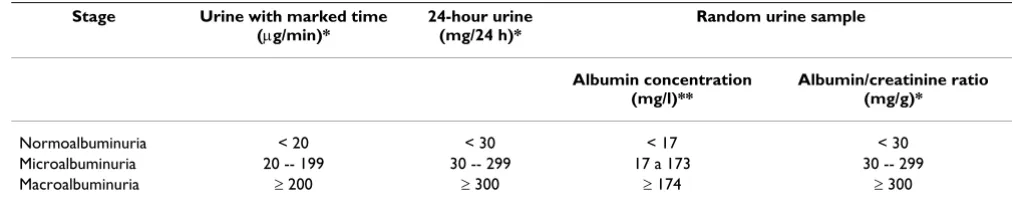
According to UAE values, DN has been didactically cate-gorized into stages. The cutoff values used [14] to charac-terize these stages are described in Table 1.
Although microalbuminuria is considered a risk factor for the development of macroalbuminuria, not all patients progress to this stage, and some may regress to nor-moalbuminuria [15,16]. The initial studies suggested that about 80% of type 1 diabetic patients with microalbu-minuria would progress to proteinuria over a period of 6 to 14 years [4-6]. More recent studies suggest that only 30 to 45% of microalbuminuric patients will progress to pro-teinuria over 10 years of follow-up [15]. In fact, some of them will present regression to normoalbuminuria. This might be the result of more intensive glucose and BP con-trol strategies employed in the last decade than in the ini-tial studies. This regression of microalbuminuria is more frequent among subjects with short duration of micro-albuminuria, glicohemoglobin A1c (HbA1c) below 8%, systolic BP <115 mm Hg, and favorable lipid profile (serum total cholesterol <198 mg/dl and triglycerides <145 mg/dl). Independent of the role as a prognostic fac-tor for macroalbuminuria, the presence of microalbu-minuria, reflecting a state of generalized endothelial dysfunction, is a risk factor for cardiovascular disease and mortality [17,18].
The cut-off values of urinary albumin to define the stages of DN are arbitrary (Table 1). On the one hand, not all
Table 1: Diabetic nephropathy stages based on urinary albumin excretion
Stage Urine with marked time (μg/min)*
24-hour urine (mg/24 h)*
Random urine sample
Albumin concentration (mg/l)**
Albumin/creatinine ratio (mg/g)*
Normoalbuminuria < 20 < 30 < 17 < 30 Microalbuminuria 20 -- 199 30 -- 299 17 a 173 30 -- 299 Macroalbuminuria ≥ 200 ≥ 300 ≥ 174 ≥ 300
subjects will progress to overt DN, and some might even regress as stated before. On the other hand subjects in the upper-normal range of albuminuria seem to be at high risk for complications. In patients with type 2 DM, the progression to micro- or macroalbuminuria is more fre-quent in individuals whose baseline UAE was normal but above 2.5 mg/24 h [19]. Furthermore, in another study after 10 years of follow-up, patients with type 2 DM and UAE values above 10 μg/min were at 29 times higher risk of developing DN [20]. Similar results were observed in patients with type 1 DM [21]. Another interesting obser-vation is that patients with type 2 DM and UAE in the upper-normal range had higher BP than normoalbuminu-ric patients in the lower UAE range [22]. This favors the concept that UAE is a continuum similar to what has been demonstrated for BP and cholesterol levels.
In the microalbuminuric stage, no decline in GFR is expected. Once the subject has developed macroalbu-minuria, the expected GFR decline is 1.2 ml/min/month in type 1 DM [23]. This could be decreased by BP treat-ment. In type 2 DM, the rate of GFR decline is less predict-able. A mean decline of approximately 0.5 ml/min/ month [24] has been described, but in some patients GFR may remain stable for long periods of time [25]. The greater GFR decline is associated with more advanced dia-betic glomerulopathy and worse metabolic control [26].
Screening and Diagnosis
The first step in screening for DN is to measure albumin in an isolated urine sample [27]. The results of albuminu-ria in an isolated sample can be expressed as albumin con-centration (mg/l) or as albumin/creatinine ratio (mg/g) [28]. Although albumin concentration may be influenced by urine dilution/concentration, this measure appears to be the best choice, considering its cost and accuracy [29]. Every abnormal albuminuria test should be confirmed in two of three samples collected at a three to six-months interval, due to the daily variability of UAE [30]. Screening should not be performed under conditions that may increase UAE, such as hematuria, acute systemic diseases or fever, vigorous physical exercise, poor glycemic control, uncontrolled arterial hypertension and decompensated cardiac failure [31]. Bacteriuria had also been considered a factor that could interfere in the measure of urinary albumin [31-33]; but in a recent study this finding was not confirmed, suggesting that it is not necessary to exclude bacteriuria to measure albuminuria [34].
In situations in which UAE measurement is not available, semiquantitative dipstick measurements of albuminuria (for instance: Micral® Test II) can be used, although these
tests are less accurate [29].
The quantitative methods most commonly used to meas-ure albuminuria are immunoturbidimetry, immunon-ephelometry and radioimmunoassay. However, recently it has been observed that an appreciable quantity of albu-min is not detected by routine immunoassay methods, defined as non-immunoreactive fraction, which results in an underestimate of UAE [35-37]. On the other hand the HPLC (high performance liquid chromatography) method measures the immunoreactive and non-immunoreactive fractions which compose the total intact albumin, allow-ing the detection of earlier albumin elevations [37,38]. However, this method can overestimate UAE as observed in some community studies, possibly due to the fact that albumin peaks measured using this method can be con-founded with other proteins [39]. Therefore, the signifi-cance of total intact albumin, both to diagnose DN, and for its association with cardiovascular disease has not yet been well established.
DN screening must be performed when DM is diagnosed in patients with type 2 DM, since these individuals may have had a silent form of DM for some time already. For patients with type 1 DM, it is recommended that screen-ing be performed beginnscreen-ing in the fifth year after DM diagnosis or earlier if the DM is chronically poorly com-pensated, or if the patient is an adolescent. In all cases, if albuminuria is normal, screening must be repeated annu-ally [27].
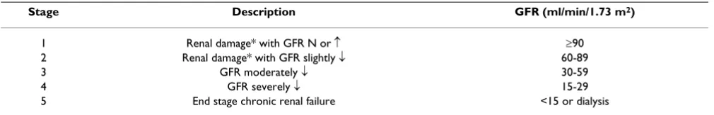
Although the measurement of albuminuria is essential to diagnose DN, there are some patients who present decreased GFR when UAE values are normal. Based on this, the classification of the National Kidney Foundation
can also be used to stage chronic kidney disease in these patients (Table 2) [27]. It is recommended that GFR be routinely estimated for appropriate screening of DN. GFR can be measured using specific techniques such as inulin clearance, 51Cr-EDTA, 125I-iothalamate, and iohexol. However, in clinical practice, GFR is estimated by equa-tions that take into account serum creatinine concentra-tion and some or all of the following variables: age, sex, body weight and race. The equation recommended by the
National Kidney Foundation is that of the study on Modifi-cation of Diet in Renal Disease (MDRD): GFR (ml/min/ l.73 m2) = 186 × [serum creatinine (mg/dl) -1,154 × age
(years) -0,203 × (0.742 if a woman) × (1.21 if
African-Amer-ican)]. If the creatinine measurement method is cali-brated, the formula should use factor "175" in place of the value "186". The Cockroft-Gault formula, creatinine depuration (ml/min) = [140 - age (years)] × weight (kg)/ [72 × serum creatinine (mg/dl)] × 0.85 (if a woman) is less precise [40]. The reference values of GFR for young individuals are 90 to 130 ml/min/l.73 m2, with the
Serum creatinine concentration should not be used as an isolated index for the evaluation of renal function, since its measure is affected by other factors besides GFR itself, such as tubular secretion, extrarenal generation and pro-tein ingestion.
Special Situations
Patients with micro- or macroalbuminuria, after the con-firmation of diagnosis (2 measurements), should undergo a complete evaluation concerning differential diagnosis and assessment of renal function. DN is associated with several other conditions that need to be addressed making the management of these patients very complex. However this is not within the scope of the present manuscript and a detailed approach could be found in a recent review [30].
Diabetic patients can have other kidney diseases. The dif-ferential diagnosis is usually based on the history, physi-cal examination, laboratory evaluation and kidney imaging. Renal biopsy has only been recommended in special situations. In the presence of micro or microalbu-minuria and diabetic retinopathy in a patient with long term DM (e.g. >10 years) the assumption that DM is caus-ing the renal disease is generally correct. Conversely, in diabetic patients with type 2 DM with a fast increment in albuminuria and in patients with type 1 DM where mac-roalbuminuria develop in the absence of diabetic retinop-athy a differential diagnosis should be carried out. However, in type 2 diabetic patients the time of DM onset is usually unknown and retinopathy could be absent in a significant proportion (28%) of patients with albuminu-ria [42]. In summary, absent retinopathy, short duration of DM and faster decline in GFR and/or albuminuria increment are indications to suspect of nondiabetic renal disease [43]. If after a non-invasive evaluation the diagno-sis is still unclear, a kidney biopsy should be discussed. In type 2 DM the prevalence of nondiabetic renal disease could vary from 12 to 38% [42,44,45]. All the kidney biopsy data are derived from retrospective studies. The dif-ferences in the prevalence of non-diabetic lesion observed in the studies probably reflect different criteria used to indicate renal biopsies. In one study, subjects with type 2 DM, gross proteinuria (>1 g) without retinopathy, and
hematuria and no retinopathy, 19% of patients with con-firmed DN had another glomerulopathy associated [45]. In this study, patients without diabetic glomerulosclerosis had a better prognosis than those with diabetic glomeru-losclerosis [45]. Another aspect that needs to be addressed is that it is not clear if there is additional benefit of detect-ing other nephropathies in the management of these patients.
Risk Factors
The two main risk factors for DN are hyperglycemia and arterial hypertension. However, DN develops in only about 40% of patients, even in the presence of hyperglyc-emia and elevated BP for long periods of time. This obser-vation raised the concept that DN will develop only in a susceptible subset of patients [46-48]. Furthermore, fam-ily studies have confirmed a genetic contribution for the development of DN in both type 1 and type 2 DM [49-53]. Once DN is present, progression factors may act, favoring evolution to more advanced stages. There is evi-dence that some factors involved in the development of proteinuria are also common to the loss of GFR, but oth-ers are unique to each one of them [54].
Hyperglycemia
Hyperglycemia is a significant risk factor for the develop-ment of microalbuminuria, both in type 1 and in type 2 DM [21,55,56]. A reduction of 1% in HbA1c is associated with a 37% decrease in microvascular endpoints [57]. In the presence of micro- and macroalbuminuria the role of metabolic control is less defined, even though some stud-ies showed a deleterious effect of high glucose levels on GFR [58,59]. Moreover, it was demonstrated that pancreas transplantation reversed renal damage in type 1 DM patients with mild to advanced DN lesions [60]. Recently a large trial also reinforced the importance of intensive treatment of DM to decrease the microvascular complica-tions [61].
Arterial Hypertension
Arterial hypertension is a main risk factor for the develop-ment of DN [56,62], and probably the best known rele-vant factor related to its progression. Analysis of UKPDS showed that every 10 mmHg reduction in systolic BP is
Table 2: Chronic kidney disease stages
Stage Description GFR (ml/min/1.73 m2)
1 Renal damage* with GFR N or ↑ ≥90 2 Renal damage* with GFR slightly ↓ 60-89
3 GFR moderately ↓ 30-59
4 GFR severely ↓ 15-29
5 End stage chronic renal failure <15 or dialysis
associated with a 13% reduction in the risk of microvascu-lar complications, with the smallest risk among those patients with systolic BP <120 mm Hg [63].
Smoking
Smoking is a risk factor for DN [19,56] and might contrib-ute to its progression [64]. Although some studies did not confirm these observations [55,59,65], it is strongly rec-ommended to quit smoking in any phase of DN, also aim-ing to reduce the associated cardiovascular and cancer risk.
Dyslipidemia
In type 2 DM, elevated serum cholesterol is a risk factor for the development of DN [55,56]. In type 1 DM patients increased serum triglycerides, total and LDL-cholesterol were associated with micro- and macroalbuminuria [66,67]. High serum cholesterol also seems to be a risk factor for GFR loss in macroalbuminuric type 1 diabetic subjects [68].
Proteinuria
Proteinuria itself could lead to progression of DN. Pro-teinuria >2 g/24 h is associated with a greater risk of ESRD [69]. Increased leakage of albumin may induce glomeru-lar damage probably through activation of inflammatory cascades [70]. This would be a reason to target decreased urinary albumin excretion in DN treatment.
Glomerular hyperfiltration
Elevated GFR values are present in about one third of type 2 DM patients [71,72] and theoretically it could cause DN due to glomerular damage [73]. Studies led to controver-sial findings regarding its role as a risk factor for the devel-opment of DN [20,71,74]. Type 2 DM patients with a single-kidney more often present increased UAE levels [75,76]. On the other hand, type 1 DM patients with only one kidney do not have a more aggressive disease [77]. Glomerular hyperfiltration probably plays a small role, if any, in the development of DN [78].
Dietary factors
Increased dietary protein intake seems to be associated with the presence of higher UAE values, at least in patients with type 1 DM [79]. In patients with type 2 DM this asso-ciation has not been documented. The source of proteins in the diet also seems to be related to the presence of DN. A higher intake of fish protein is related to a lower risk of microalbuminuria in type 1 DM patients [80]. The mech-anisms involved in these findings are unknown but prob-ably related to hemodynamic factors [81].
Regarding the dietary lipid content, an association has been observed between the higher intake of saturated fat and the presence of microalbuminuria in patients with
type 1 DM [82]. In patients with type 2 DM, very recently, it was observed that the presence of microalbuminuria was associated with the lower content of polyunsaturated fatty acids, especially those of vegetal origin [83]. In a study performed with patients with type 1 and type 2 DM, followed for 6 years, it was also demonstrated that those who evolved with regression of the DN presented a higher intake of polyunsaturated fatty acids and a lower intake of saturated fatty acids [84].
Genetic risk factors
The exact genetic model underlying DN susceptibility is uncertain, but theoretically few genes with a major contri-bution and some with minor interaction with the envi-ronment could cause DN [47,48]. Unfortunately, no gene with a major effect had been identified so far. The knowl-edge of which gene(s) predisposes to DN will allow the identification of patients at high risk for this complica-tion, and adoption of preventive measures.
In genetic studies the clear definition of the phenotype, DN, is very important. DN could be defined by different parameters: for instance, the presence of microalbuminu-ria, macroalbuminumicroalbuminu-ria, ESRD or decreased GFR. Some genes probably are involved in the development of pro-teinuria, others with decline in GFR and some will be involved in both situations [47,48]. Therefore, a more comprehensive definition of DN used in the genetic stud-ies is important to make the results more comparable.
A familial aggregation of DN has been demonstrated in studies of sibling-pairs [49,53,85], parent-offspring pairs or studies of extended families [51,52]. One practical application of the studies with diabetic siblings is that the chance of having DN increases 2-3 times if the subject's sibling has DN when compared to the subject who has a normoalbuminuric sibling, either in type 1 or type 2 dia-betes [49,53].
Recent advances in technology make easier to look for regions in whole genome linked to different DN pheno-types [86-88]. This approach identified regions and puta-tive genes not previously known to be associated with DN and it could raise new candidate genes. Moreover, new tar-gets for drug development may come into sight, since some of the genes found are novel and have not been pre-viously implicated in the pathogenesis of DN.
homeosta-sis, and in insulin sensitivity, have been considered candidates for the development of DN [9,30,89]. How-ever, the studies have not been successful in identifying genes that consistently show an association with DN. Rep-lication studies have demonstrated conflicting results [90]. The evaluation of 360 thousand polymorphisms in patients with type 1 DM, with and without DN, showed a total of 13 polymorphisms located at 4 loci in two inde-pendent cohorts of subjects strongly associated with the presence of DN [87]. Some of these polymorphisms are located in genes highly expressed in the kidney with DN, and its development over time [87].
Another approach that has been used to investigate the genetics of DN involves the study of microRNAs role on this process. These are non-encoding short RNAs that induce post-transcriptional protein modifications. Little is known about these molecules and their role in DN. In a study, microRNA mirR-192 expression was increased in the glomeruli of rodents with DM [91]. Their induction by TGF-β in mesangial cells caused increased collagen syn-thesis and suggests that this type of molecule may be implicated in the development of DN, opening up a new prospect of research in elucidating the pathogenesis of this DM complication. The replication of this finding and this type of approach must be better explored in studied conducted in human beings.
As previously stated, Brazilians of African descent have more aggressive renal disease than people of European ancestry [13]. This could be due to several reasons, such as the presence of different risk factors, different access to medical attention, and socioeconomic differences. How-ever, none of the assessed known risk factors were differ-ent between African and Europeans [13,92] make unclear an explanation for the different rates of DN between black and white subjects. Unfortunately, data on socioeco-nomic status were unavailable. An alternative explanation for this observation, but hard to prove, would be a differ-ent genetic susceptibility.
Pathology
DN in individuals with type 1 DM is initially character-ized by a thickening of the glomerular and tubular basal membrane, with progressive mesangial expansion leading to the progressive reduction of the glomerular filtration surface [93]. Concurrent interstitial morphological altera-tions also occur, as well as hyalinization of the afferent and efferent glomerular arterioles [93]. Mesangial expan-sion can be diffuse (diabetic glomerulosclerosis) or with areas of marked mesangial expansion, forming roundish and fibrillar zones, with nuclei in palisade (nodular glomerulosclerosis, Kimmelstiel-Wilson nodes). While mesangial expansion is the critical lesion which leads to progression to loss of renal function, damage to the
tubu-lar glomerutubu-lar junction, to the tubules and to the inter-stice determines progression to ESRD [87,91].
Podocytes damage also appears to be involved in the glomerulosclerosis process. In a study conducted in Pima Indians, highly susceptible to developing DN, a smaller number of podocytes per glomerulus was the greatest pre-dictor of increased UAE and progression to clinical DN [93]. When this finding was present, normoalbuminuric individuals had a higher risk of progressing to renal dis-ease than those who did not have a podocyte lesion [93]. In addition, nephrine, a protein synthesized by the podo-cyte and considered vital to the stability of the glomerular barrier, has its expression reduced in DN [94]. The admin-istration of ACE inhibitors results in the expression of nephrine at levels similar to those of individuals with DM without DN [95].
In a subgroup of patients with DM, loss of renal function precedes the development of microalbuminuria. This group presents more advanced glomerular lesions than those that present microalbuminuria [93].
Renal lesions in individuals with type 2 DM are more complex than in individuals with type 1 DM. The preva-lence of a renal lesion that is non-typical for DM in indi-viduals with type 2 DM is high, reaching 10 - 30% of subjects with proteinuria [91,93]. In a minority, the his-topathological aspects are similar to the typical lesion of subjects with type 1 DM. The rest presents only mild or absent DN, with or without tubulointerstitial alterations, arteriolar alterations or diffuse glomerulosclerosis [93]. The tubulopathy is possibly related to persistent hypergly-cemia and changes related to age, atherosclerosis and arte-rial hypertension [91]. Despite the heterogeneity of the lesions and the impact of diseases such as arterial hyper-tension on individuals with type 2 DM, in a large cohort of individuals with type 2 DM, the severity of the lesions was correlated with the progression of DN and the veloc-ity of GFR loss [26].
Pathophysiological Mechanisms Hemodynamic factors
perfusion makes it easier for albumin to leak from capil-laries to renal glomerulus, and leads to compensatory increase of mesangial matrix, thickening of the glomerular basement membrane and podocyte damage. Albuminuria also activates a series of inflammatory pathways through tubular cells and feeds this process [91]. In addition, the mechanical stress resulting from renal hyperperfusion induces the release of cytokines (TNF-α), growth factors (VEGF, TGF-β1), cholesterol and local triglycerides that induce the accumulation of proteins from extracellular matrix, leading to mesangial expansion and glomerulo-sclerosis. A reduction of TGF-β1 by blocking the renin-angiotensin-aldosterone system retards the progression of DN and preserves glomerular morphology [99].
Hyperglycemia and advanced products of non-enzymatic glycosilation
Persistent hyperglycemia is a strong risk factor for DN and causes the proliferation of mesangial cells and their matrix, as well as the thickening of the basement mem-brane. Hyperglycemia increases the expression of vascular endothelial growth factor (VEGF) in podocytes causing increased vascular permeability. Hyperglycemia also increases the generation of advanced products of non-enzymatic glycosilation of proteins through activation of aldol reductase pathway and protein kinase C (PKC). The final products of non-enzymatic glycosilation are bound to collagen and proteins that constitute the glomerular basement membrane and make the glomerular barrier more permeable to the passage of proteins, resulting in increased UAE [94,100-104].
Cytokines
A series of circulating markers of inflammation such as C reactive protein and interleukin 1, 6 and 18, and tumor necrosis factor are increased in DN and their levels corre-late with albuminuria and progression to ESRD. In addi-tion, hyperglycemia, TGF-β1 and angiotensin II stimulate the secretion of VEGF, causing the production of endothe-lial nitric oxide, vasodilation and glomerular hyperfiltra-tion [94]. Hyperglycemia, possibly mediated by oxidative stress, also induces angiotensin II to the synthesis of TGF-β, type IV collagen and fibronectin, contributing to pro-gressive glomeruloesclerosis [87].
Inflammatory factors are also involved in the develop-ment of tubulointerstitial lesion, and appear to lead to accumulation of macrophages in the tubular interstice in animal models designed to study DN. Macrophages also produce free radicals, inflammatory cytokines and pro-teases that induce tubular damage [91]. Furthermore, glomerular and renal cells also produce a series of inflam-matory factors when they are exposed to glomerular hyperfiltration and increased UAE, intensifying this proc-ess [91].
Treatment
The principles of prevention and treatment of DN are the same. However, the role of each factor could be different in each stage of disease. It is important to define the DN stage that is the target of intervention (microalbuminuria, proteinuria or GFR) and the outcome of interest. Two recent meta-analyses have demonstrated different results when evaluating different outcomes, such as proteinuria, GFR decline or progression to ESRD [105,106]. Both, ACE inhibitors and angiotensin receptor blockers (ARBs) seem to be effective reducing proteinuria and decreasing the cre-atinine doubling rate, but not decreasing mortality [106]. Probably the best treatment is a multiple risk factor inter-ventional approach, but due to a practical point of view each aspect will be addressed individually. The goal to be pursued is retarding the development or progression of DN and to decrease the subject's cardiovascular risk and mortality.
In normo- or microalbuminuric subjects, the aim of treat-ment is to intervene at arterial hypertension, hyperglyc-emia, smoking habit and probably dyslipidemia. Even in the absence of clear data showing that the management of these risk factors individually is beneficial to DN, they are also risk factors for cardiovascular disease and should be aggressively treated [30].
Clinical trials have demonstrated that intensive treatment of hyperglycemia is associated with a decreased risk for the development of DN in type 1 and type 2 diabetic patients [107-110]. In type 1 and type 2 subjects the effect of intensive therapy could be seen many years later [108,111]. The effect of the intervention in hyperglycemia in type 1 macroalbuminuric subjects is not so clear [107,112,113]. This became more evident in the EDIC/ DCCT follow up study [108]. In the Kumamoto study, prevention of macroalbuminuria was observed in type 2 DM patients intensively treated [110].
val-ues [114] and a minor effect on albuminuria levels was observed [114].
Treatment of hypertension leads to an important risk reduction in cardiovascular and microvascular events. In the UKPDS, a reduction from 154 to 144 mm Hg on systo-lic BP reduced the risk for the development of microalbu-minuria by 29% [115]. BP targets for patients with DM are lower (130/80 mm Hg) than those for patients without DM [116]. In the Hypertension Optimal Treatment (HOT) study a reduction of diastolic BP from 85 to 81 mm Hg resulted in 50% reduction in the risk of cardiovas-cular events in diabetic but not in non-diabetic patients [117]. In the presence of microalbuminuria the treatment of hypertension, irrespective of the agent used, produced a beneficial effect on albuminuria [118].
Aggressive treatment of hypertension should be estab-lished in subjects with DM. A discussion of agents used to treat hypertension in patients with DN are beyond the scope of this manuscript, and recent guidelines [108,116] and excellent reviews in this subject are available [30,119,120].
In order to reach the BP goal of 130/80 mmHg in diabetic patients in general [116] or 125/75 mmHg in patients with proteinuria >1.0 g/24 h and increased serum creati-nine, three to four antihypertensive agents are usually nec-essary [121].
The choice of anti-hypertensive agents to use is in some way not a problem in clinical practice, because to reach the BP goals the majority of patients will need several agents. However, due to the known renoprotective effect of ACE inhibitors and ARB, these agents (see below) should be used initially associated with a diuretic.
Renin-Angiotensin System (RAS) Blockade
ACE inhibitors could be used in normotensive subjects to prevent or postpone the development of microalbuminu-ria [122]. The aim of ACE inhibitors and ARBs use is not only to diminish the risk for the development of micro-and macroalbuminuria [123-125] but also to decrease the occurrence of cardiovascular events [124]. However, a recent 5 year multicenter randomized controlled trial involving 285 normoalbuminuric, normotensive patients with type 1 DM failed to show any improvement in biopsy parameters with losartan (100 mg daily) or enal-april (20 mg daily) compared to placebo [126]. Surpris-ingly, the 5-year cumulative incidence of microalbuminuria was 17% with losartan, significantly greater than with placebo (6%, P = 0.01). The enalapril group had a similar incidence of microalbuminuria (4%, P = 0.96) in comparison to the placebo group [126].
RAS blockade with ACE inhibitors or ARB confers an addi-tional benefit on renal function. This renoprotective effect is independent of BP reduction [118,127]. These drugs decrease UAE and the rate of progression from microalbu-minuria to more advanced stages of DN. A meta-analysis of 12 trials in non-hypertensive microalbuminuric type 1 diabetic patients showed that ACE inhibitors decreased the risk of progression to macroalbuminuria by 60%, and increased the chances of regression to normoalbuminuria [128]. Therefore, the use of ACE inhibitors or ARB is rec-ommended for all microalbuminuric patients, even if nor-motensive [14]. ARBs were also effective in reducing the development of macroalbuminuria in microalbuminuric type 2 diabetic patients [127,129].
The aggressive treatment of hypertension has a strong beneficial effect in reducing GFR decline in proteinuric type 1 diabetic patients [130]. This reduction in GFR decline was predicted by reduction in albuminuria [131]. According to the MDRD trial, the lower the BP the greater the preservation of renal function in non-diabetic patients [132]. Patients with proteinuria >1 g/day and renal insuf-ficiency had a slower decline in renal function when BP was <125/75 mm Hg [132]. Addition of ACE inhibitors in proteinuric type 1 [133] or ARB in macroalbuminuric type 2 [134,135] diabetic patients has a beneficial effect in decreasing proteinuria and reducing renal function decline. The effect of ARBs on protein excretion could be noted within 7 days after starting the treatment, and may persist after [136]. It seems to be independent of BP reduc-tion [127] and has a dose response effect beyond the doses needed to control BP [137]. An acute increase in serum creatinine up to 30 to 35% that stabilizes within 2 months might occur and it is not a reason to stop the treat-ment [138]. Increase in serum creatinine above these val-ues should raise the possibility of renal-artery stenosis [138,139]. Another limitation to the use of ACE inhibitors is hyperkalemia, especially among those with renal insuf-ficiency [138]. Acute hyperkalemia (>5.5 mEqL) is an indication to stop these medications. Therefore, albu-minuria, serum creatinine and potassium should be checked monthly in the first 2 to 3 months after starting treatment with ACE inhibitors or ARB [138,139].
the association of the two classes of drugs had a major effect on decreasing proteinuria but not on GFR decline or mortality [142]. In fact, a worse effect on GFR and mortal-ity was observed. Analyzing the subgroups, the increased mortality came from the less sick subjects. Among diabetic subjects no increased mortality was observed, but also no benefit from the dual blockage was observed [142]. The VA NEPRHON-D study aimed to evaluate this issue is patients with type 2 DM [143].
Another step that has been proposed to be blocked is the aldosterone action. Adding the aldosterone antagonist -spironolactone - to ARBs [144] or ACE inhibitor [145] is also more effective in reducing UAE and BP in type 2 dia-betic patients than each drug alone. A recent meta-analy-ses that included diabetic and non-diabetic subjects demonstrated that the addition of aldosterone antago-nists in patients already on ACE inhibitors and ARBs reduces proteinuria in chronic kidney disease [146]. This was not associated with an improvement on GFR, but increases the risk of hyperkalemia. Long-term effects of these agents on renal outcomes, mortality, and safety need to be determined [146].
More recently, the dual blockage of the renin-angiotensin-aldosterone system with aliskiren, a direct renin inhibitor, and losartan at maximal recommended dose (100 mg daily) showed a greater reduction in proteinuria (20%) compared to losartan and placebo [147]. The effect does not seem to be due to anti-hypertensive effect. However, this was a short duration study (12 weeks) and long term studies are needed. The ongoing trial ALTITUDE might answer some of these questions [148]. This placebo con-trolled, randomized trial intends to follow-up about 8600 subjects during two years and compare the effect of aliskiren added to standard treatment (ACE or ARBs) [148]. The results will be available by 2012.
Hyperglycemia treatment peculiarities
The treatment of DM is not the aim of the present review, but a few special remarks could be made regarding the treatment of hyperglycemia in a patient with renal disease (Table 3).
Metformin is the standard therapy for patients with type 2 DM and will only be briefly discussed here. Metformin is contraindicated when serum creatinine is above 1.5 mg/dl in men and 1.4 mg/dl in women due to the increased risk of lactic acidosis [149]. However, these values are being questioned [150]. In these creatinine ranges, some sub-jects will be using metformin on chronic renal disease stages II and III [151].
Sulfonylureas and their metabolites, except glimepiride, are eliminated via renal excretion and should be used with
caution in patients with GFR [152]. Glibenclamide is a potent drug and has been known for a long time. It is low cost and available in the public health system. However, it presents a high risk of hypoglycemia. It has active metabolites that increase in patients with decreased GFR, and its pharmacological action is such that the use of glib-enclamide is not recommended from stage 3 onwards [153,154]. Among the sulfonylureas there is also glipizide which carries a lower risk of hypoglycemia being and alternative on this situation [154]. Glipizide can be used in chronic renal disease stages 3 and 4. It could still be used in stage 5, with a therapeutic adjustment. Glimepir-ide is a third generation sulfonylurea with a slightly higher cost and a lower risk of hypoglycemia. However, it is believed that it has a few active metabolites filtered by the kidneys what could be potentially related to higher risk of hypoglycemia compared to glipizide.
Repaglinide [155] and nateglinide [156] have a short duration of action, are excreted independently of renal function and have a safety profile in patients with renal impairment. These drugs, like the sulfonylureas, are insu-lin secretagogues, but they act in different cellular mem-brane channels, and this brings some pharmacological properties such as quick initial action, non-prolonged action and greater effect on post-prandial glycemia. A flex-ible aspect of this drugs making therapeutic management easier is the lower risk of hypoglycemia because of the dif-ferent connection of the membrane channels. But one side effect described similar to observed with the sulfony-lureas is weight gain. Its cost is higher than that of sulfo-nylureas, but theoretically it has a less deleterious effect on beta cells. Among the glinides, the first choice would be repaglinide because of the low risk of hypoglycemia, and it can be used in stage 3 and stage 4 [155,157]. Data in the literature are not sufficient to indicate the use of this drug in chronic renal disease stage 5. Nateglinide would be at a disadvantage because it is less potent, and it has active metabolites that can increase the risk of hypoglyc-emia in subjects with decreased GFR [158,159].
Table 3: Treatment of hyperglycemia in the patient with type 2 diabetes mellitus and chronic kidney disease
Stage of Renal Disease Clearance Reduction of
HbA1c
Risk of hypoglycemia
III IV V
Glibenclamide [153,154]
Hepatic metabolism: 100%.
Excretion: bile and feces 50% and urine 50%
-1.5% High (active metabolites)
Avoid Avoid Avoid
Glipizide [154] Excretion: metabolites 90% in urine and feces. 10% excreted without metabolization
-1.5% Low Can be used Can be used Can be used (adjustments)
Glimepyride Hepatic metabolism 100%.
Excretion: urine 60% and feces 40%
-1.5% Low Can be used Can be used Use with care
Repaglinide [155,157]
Hepatic metabolism: 100%.
Excretion: 10% urine and 90% feces
-1.0% Low Can be used Can be used Use with care. Adjust dose
Nateglinide [156,158,159]
Hepatic metabolism: 85%.
Excretion: urine 83% and feces 10%. 15% excreted inactive in urine
-0.7% High (active metabolites)
Use with care Use with care Avoid if possible
Acarbose* [153,160]
Excretion: urine 34%, feces 51% and <2% in urine in the free or active metabolic form
-0.6% Low Can be used Can be used Avoid
Rosiglitazone [162] Hepatic metabolism and excretion in the urine, of rather inactive metabolites in the urine 64% and feces 23%
-0.6 to 1.5% Low Can be used Can be used Can be used
Pioglitazone [162] Hepatic metabolism and excretion in urine of rather inactive metabolites in the urine 15% and feces 85%
-0.6 to 1.5% Low Can be used Can be used Can be used
Sitaglipitine [171,172]
Excretion: urine 87% and feces 13%, in an unaltered form.
-0.7% Low Can be used Can be used. Reduce dose 50%
Can be used. Reduce dose 75%
Vildagliptine Excretion: urine: 85% and feces 15%.
-0.7% Low Can be used Can be used Not recommended
Exanetide [173] Metabolism and renal excretion
than 2 mg/dl. It could be considered up to stage 3, and it should be avoided in stages 4 and 5 [153,160].
Glitazones, represented by rosiglitazone and pioglita-zone, act through the PPR gamma system and are insulin sensitizer drugs that increase the muscle uptake of glucose and diminish the atherogenic profile of the DM patient, and could be used in renal failure [161,162]. Rosiglita-zone has been shown to decrease UAE in type 2 diabetic patients as compared to glyburide, suggesting a beneficial effect in the prevention of renal complications of type 2 DM [163]. This antiproteinuric effect occurs also in nondiabetic disease [161,164,165]. The side effects include anemia, water retention, weight gain and poten-tial hepatotoxicity due to the accumulation of its metabo-lites. Recently, cardiovascular safety and the risk of increased incidence of fractures have been discussed [166,167]. Both would present a low risk of hypoglycemia and could theoretically be used in the different stages of chronic renal disease without adjusting the dose [168].
A recent meta-analysis suggests beneficial effects of glita-zones, with improvement of dyslipidemia in DM, internal carotid intima layer thickness reduction, improved fibri-nolysis, and a direct action of the PPR gamma system at glomerular, tubular and vascular levels [169]. In theory, all these actions (hemodynamic, inflammatory, anti-proliferative and metabolic) would be beneficial actions in nephropathy [169].
In relation to fractures, a recent meta-analysis showed that in the female population there has been up to two-fold increase in the incidence of fractures including both hip and vertebral fractures [170]. Since a uremic patient has already an increased osteometabolic risk, a drug that would increase the incidence of fractures should be ques-tioned in these patients.
Two representatives of the DPP-4 inhibitors are available, vildagliptin and sitagliptin. These drugs inhibit the dipep-tidyl peptidase-4 enzyme which, in turn, prevents degra-dation of the GLP-1 which remains active longer. Thus they lead to the reduction of fasting and post-prandial gly-cemia, without a risk of hypoglycemia. The gliptins sup-press the high release of glucagon and are neutral as regards weight. The side effects include airway infection and transaminases elevation. The standard dose is 100 mg, orally, in a single daily dose. Sitagliptin secretion occurs mostly in urine and an adjustment in the dose is recommended according to the stage of renal disease: 50 mg for stage 3 and 25 mg for stages 4 and 5 [171,172]. Vil-dagliptin also is predominantly excreted in the urine. It is unnecessary to adjust the dose in patients with mild or moderate renal failure (50 mg orally, bid). The use of vil-dagliptin is not recommended, according to the directions
that accompany medications, in patients with severe renal failure, patients who are already on dialysis or some other renal substitution therapy.
Exenatide is a GLP-1 analog. Subcutaneous applications (beginning at 5 μg bid for 30 days and then 10 μg bid) should be performed up to one hour before meals twice a day. It is a drug that reduces weight, which may be an advantage in managing the diabetic patient. The major side effects are nausea and vomiting, what occasionally an individual cannot tolerate using it. It is metabolized and excreted by the kidneys. It presents a low risk of hypogly-cemia and can be used in stage 3, and it is not recom-mended in stages 4 and 5 due to the increased risk of side effects [173].
However, when the renal function is highly compro-mised, metformin, exenatide and gliptins are contraindi-cated, and insulin secretagogues are usually not very effective, since these patients have low endogenous pro-duction of insulin. Therefore, most patients should be treated with insulin [153]. We should remember that the half life of insulin is changed as soon as the individual begins to have a major renal function impairment. Phar-macokinetics is modified, and the insulin will have a slightly longer profile. This may make it difficult to man-age the day to day situation, in peculiar situations on dif-ferent days, i.e., the individual who is undergoing a dialysis session may feel bad and change his diet on that day. We should be able to rationalize more with the flexi-bility of doses here, which is often rather difficult for the patient and the physician.
These individuals certainly will have a greater propensity to hypoglycemia, so we have to be more careful and remember that hypoglycemia may be one of the compli-cations implicated in the increased cardiovascular mortal-ity of these patients. We should also keep in mind that therapeutic goals should be individualized.
Dietary intervention
There are several modalities for a dietary intervention in DN, whether changing protein content or through the manipulation of lipid content. However, few have their efficacy shown based on long term randomized clinical trials.
months. Besides, in these studies there was no evidence of benefit on hard outcomes such as mortality or risk of end stage chronic renal failure. A randomized controlled clin-ical trial with patients with type 1 DM and DN followed for four years, showed that a diet with a moderate protein restriction (0.9 g/kg/day) was associated with a 76% reduction of the risk of end stage chronic renal failure or death [175].
In patients with type 2 DM this benefit has not been well established. There are few studies with type 2 DM patients addressing this issue, showing no benefit on renal func-tion, probably due to lack of compliance with the diet and short follow-up [176]. A recent meta-analysis performed with eight studies including patients with type 1 and 2 DM showed a benefit of protein restriction on proteinuria reduction, but not on GFR reduction [177]. The American Diabetes Association recommends moderate protein restriction (0.8-1.0 g/kg/day) for patients in the initial stages of DN, and a reduction to 0.8 g/kg/day for patients in a more advanced stage of this complication [27].
Interventions in the dietary lipid content has also been suggested, especially by manipulating the type of meat in the diet. Substituting red meat by chicken meat in the diet over the short term proved be able to reduce UAE, and also the serum levels of total cholesterol, LDL and apoli-poprotein B in patients with type 2 DM and micro and macroalbuminuria [89,178]. Recently it was also observed that the beneficial effect of this dietary interven-tion on renal funcinterven-tion was similar to the use of enalapril for a 12-month period in patients with type 2 DM [179]. This effect is probably related to the lower saturated fat content and greater proportion of polyunsaturated fatty acids, observed in chicken meat compared to red meat. Long term studies are needed to confirm this favorable effect.
Dyslipidemia
The desired target of LDL is <100 mg/dl for patients with DM in general, and <70 mg/dl when cardiovascular dis-ease is present. No data based on a large clinical trial is available showing that the treatment of dyslipidemia is able to prevent the development or progression of DN. In the Heart Protection Study (HPS), sinvastatin, 40 mg, reduced vascular event rates and GFR decline in patients with DM by 25%, independent of baseline cholesterol lev-els. Furthermore the results of the Collaborative Atorvas-tatin Diabetes Study (CARDS) demonstrated a marked reduction in cardiovascular events in DM patients, and at least one additional risk factor for coronary disease, sug-gesting that all DM patients should use statins. A recent publication of CARDS showed a modest beneficial effect of atorvastatin on eGFR, particularly in those with
albu-minuria [180]. However, atorvastatin did not influence albuminuria incidence [180].
Multifactorial intervention
As stated before, probably the best approach to a subject with DN is a multifactorial intervention. However, only one study, with a small number of patients (n = 160) addressed this aspect [181]. In this study the targets were: BP levels <130/80 mm Hg, fasting serum cholesterol <175 mg/dl, fasting serum triglycerides <150 mg/dl, and HbA1c <6.5%. The intervention consisted of a stepwise imple-mentation of lifestyle changes and pharmacological ther-apy including low-fat diet, three to five times a week light-to-moderate exercise program, smoking-cessation course, and prescription of ACE inhibitors or ARB and aspirin. The multiple intervention group had a 61% reduction in the risk of macroalbuminuria, and a 58% and 63% reduc-tion in the risk of retinopathy and autonomic neuropathy, respectively. Most importantly, a 55% reduction in the risk for the development of a composite end-point con-sisting of death from cardiovascular causes, non-fatal myocardial infarction, revascularization procedures, non-fatal stroke and amputation was also associated with the multifactorial intervention. It is important to point out that even among highly motivated subjects only a small number reached the proposed goals. Less than 20% in the intensive arm reached the HbA1c goal and less than 50% the systolic BP goal [181].
Conclusion
Diabetic nephropathy is a chronic complication of DM with a growing incidence. Therefore it is essential to have a better understanding of it, especially in relation to pre-vention and aggressive management to avoid progression to ESRD. Besides, its direct association with cardiovascular complications makes it imperative to perform intensive, early management of the risk factors. The study of DN has evolved a lot as regards its pathophysiology, stages of renal involvement and, especially, the therapeutic instru-ments available. Early detection of DN, the multifactorial approach targeting the main risk factors (hyperglycemia, hypertension, dyslipidemia and smoking), and the use of renoprotective agents such as the drugs that act on the renin-angiotensin-aldosterone system, may delay progres-sion of kidney disease in DM, besides reducing cardiovas-cular mortality.
Abbreviations
ACE: angiotensin convertin enzyme; ARB: angiotensin receptor blocker; BP: blood pressure; DM: diabetes melli-tus; DN: diabetic neprhopathy; ESRD: end-stage renal dis-ease; ET-1: endothelin-1; GFR: glomerular filtration rate;
HbA1c: glicohemoglobin A1c; MDRD: Modification of Diet in Renal Disease; RAS: renin-angiotensin system;
Competing interests
The authors declare that they have no competing interests.
Authors' contributions
All authors contributed in the same way.
Acknowledgements
This study was partially supported by Projeto de Núcleos de Excelência do Ministério de Ciência e Tecnologia, Conselho Nacional de Desenvolvi-mento Científico e Tecnológico (CNPq) and Fundo de Incentivo a Pesquisa e Eventos (FIPE) do Hospital de Clínicas de Porto Alegre.
References
1. U.S. Renal Data System, USRDS 2003 Annual Data Report: Atlas of End-Stage Renal Disease in the United States, National Institute of Health, National Institute of Diabetes and Digestive and Kidney Diseases, Bethesda, MD, 2003. . 2. Bruno RM, Gross JL: Prognostic factors in Brazilian diabetic
patients starting dialysis: a 3.6-year follow-up study. J Diabetes Complications 2000, 14:266-271.
3. Valmadrid CT, Klein R, Moss SE, Klein BE: The risk of cardiovas-cular disease mortality associated with microalbuminuria and gross proteinuria in persons with older-onset diabetes mellitus. Arch Intern Med 2000, 160:1093-1100.
4. Mogensen CE, Christensen CK: Predicting diabetic nephropathy in insulin-dependent patients. N Engl J Med 1984, 311:89-93. 5. Parving HH, Oxenboll B, Svendsen PA, Christiansen JS, Andersen AR:
Early detection of patients at risk of developing diabetic nephropathy. A longitudinal study of urinary albumin excre-tion. Acta Endocrinol (Copenh) 1982, 100:550-555.
6. Viberti GC, Hill RD, Jarrett RJ, Argyropoulos A, Mahmud U, Keen H: Microalbuminuria as a predictor of clinical nephropathy in insulin- dependent diabetes mellitus. Lancet 1982, 1:1430-1432. 7. Mogensen CE: Microalbuminuria predicts clinical proteinuria and early mortality in maturity-onset diabetes. N Engl J Med 1984, 310:356-360.
8. MacIsaac RJ, Tsalamandris C, Panagiotopoulos S, Smith TJ, McNeil KJ, Jerums G: Nonalbuminuric renal insufficiency in type 2 diabe-tes. Diabetes Care 2004, 27:195-200.
9. Caramori ML, Fioretto P, Mauer M: Low glomerular filtration rate in normoalbuminuric type 1 diabetic patients: an indica-tor of more advanced glomerular lesions. Diabetes 2003, 52:1036-1040.
10. Kramer CK, Leitao CB, Pinto LC, Silveiro SP, Gross JL, Canani LH: Clinical and laboratory profile of patients with type 2 diabe-tes with low glomerular filtration rate and normoalbuminu-ria. Diabetes Care 2007, 30:1998-2000.
11. Perkins BA, Krolewski AS: Early nephropathy in type 1 diabetes: the importance of early renal function decline. Curr Opin Neph-rol Hypertens 2009, 18:233-240.
12. Young BA, Maynard C, Boyko EJ: Racial differences in diabetic nephropathy, cardiovascular disease, and mortality in a national population of veterans. Diabetes Care 2003, 26:2392-2399.
13. Gerchman F, Zanatta CM, Burttet LM, Picon PX, Lisboa HR, Silveiro SP, Gross JL, Canani LH: Vascular complications of black patients with type 2 diabetes mellitus in Southern Brazil. Braz J Med Biol Res 2008, 41:668-673.
14. American Diabetes Association: Nephropathy in Diabetes. Diabe-tes Care 2004, 27:S79-83.
15. Caramori ML, Fioretto P, Mauer M: The need for early predictors of diabetic nephropathy risk: is albumin excretion rate suffi-cient? Diabetes 2000, 49:1399-1408.
16. Perkins BA, Ficociello LH, Silva KH, Finkelstein DM, Warram JH, Krolewski AS: Regression of microalbuminuria in type 1 diabe-tes. N Engl J Med 2003, 348:2285-2293.
17. Dinneen SF, Gerstein HC: The association of microalbuminuria and mortality in non-insulin-dependent diabetes mellitus. A systematic overview of the literature. Arch Intern Med 1997, 157:1413-1418.
18. Stehouwer CD, Gall MA, Twisk JW, Knudsen E, Emeis JJ, Parving HH: Increased urinary albumin excretion, endothelial
dysfunc-tion, and chronic low-grade inflammation in type 2 diabetes: progressive, interrelated, and independently associated with risk of death. Diabetes 2002, 51:1157-1165.
19. Forsblom CM, Groop PH, Ekstrand A, Totterman KJ, Sane T, Sal-oranta C, Groop L: Predictors of progression from normoalbu-minuria to microalbunormoalbu-minuria in NIDDM. Diabetes Care 1998, 21:1932-1938.
20. Murussi M, Baglio P, Gross JL, Silveiro SP: Risk factors for micro-albuminuria and macromicro-albuminuria in type 2 diabetic patients: a 9-year follow-up study. Diabetes Care 2002, 25:1101-1103.
21. Predictors of the development of microalbuminuria in patients with Type 1 diabetes mellitus: a seven-year prospec-tive study. The Microalbuminuria Collaboraprospec-tive Study Group. Diabet Med 1999, 16:918-925.
22. Leitao CB, Canani LH, Polson PB, Molon MP, Pinotti AF, Gross JL: Urinary albumin excretion rate is associated with increased ambulatory blood pressure in normoalbuminuric type 2 dia-betic patients. Diabetes Care 2005, 28:1724-1729.
23. Viberti GC, Bilous RW, Mackintosh D, Keen H: Monitoring glomerular function in diabetic nephropathy. A prospective study. Am J Med 1983, 74:256-264.
24. Gall MA, Nielsen FS, Smidt UM, Parving HH: The course of kidney function in type 2 (non-insulin-dependent) diabetic patients with diabetic nephropathy. Diabetologia 1993, 36:1071-1078. 25. Friedman R, Gross JL: Evolution of glomerular filtration rate in
proteinuric NIDDM patients. Diabetes Care 1991, 14:355-359. 26. Nosadini R, Velussi M, Brocco E, Bruseghin M, Abaterusso C, Saller
A, Dalla Vestra M, Carraro A, Bortoloso E, Sambataro M, Barzon I, Frigato F, Muollo B, Chiesura-Corona M, Pacini G, Baggio B, Piarulli F, Sfriso A, Fioretto P: Course of renal function in type 2 diabetic patients with abnormalities of albumin excretion rate. Diabe-tes 2000, 49:476-484.
27. Standards of medical care in diabetes--2009. Diabetes Care 2009, 32(Suppl 1):S13-61.
28. Zelmanovitz T, Gross JL, Oliveira JR, Paggi A, Tatsch M, Azevedo MJ: The receiver operating characteristics curve in the evalua-tion of a random urine specimen as a screening test for dia-betic nephropathy. Diabetes Care 1997, 20:516-519.
29. Incerti J, Zelmanovitz T, Camargo JL, Gross JL, de Azevedo MJ: Eval-uation of tests for microalbuminuria screening in patients with diabetes. Nephrol Dial Transplant 2005, 20:2402-2407. 30. Gross JL, de Azevedo MJ, Silveiro SP, Canani LH, Caramori ML,
Zel-manovitz T: Diabetic nephropathy: diagnosis, prevention, and treatment. Diabetes Care 2005, 28:164-176.
31. Mogensen CE, Vestbo E, Poulsen PL, Christiansen C, Damsgaard EM, Eiskjaer H, Froland A, Hansen KW, Nielsen S, Pedersen MM: Micro-albuminuria and potential confounders. A review and some observations on variability of urinary albumin excretion. Dia-betes Care 1995, 18:572-581.
32. Levey AS, Eckardt KU, Tsukamoto Y, Levin A, Coresh J, Rossert J, De Zeeuw D, Hostetter TH, Lameire N, Eknoyan G: Definition and classification of chronic kidney disease: a position statement from Kidney Disease: Improving Global Outcomes (KDIGO). Kidney Int 2005, 67:2089-2100.
33. Beetham R, Cattell WR: Proteinuria: pathophysiology, signifi-cance and recommendations for measurement in clinical practice. Ann Clin Biochem 1993, 30(Pt 5):425-434.
34. Kramer CK, Camargo J, Ricardo ED, Almeida FK, Canani LH, Gross JL, Azevedo MJ: Does bacteriuria interfere with albuminuria measurements of patients with diabetes? Nephrol Dial Trans-plant 2009, 24:1193-1196.
35. Comper WD, Osicka TM, Jerums G: High prevalence of immuno-unreactive intact albumin in urine of diabetic patients. Am J Kidney Dis 2003, 41:336-342.
36. Brinkman JW, Bakker SJ, Gansevoort RT, Hillege HL, Kema IP, Gans RO, de Jong PE, de Zeeuw D: Which method for quantifying uri-nary albumin excretion gives what outcome? A comparison of immunonephelometry with HPLC. Kidney Int Suppl 2004:S69-75.
37. Comper WD, Osicka TM, Clark M, MacIsaac RJ, Jerums G: Earlier detection of microalbuminuria in diabetic patients using a new urinary albumin assay. Kidney Int 2004, 65:1850-1855. 38. Comper WD, Osicka TM: Detection of urinary albumin. Adv
39. Jerums G, Premaratne E, Panagiotopoulos S, Clarke S, Power DA, MacIsaac RJ: New and old markers of progression of diabetic nephropathy. Diabetes Res Clin Pract 2008, 82(Suppl 1):S30-37. 40. Levey AS, Coresh J, Balk E, Kausz AT, Levin A, Steffes MW, Hogg RJ,
Perrone RD, Lau J, Eknoyan G: National Kidney Foundation practice guidelines for chronic kidney disease: evaluation, classification, and stratification. Ann Intern Med 2003, 139:137-147.
41. Granerus G, Aurell M: Reference values for 51Cr-EDTA clear-ance as a measure of glomerular filtration rate. Scand J Clin Lab Invest 1981, 41:611-616.
42. Christensen PK, Larsen S, Horn T, Olsen S, Parving HH: Renal func-tion and structure in albuminuric type 2 diabetic patients without retinopathy. Nephrol Dial Transplant 2001, 16:2337-2347. 43. Mauer M, Fioretto P, Woredekal Y, Friedman EA: Diabetic Neph-ropathy. In Diseases of the Kidney and Urinary Tract 7th edition. Edited by: Schrier RW. Lippincott Williams & Wilkins; 2001:2083-2116. 44. Huang F, Yang Q, Chen L, Tang S, Liu W, Yu X: Renal pathological
change in patients with type 2 diabetes is not always diabetic nephropathy: a report of 52 cases. Clin Nephrol 2007, 67:293-297.
45. Wong TY, Choi PC, Szeto CC, To KF, Tang NL, Chan AW, Li PK, Lai FM: Renal outcome in type 2 diabetic patients with or with-out coexisting nondiabetic nephropathies. Diabetes Care 2002, 25:900-905.
46. Krolewski AS, Warram JH, Christlieb AR, Busick EJ, Kahn CR: The changing natural history of nephropathy in type I diabetes. Am J Med 1985, 78:785-794.
47. Krolewski AS: Genetics of diabetic nephropathy: evidence for major and minor gene effects. Kidney Int 1999, 55:1582-1596. 48. Krolewski AS, Ng DP, Canani LH, Warram JH: Genetics of diabetic
nephropathy: how far are we from finding susceptibility genes? Adv Nephrol Necker Hosp 2001, 31:295-315.
49. Quinn M, Angelico MC, Warram JH, Krolewski AS: Familial factors determine the development of diabetic nephropathy in patients with IDDM. Diabetologia 1996, 39:940-945.
50. Pettitt DJ, Saad MF, Bennett PH, Nelson RG, Knowler WC: Familial predisposition to renal disease in two generations of Pima Indians with type 2 (non-insulin-dependent) diabetes melli-tus. Diabetologia 1990, 33:438-443.
51. Freedman BI, Tuttle AB, Spray BJ: Familial predisposition to nephropathy in African-Americans with non-insulin-depend-ent diabetes mellitus. Am J Kidney Dis 1995, 25:710-713. 52. Clustering of long-term complications in families with
diabe-tes in the diabediabe-tes control and complications trial. The Dia-betes Control and Complications Trial Research Group. Diabetes 1997, 46:1829-1839.
53. Canani LH, Gerchman F, Gross JL: Familial clustering of diabetic nephropathy in Brazilian type 2 diabetic patients. Diabetes 1999, 48:909-913.
54. Placha G, Canani LH, Warram JH, Krolewski AS: Evidence for dif-ferent susceptibility genes for proteinuria and ESRD in type 2 diabetes. Adv Chronic Kidney Dis 2005, 12:155-169.
55. Gall MA, Hougaard P, Borch-Johnsen K, Parving HH: Risk factors for development of incipient and overt diabetic nephropathy in patients with non-insulin dependent diabetes mellitus: prospective, observational study. BMJ 1997, 314:783-788. 56. Ravid M, Brosh D, Ravid-Safran D, Levy Z, Rachmani R: Main risk
factors for nephropathy in type 2 diabetes mellitus are plasma cholesterol levels, mean blood pressure, and hyperg-lycemia. Arch Intern Med 1998, 158:998-1004.
57. Stratton IM, Adler AI, Neil HA, Matthews DR, Manley SE, Cull CA, Hadden D, Turner RC, Holman RR: Association of glycaemia with macrovascular and microvascular complications of type 2 diabetes (UKPDS 35): prospective observational study. BMJ 2000, 321:405-412.
58. Alaveras AE, Thomas SM, Sagriotis A, Viberti GC: Promoters of progression of diabetic nephropathy: the relative roles of blood glucose and blood pressure control. Nephrol Dial Trans-plant 1997, 12(Suppl 2):71-74.
59. Hovind P, Rossing P, Tarnow L, Parving HH: Smoking and progres-sion of diabetic nephropathy in type 1 diabetes. Diabetes Care 2003, 26:911-916.
60. Fioretto P, Steffes MW, Sutherland DE, Goetz FC, Mauer M: Reversal of lesions of diabetic nephropathy after pancreas transplantation. N Engl J Med 1998, 339:69-75.
61. Patel A, MacMahon S, Chalmers J, Neal B, Billot L, Woodward M, Marre M, Cooper M, Glasziou P, Grobbee D, Hamet P, Harrap S, Hel-ler S, Liu L, Mancia G, Mogensen CE, Pan C, Poulter N, Rodgers A, Williams B, Bompoint S, de Galan BE, Joshi R, Travert F: Intensive blood glucose control and vascular outcomes in patients with type 2 diabetes. N Engl J Med 2008, 358:2560-2572. 62. Park JY, Kim HK, Chung YE, Kim SW, Hong SK, Lee KU: Incidence
and determinants of microalbuminuria in Koreans with type 2 diabetes. Diabetes Care 1998, 21:530-534.
63. Adler AI, Stratton IM, Neil HA, Yudkin JS, Matthews DR, Cull CA, Wright AD, Turner RC, Holman RR: Association of systolic blood pressure with macrovascular and microvascular complica-tions of type 2 diabetes (UKPDS 36): prospective observa-tional study. BMJ 2000, 321:412-419.
64. Sawicki PT, Didjurgeit U, Muhlhauser I, Bender R, Heinemann L, Berger M: Smoking is associated with progression of diabetic nephropathy. Diabetes Care 1994, 17:126-131.
65. Smulders YM, Rakic M, Stehouwer CD, Weijers RN, Slaats EH, Silber-busch J: Determinants of progression of microalbuminuria in patients with NIDDM. A prospective study. Diabetes Care 1997, 20:999-1005.
66. Chaturvedi N, Fuller JH, Taskinen MR: Differing associations of lipid and lipoprotein disturbances with the macrovascular and microvascular complications of type 1 diabetes. Diabetes Care 2001, 24:2071-2077.
67. Jenkins AJ, Lyons TJ, Zheng D, Otvos JD, Lackland DT, McGee D, Garvey WT, Klein RL: Lipoproteins in the DCCT/EDIC cohort: associations with diabetic nephropathy. Kidney Int 2003, 64:817-828.
68. Mulec H, Johnsen SA, Wiklund O, Bjorck S: Cholesterol: a renal risk factor in diabetic nephropathy? Am J Kidney Dis 1993, 22:196-201.
69. Ruggenenti P, Remuzzi G: Nephropathy of type-2 diabetes mel-litus. J Am Soc Nephrol 1998, 9:2157-2169.
70. Remuzzi G, Ruggenenti P, Benigni A: Understanding the nature of renal disease progression. Kidney Int 1997, 51:2-15.
71. Caramori ML, Gross JL, Pecis M, de Azevedo MJ: Glomerular filtra-tion rate, urinary albumin excrefiltra-tion rate, and blood pres-sure changes in normoalbuminuric normotensive type 1 diabetic patients: an 8-year follow-up study. Diabetes Care 1999, 22:1512-1516.
72. Silveiro SP, Friedman R, Gross JL: Glomerular hyperfiltration in NIDDM patients without overt proteinuria. Diabetes Care 1993, 16:115-119.
73. Brenner BM, Lawler EV, Mackenzie HS: The hyperfiltration the-ory: a paradigm shift in nephrology. Kidney Int 1996, 49:1774-1777.
74. Dahlquist G, Stattin EL, Rudberg S: Urinary albumin excretion rate and glomerular filtration rate in the prediction of dia-betic nephropathy; a long-term follow-up study of childhood onset type-1 diabetic patients. Nephrol Dial Transplant 2001, 16:1382-1386.
75. Silveiro SP, da Costa LA, Beck MO, Gross JL: Urinary albumin excretion rate and glomerular filtration rate in single-kidney type 2 diabetic patients. Diabetes Care 1998, 21:1521-1524. 76. Ficociello LH, Perkins BA, Roshan B, Weinberg JM, Aschengrau A,
Warram JH, Krolewski AS: Renal hyperfiltration and the devel-opment of microalbuminuria in type 1 diabetes. Diabetes Care 2009, 32:889-893.
77. Chang S, Caramori ML, Moriya R, Mauer M: Having one kidney does not accelerate the rate of development of diabetic nephropathy lesions in type 1 diabetic patients. Diabetes 2008, 57:1707-1711.
78. Yip JW, Jones SL, Wiseman MJ, Hill C, Viberti G: Glomerular hyperfiltration in the prediction of nephropathy in IDDM: a 10-year follow-up study. Diabetes 1996, 45:1729-1733.
79. Toeller M, Buyken A, Heitkamp G, Bramswig S, Mann J, Milne R, Gries FA, Keen H: Protein intake and urinary albumin excretion rates in the EURODIAB IDDM Complications Study. Diabet-ologia 1997, 40:1219-1226.