Neuropsychiatric Disease and Treatment 2017:13 2691–2703
Neuropsychiatric Disease and Treatment
Dove
press
submit your manuscript | www.dovepress.com 2691
R e v i e w
open access to scientific and medical research
Open Access Full Text Article
Transient global amnesia: current perspectives
David R Spiegel Justin Smith Ryan R wade Nithya Cherukuru Aneel Ursani Yuliya Dobruskina Taylor Crist Robert F Busch Rahim M Dhanani Nicholas Dreyer Department of Psychiatry and Behavioral Sciences, eastern virginia Medical School, Norfolk, vA, USA
Abstract: Transient global amnesia (TGA) is a clinical syndrome characterized by the sudden onset of an extraordinarily large reduction of anterograde and a somewhat milder reduction of retrograde episodic long-term memory. Additionally, executive functions are described as diminished. Although it is suggested that various factors, such as migraine, focal ischemia, venous flow abnormalities, and epileptic phenomena, are involved in the pathophysiology and differential diagnosis of TGA, the factors triggering the emergence of these lesions are still elusive. Recent data suggest that the vulnerability of CA1 neurons to metabolic stress plays a pivotal part in the pathophysiological cascade, leading to an impairment of hippocampal func-tion during TGA. In this review, we discuss clinical aspects, new imaging findings, and recent clinical–epidemiological data with regard to the phenotype, functional anatomy, and putative cellular mechanisms of TGA.
Keywords: transient global amnesia, vascular, migraines, psychiatric
Introduction
More than 50 years after its initial description, transient global amnesia (TGA) remains one of the most enigmatic syndromes in clinical neurology. TGA is defined as a sudden onset of an anterograde and retrograde amnesia that lasts up to 24 hours, although mild subclinical neuropsychological deficits with concomitant vegetative symptoms can last for days after the episode. The memory impairment of patients with acute TGA shows a profound reduction of anterograde and a milder reduction of retrograde episodic memory, including executive functions and recognition. In this review of TGA, we summarize the epidemiology, symptomatology, pathophysiology, assessment, differential diagnosis, longer-term outcome, and possible treatment recommendations
associated with this condition.1
Literature review
Full-text articles and abstracts were selected for this review and were identified through searches of PubMed with the search terms “transient amnesia”, “transient global amnesia”, “hippocampus”, and “amnestic syndromes” between 1985 and 2017. Additionally, more distant seminal articles on this subject matter were included. Only articles published in English were reviewed. The final reference list was chosen on the basis of relevance to the topics covered in this review (eg, their originality, contribution to hippocampal and TGA epidemiology, anatomy and physiology, pathophysiology, diagnosis, and treatment).
Epidemiology
The following prospective and retrospective systematic studies identify TGA cases from either hospitalized or national health registry samples.
Correspondence: David R Spiegel Department of Psychiatry and Behavioral Sciences, eastern virginia Medical School, 825 Fairfax Avenue, Norfolk, vA 23507, USA
email [email protected]
Journal name: Neuropsychiatric Disease and Treatment Article Designation: Review
Year: 2017 Volume: 13
Running head verso: Spiegel et al
Running head recto: Transient global amnesia DOI: http://dx.doi.org/10.2147/NDT.S130710
Neuropsychiatric Disease and Treatment downloaded from https://www.dovepress.com/ by 118.70.13.36 on 25-Aug-2020
For personal use only.
Number of times this article has been viewed
This article was published in the following Dove Press journal: Neuropsychiatric Disease and Treatment
Dovepress Spiegel et al
incidence
Recent epidemiological data put the annual incidence of TGA
between 3.4 and 10.4/100,000.2,3
Age and gender
TGA most commonly presents in the seventh decade of life. Across studies, the mean age of an episode ranges from 61
to 67.3 years.2,4–6 In a 2006 study, 96% of subjects with TGA
(n=142) presented between the ages of 51 and 80 years.
Epidemiological studies fail to identify any subjects under the age of 55 years. Several small-scale studies show a slight
female predominance.2 However, a study on 5,097 TGA cases
shows gender distribution to be 50.7% females and 49.3%
males,4 consistent with one previous analysis.5
Recurrence
Depending on the length of follow-up, the annual rate of TGA
recurrence varies from 2.9 to 26.3%.5 A 2005 study with a
7-year follow-up period, which is the longest systematic
follow-up period reported, found a recurrence rate of 8%.6
Another study that recruited subjects over a 10-year period
(n=142), however, with no designated follow-up period after
the first occurrence, found a recurrence rate of 6.3%, if
prob-able episodes were taken into account.5
Risk factors
A migraine history is one of the more notable risk factors associated with developing TGA. In a 2014 population-based
study (n=316,602), migraine patients were significantly more
likely to develop TGA than their matched controls, with the
incidence rate ratio of 2.48.7 Additionally, of the subjects who
developed TGA after the age of 40 years, those with a history of migraine had a significantly younger age of onset (56.6) compared to the control group (61.4). No associations were
found between various migraine subtypes and TGA.7
Cardiovascular risk factors are also well studied in TGA. A retrospective case–control study found age- and
sex-matched control subjects (n=293) to have significantly
decreased odds of having hyperlipidemia and ischemic heart disease when compared with those subjects with TGA
(n=293). Within this same study, 632 transient ischemic
attack (TIA) subjects had greater rates of hypertension, diabetes mellitus, ischemic stroke, and atrial fibrillation when compared with TGA subjects, likely indicating differing risk
factors between TGA and TIA.8 Furthermore, a retrospective
study of 85 TGA subjects revealed that those with history of two episodes of TGA showed a higher frequency of carotid atheromasia and ischemic heart disease than those with a
history of just one episode of TGA. Also, of note is that
cancer diagnosis carries no increased risk of TGA, according to a prospective cohort study with 5,365,608 subjects running
between 2001 and 2009.4
Psychiatric comorbidity
One study compared psychiatric disease in 51 subjects who experienced a TGA to 51 subjects who experienced a TIA. Psychiatric disease was defined as having “a diagnosis of depression or anxiety disorder” or having received “treatment with specific drugs for at least 3 months”. TGA subjects had a significantly higher percentage of psychiatric disease compared to TIA controls (39.2% vs 13.7%, age- and sex-adjusted odds
ratio [OR] =2.86).6 Additionally, a significantly higher
per-centage of TGA subjects (33.3%) reported a family history of psychiatric disease as compared with TIA subjects (13.7%).
In summary, according to community-based studies, the annual incidence of TGA is 5–10/100,000 and 23.5– 32/100,000 for people aged 50 years and older. Peak inci-dence is around the age of 62 years (standard deviation [SD] 10 years). A total of 54%–67% of TGA patients are female. Chances of recurrence are reported variedly from 2.9 to 25%. Other than migraine headaches, there are no definitive risk
factors for the development of TGA.10
Clinical diagnosis
Development of TGA criteria
TGA is a clinical diagnosis. It was first described in 1956 as an “isolated episode of confusion with amnesia” not
other-wise associated with other neurological deficits.11,12 Subjects
were described as becoming repetitious and asking the same questions, although mostly revolved around the memory loss
itself. Fisher and Adams13 coined the term TGA in 1958;
however, it was not until 1964 that they detailed a report of 17 patients with sudden onset anterograde amnesia and
confusion that resolved within a few hours.14
Hodges and Warlow15 later developed criteria for the
clinical syndrome in 1990 (Table 1), and since then, this has been used as the foundation of TGA diagnosis. They
Table 1 Hodges and warlow criteria for TGA Diagnostic criteria of TGA
– Attacks must be witnessed
– There must be anterograde amnesia during the attack – Cognitive impairment is limited to amnesia
– No clouding of consciousness or loss of personal identity – No focal neurological signs/symptoms
– No epileptic features
– Attack must resolve within 24 hours – No recent head injury or active epilepsy
Note: Data from Hodges and warlow.15
Neuropsychiatric Disease and Treatment downloaded from https://www.dovepress.com/ by 118.70.13.36 on 25-Aug-2020
Dovepress Transient global amnesia
divided patients into the following three categories: pure TGA, probably epileptic amnesia, and probably TIA. Exclu-sionary criteria for pure TGA included focal neurological symptoms, such as ataxia, limb weakness, and sensory dis-turbances. Of the 153 patients reviewed between 1984 and 1987, 114 patients met the proposed criteria for pure TGA. The majority of attacks in this group lasted between 1 and 8 hours. Both TGA and non-TGA groups showed disorien-tation in time and repetitive questioning, although the TGA group demonstrated more repetitive questioning with 92% vs 71% in non-TGA groups. Most patients with TGA had a permanent retrograde amnesic gap for the events immediately prior to and during the attack, although this was also seen
in non-TGA cases.15
Precipitating events
A study of 142 cases of TGA found precipitating factors in
131 of these episodes (89.11%).5 Emotional stress (ie,
trig-gered by gastric endoscopy, birth/death announcement, and difficult/exhausting work day), physical effort (ie, gardening, house work, and sawing wood), and water contact/ temperature change (ie, hot bath/shower and cold swim) were observed most frequently immediately before an attack and are considered “close events”. Anxiety triggered by conflict at home or work, health problems, and financial stressors were often reported weeks prior to TGA and are considered “remote events”. In the TGA series, the percentage of emo-tional stress, physical effort, and water contact/temperature change was 29, 25, and 14%, respectively, compared to 48, 9, and 0% in control groups, respectively. Remote events of anxiety and exhaustion were reported in 24 and 33% of TGA cases and 6 and 90% in controls, respectively.
Differ-ences from control subjects in both close (P0.000) and
remote precipitating events were found to be statistically significant, providing strong evidence that TGA occurs in certain contexts.
Associated symptoms
Although focal neurological deficits exclude a diagnosis of TGA, there are several nonfocal symptoms, which are often observed. Headache and nausea/vomiting are the most common and were each present in 10% of TGA cases
immediately after the attack.15 Dizziness, chills or hot flushes,
fear of dying, cold extremities, paresthesias, emotionalism,
trembling, chest pain, and sweating have also been reported.5
Quinette et al proposed that such associated symptoms were somatic manifestations of anxiety and found that, compared to controls, TGA episodes were more frequently related to the Diagnostic and Statistical Manual of Mental
Disorders, fourth edition criteria of panic attacks. While
these symptoms have been useful in identifying areas of dysfunction, TGA diagnosis should be made based on criteria listed in Table 1.
In summary, while multiple exclusionary criteria exist, heteroanamnestic confirmed anterograde amnesia in a clear sensorium and cognitive impairment limited to amnesia is the main “rule-in” criteria for TGA. Additionally, the amnesia
must last 24 hours. Headache, dizziness, and nausea are
the most common accompanying complaints. Finally, in 89% of cases, some provoking activity can be pointed out immediately before the attack occurred. Physical exertion (including sexual activity) is the most common precipitat-ing event, followed by emotional stress and sudden change
of temperature.10
Pathophysiology
Relevant neuroanatomy
The clinical picture of TGA has led researchers to inves-tigate focal injury to the neurological circuits involved in memory as a potential etiology of the syndrome. Hodges
and Warlow15 performed computed tomography (CT) scans
on 83% of the pure TGA cases in their study (n=95), finding
small white matter changes, basal ganglia lesions, or
periven-tricular lucencies in 12% of these patients (n=11). These
areas, however, are not located in known memory-related structures. Current evidence points to the formation of early memory within the hippocampal/entorhinal cortex network and its eventual transference to remote memories stored in
the neocortex network.16 Particular attention has been given
to the cornu ammonis (CA1) field of the hippocampus,17
which has been hypothesized to play a central role in the pathophysiology of TGA given its extraordinary sensitivity
to cell stress.18
More recent studies have found imaging changes strongly related to TGA using magnetic resonance diffusion-weighted imaging (DWI). These findings are typically unilateral and tend to be small (1–3 mm), high-signal foci found in the
CA1 field of the hippocampus.17–20 These lesions are most
prominent 24–48 hours after the initial TGA episode,21 which
matches the peak activation period for microglia in stroke
models.22 Alternatively, there are cases of TGA with
bilat-eral and even multifocal hippocampal involvement and also cases with ischemic or hemorrhagic damage to other brain
regions.22–27 These conflicting reports do not help to clarify
the initial neuronal insult in TGA.
Similarly, the underlying etiology of TGA remains obscure with multiple proposed mechanisms, such as arte-rial ischemia, venous congestion, migraine, and psychogenic
Neuropsychiatric Disease and Treatment downloaded from https://www.dovepress.com/ by 118.70.13.36 on 25-Aug-2020
Dovepress Spiegel et al
disorders. Early researchers hypothesized that atherosclerotic or thromboembolic events disrupt blood flow to the hip-pocampus, which is supplied by the posterior cerebral artery
and the hippocampal arteries.20,23,24 Venous congestion and
jugular vein valve insufficiency are also hypothesized etiolo-gies, given that many patients report Valsalva-associated
maneuvers prior to a TGA event.19,28 In addition, severe
emotional reactions and migraines may contribute to the destabilization of the CA1 sector of the hippocampus via
massive glutamate release.28 The following sections explore
these postulated mechanisms in greater detail.
Arterial ischemia and venous congestion
There are many parallels between TGA and TIA, which support the arterial ischemia hypothesis. TGA and TIA are characterized by an abrupt onset of reversible loss of function occurring in patients within similar age group demographics. Reported mean age of onset is 60–66 years for TGA and 69–71 years for TIA, with modal onset, for both, over the
age of 50 years.5 Despite these similarities, statistical analysis
has reliably shown significantly reduced atherosclerotic risk factor profiles, including decreased prevalence of embolic heart disease, diabetes mellitus, hypertension, and carotid artery atherosclerotic disease, in patients with TGA in
com-parison to those with TIA.6 Population cohort data have also
consistently indicated that a history of TGA does not put patients at a higher risk of cerebrovascular events, and in fact, recent studies suggest a more favorable prognosis for TGA patients compared to TIA patients in regard to risk of
future cerebrovascular events.8 Even more, one retrospective
analysis showed no heightened risk of future cerebrovas-cular event following a TGA event when compared with healthy matched controls, further refuting an atherosclerotic
hypothesis.8
Several prominent clinical features of TGA additionally argue against arterial ischemia as a likely mechanism. The mean duration of a TGA episode largely exceeds that seen in TIA, with a documented mean duration of 4–8 hours
and 97% of episodes lasting 1 hour.5 For TIA, in
con-trast, the vast majority of episodes last 60 minutes, with
the bulk of these lasting only a few minutes.29 Also, the
absence of associated focal neurologic dysfunction during the TGA episode, such as lateralizing weakness and visual field deficits, is inconsistent with the ischemic hypothesis, which would be expected in cases of an acute ischemic event involving bordering neuroanatomy.
The pathophysiology behind frequently cited precipitat-ing events and comorbidities accompanyprecipitat-ing TGA (includprecipitat-ing
Valsalva-like activities, anxious and phobic personality traits, emotional stressors, and immersion in hot or cold
water) has supported an arterial mechanism.5,30 It has been
postulated that an increased prevalence of patent foramen ovale among patients with TGA is responsible for its asso-ciation with Valsalva-like activity by means of paradoxical embolism; however, further attempts to reproduce this
association have since failed.31,32 Increased incidence of
TGA among particular personality traits and precipitation by emotional stressors suggest that reactive hyperventilation-induced cerebral vasoconstriction can result in changes in the cerebrovascular hemodynamics within the hippocampal
region.20 Functional imaging technology supports such
reactive changes, demonstrating relative hypoperfusion within the medial temporal lobes (MTLs) in patients with
TGA.33 Results among other studies, however, have been
inconsistent, and it is unclear whether the findings are causative in nature or reactive to derangements in cerebral
metabolism.33,34
Structural magnetic resonance imaging (MRI) also provides support for an ischemic mechanism, with DWI– MRI showing evidence of abnormal lateral hippocampal punctate hyperintensities in patients with TGA. Analysis of the appearance and evolution of these lesions over time have displayed similarities to previously described lesions from cerebral ischemic injury, with reported detection rates
ranging from 57 to 100% in patients with TGA.19,35–38 These
lesions primarily involve the hippocampal CA1 neuronal field, a region known to be critically involved in the process of memory consolidation and to be vulnerable to stress.
However, several temporal and anatomical aspects of these lesions are fundamentally inconsistent with conven-tional ischemic lesions. Lesions associated with TGA were most reliably seen 24–72 hours after symptom onset and were shown to disappear soon after. Additionally, the typical dura-tion of TGA episodes (4–6 hours) is inconsistent with this delayed and reversible nature of DWI–MRI lesions, which when present in the case of TIA are detected much earlier
and persist longer.39
Venous congestion with retrograde cerebral flow is another prominent hypothesis for the pathophysiology behind
TGA.30 Valsalva-like activity causes transient elevation of
intrathoracic pressure with obstruction of venous return, potentially resulting in retrograde transmission of pressure to the cerebral venous vasculature draining the involved struc-tures. Other frequently cited precipitating events (cold water immersion, exercise, and emotional stressors) are mechanisti-cally analogous by the way of increasing sympathetic tone
Neuropsychiatric Disease and Treatment downloaded from https://www.dovepress.com/ by 118.70.13.36 on 25-Aug-2020
Dovepress Transient global amnesia
with central diversion of peripheral venous volume in effort to preserve flow to vital organs, which similarly results in
elevated central venous pressures.39 Proponents also
recog-nize the anatomy of cerebral venous drainage as supportive evidence, as venous outflow from bilateral hippocampal regions converge to a common great vein of Galen before later diverging at the confluence of venous sinuses supporting perturbation of bilateral hippocampal function previously
unexplained by ischemic hypotheses.40
Numerous studies have demonstrated a significant increase in the prevalence of jugular venous valve insuf-ficiency with jugular vein retrograde flow among TGA
patients when compared with healthy matched controls.40–42
This association was detected via MRI and Doppler and was particularly common among patients reporting a concomitant precipitating event. The largest study dem-onstrates 80% prevalence of internal jugular venous valve insufficiency with retrograde flow among 142 TGA subjects
compared to only 25% among control subjects.41 However,
several recent studies have brought the significance of this finding into question. One study using transcranial Doppler sonography of intracranial vessels to record blood flow direction and velocity at the internal jugular veins, basal veins of Rosenthal, and great vein of Galen compared findings at rest and during Valsalva-like maneuvers. Although confirming an elevated prevalence of jugular valve insufficiency among TGA patients, intracranial
venous reflux was not seen in TGA or in control subjects.43
This was confirmed by a study utilizing time of flight magnetic resonance angiography to observe abnormal jugular venous reflux within TGA and control patients but similarly found low rates in each study group (intracranial retrograde flow in 7/167 in TGA group, 8/167 in emergency room visitor control group, and 3/167 in healthy matched
control group).48
In summary, although the physiological findings suggest a meaningful association with TGA, there is limited evidence for venous congestion as an etiology for TGA. Current evi-dence is unable to explain the lack of association with other causes of venous congestion, including congestive heart
failure and cerebral venous thrombosis.18 It is also unclear
why such transient pressures can induce the long-lasting effects seen in TGA. Further questions surround the true association with reported precipitating events, including why episode recurrence is so uncommon when they can be prompted by a Valsalva-like activity. It is likely that vascular mechanisms play a role in the pathophysiology of TGA, but the exact role remains to be discovered.
Migraine
Another hypothesis is that TGA may be a sequela of migraines due to sweeping depression of cerebral activity that is found throughout the cortex, extending through the hip-pocampus, leading to transient dysfunction and subsequently
TGA.45 It is this same cortical depression that is thought to
give rise to the aura found in migraine sufferers and is caused by the release of massive glutamate and a subsequent wave
of short-lasting cortical depolarization.46
This mechanism has been demonstrated in animal models by local stimulation of the hippocampus, lending to the pos-sibility that a similar reaction could be elicited in humans via the experience of strong emotional events or other intense stimuli leading to a large release of glutamate from
hippocampus.47 Given this relationship between migraines
and transient hippocampal dysfunction, it is feasible to con-sider the possibility of an etiological relationship between migraines and the transient memory problems exhibited by patients with TGA. However, there is a dearth of evidence to support this theory. Currently, the evidence supportive of a connection between migraines and TGA is mainly asso-ciative and causality cannot be stated with any significant level of confidence given the observational nature of these
studies.5,28
Psychogenic causes
One of the lesser studied etiologies of TGA is of psycho-genic origin. In these cases, the precipitating trigger is often an emotional event or psychological stressor. It is encountered classically in younger populations and is often associated with a subjective indifference to the memory
loss experienced.45 In most cases, autobiographical memory
deficits, which are most often intact in TGA, are appreciated with relatively functioning anterograde memory formation capabilities. Physiologically, the insult appears to disrupt the affective learning circuit formed between amygdala, hippocampus, striatum, and prefrontal cortex. TGA could be viewed as an illness of temporary hippocampal insuf-ficiency, where its inhibitory effects to the amygdala are disrupted, which could precipitate a disruption in memory
formation.28
Other evidence suggests that approximately half of the patients with psychological disturbances as the precipitating
eti-ology of TGA also had associated phobic personality trait.28
Psychogenic amnesia can be linked to several psychi-atric disorders including posttraumatic stress disorder and dissociative disorders, where the loss of memory could be
considered a defensive psychological mechanism.28
Neuropsychiatric Disease and Treatment downloaded from https://www.dovepress.com/ by 118.70.13.36 on 25-Aug-2020
Dovepress Spiegel et al
Summary of pathophysiology
There are several potential etiologies that may be responsible for TGA, including arterial ischemia, venous congestion, migraine, and psychogenic disorders. It is possible that the mechanisms described earlier are associated with TGA by which they are involved in a common pathway that ultimately destabilizes the CA1 region of the hippocampus. At this time, discovery of a specific cause may not change the course of management or the outcome, as the nature of the disease process is self-limiting.
Differential diagnosis
The differential diagnosis of TGA includes those disease states that can present with transient anterograde amnesia (Table 2). Other causes of such disease states are ruled out
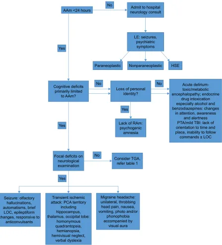
prior to reaching the diagnosis of TGA (Figure 1).110,111
Transient epileptic amnesia (TeA)
TEA is a form of adult-onset MTL epilepsy that presents
as recurrent, transient episodes of acute memory loss.7,48–50
This amnesia may be antero-retrograde or only retrograde.49
This is distinct from TGA, in which patients always have anterograde memory loss and may have permanent retro-grade amnesia that spans hours to days prior to the onset of
the episode.45 Patients with TGA are more likely to exhibit
agitation or anxiety.50 While some authors report repetitive
questioning as a feature indicative of TGA, others have found
that TEA patients also exhibit this behavior.48,49,52
Important features of TEA that differ from TGA include
occurrence upon awakening, duration 1 hour, interictal
retrograde amnesia incomplete anterograde amnesia (able to remember not being able to remember), temporal lobe features such as olfactory/gustatory hallucinations, oral automatisms,
and interictal EEG abnormalities.7,48,53 Controversy exists over
abnormal EEG being a diagnostic factor of TEA, as only a third are found to have abnormalities, while the other
two-thirds show focal slowing or normal findings.5,44,48 Subsequent
follow-up reveals that TEA has a higher recurrence rate than TGA.5,44,48 The diagnosis of TEA carries a favorable
prognos-tic response to antiepilepprognos-tic drugs49,52,53 but may yield
persist-ing interictal memory deficits.53,54 This includes accelerated
long-term forgetting and autobiographical amnesia.54
ischemic events
Ischemic events such as cerebrovascular accidents (CVAs) and TIAs must be excluded prior to arriving at a diagnosis of TGA, as the former typically requires emergent treatment. CVAs and TIAs are most commonly associated with focal
neurological deficits, which by definition preclude TGA from the working diagnosis. However, to confound matters, unilat-eral, isolated hemispheric infarction of the hippocampus or thalamus may present with amnesia as the sole manifestation of the CVA, thus becoming difficult to differentiate from TGA.55,56 Similarly, ischemia within the MTL with
involve-ment of the hippocampus, caudate, or fornix has also been
associated with a TGA-like presentation.25,57–60 Neuroimaging
is strongly recommended. For this reason, neuroimaging is strongly recommended in atypical presentations of TGA or unwitnessed episodes, due to the risk of head trauma
or CVA.43,61,62 While TGA does not typically show acute
changes on brain imaging, MRI with DWI or T2-weighted imaging may reveal hyperintense punctate lesions in the
lat-eral hippocampal regions in the subacute phase ~48–72 hours
after the onset of TGA symptoms. These lesions are visible
for up to 7–10 days after onset.17,19,36,61,62
Brain MRI is usually performed to exclude stroke at the time of symptoms; however, this may be too early to discover the findings associated with TGA. Additionally, typical risk factors associated with cerebrovascular disease should be assessed in the patient’s history. The likelihood of CVA/ TIA as the etiology of transient anterograde amnesia may be
increased with the presence of these risk factors.61,62
Migraines
Migraine is an episodic headache disorder accompanied by various neurological, gastrointestinal, and autonomic
changes. In those with migraine headaches, ~20%
experi-ence an aura, either during or before the development of the
headache.63 Similar to glutamate-mediated transient neuronal
depolarization followed by prolonged quiescence of neuronal activity, CSD may be explained by hypoperfusion that is
preceded by transient hyperperfusion in the cortex.18 CSD
has been proposed as the neurophysiological explanation of migraine with auras, as it propagates at a similar velocity as
visual scotomata during an aura.63
There are multiple case reports suggesting that TGA occurs as either an aura to or, “associated with”, migraine
headaches.64–69 Otherwise, in two larger scale studies, the
association between TGA and migraines is mixed. For instance, in an 11-year retrospective study reported in 2015, among 8,821 new migraine patients, six cases of TGA were identified during a migraine attack. For a majority of these patients, TGA occurred after the beginning of the
attack.51 Alternatively, in a 1998 case–control study, TGA
patients with migraine were identified from a group of 57 TGA patients. The former group was then compared with
Neuropsychiatric Disease and Treatment downloaded from https://www.dovepress.com/ by 118.70.13.36 on 25-Aug-2020
Dovepress Transient global amnesia
Table 2
Differential diagnosis of TGA: transient anterograde amnesia
Condition
Risk factors
Precipitating factors
Duration
Associated neuro symptoms
MRI
EEG
Recurrence of attacks Response to anti-epileptics
TGA Migraine Yes a 4–6 hours No Hippocampal D wi
hyperintensity w/o permanent lesion
Normal Low No T eA No No/yes (waking) 60 minutes
(often, a few minutes) No/yes (oral automatism, olfactory or gustatory hallucinations) Normal/hippocampal sclerosis or atrophy Abnormal (temporal or frontotemporal regions)
High Yes T iA/thrombo-embolic v ascular No
Minutes to permanent impairment
No/yes (any)
D
wi
with T2-FLA
iR
permanent or lesion
Normal Low No Dissociative amnesia b Trauma/abuse
Yes, emotional stress
v ariable No Normal Normal v aries No Migraine headache Genetic; dietary
Yes, fasting, premenstrual, emotional stress, sleep problems
4–72
hours
Auras (visual, sensory, motor, or language abnormalities) up to 30%
Normal
Normal
High
Yes, especially, valproic acid and topiramate
Hypoxic
states
(such
as
aortic
dissection with pure TGA)
increased intrathoracic pressure Underlying stress reaction triggered by the acute pain event
10–12
hours
No
Normal
Normal
None known, although death is not uncommon
No
Hypoxia inducing events of vertebrobasilar system
v ascular No 24 hours Yes
Yes/ischemic lesions in hippocampus
No
Rare
No
Notes:
avalsalva
maneuver, emotional stress, immersion in cold or hot water, sexual intercourse, or pain. bAnterograde amnesia not associated with dissociative amnesia. Data from Kumral e et al
71; April
MD
et
al
78; i
rioka
T
et
al
79;
Bonnet P et al
80; Ryoo
i et al
108; Arena J
e and Rabinstein AA.
109 Abbreviations: DWI, diffusion-weighted imaging; EEG, electroencephalography; FLAIR, fluid-attenuated inversion recovery; MRI, magnetic resonance imaging; TEA, transient epileptic amnesia; TGA, transient global amnesia; TIA, transient ischemic attack.
Neuropsychiatric Disease and Treatment downloaded from https://www.dovepress.com/ by 118.70.13.36 on 25-Aug-2020
Dovepress Spiegel et al
two groups of patients, one with TGA only and the other with normal controls. Despite the prevalence of both migraine and tension headaches being increased in those patients with TGA, there was no evidence of an increased frequency in
TGA features in those with migraines.70
Hypoxia
In addition to ischemia in the setting of TIA/CVA, hypoxia-inducing events with vasospastic, vasoconstrictive, or
arterial insufficiency etiologies have been described in the literature as causes of TGA-like symptoms. For instance, cardioembolism and large-artery disease of the vertebrobasi-lar system can lead to hippocampal infarcts. Five variants have been described including anterior, posterior, unilateral complete, bilateral, and small circumscribed (punctiform) hippocampal infarcts. Prominent clinical acute anterograde amnesia and retrograde amnesia were present in more than two-thirds of patients. In most of the patients, the lesions
7UDQVLHQWLVFKHPLF DWWDFN3&$WHUULWRU\
LQFOXGLQJ KLSSRFDPSXV WKDODPXVRFFLSLWDOOREH
KRPRQ\PRXV TXDGUDQWRSVLD
KHPLDQRSVLD KHPLYLVXDOQHJOHFW
YHUEDOG\VOH[LD
0LJUDLQHKHDGDFKH XQLODWHUDOWKUREELQJ KHDGSDLQQDXVHD YRPLWLQJSKRWRDQGRU
SKRQRSKRELD DFFRPSDQLHGE\
YLVXDODXUD
$FXWHGHOLULXP WR[LFPHWDEROLF HQFHSKDORSDWK\HQGRFULQH
GUXJLQWR[LFDWLRQ HVSHFLDOO\DOFRKRODQG EHQ]RGLD]HSLQHVFKDQJHV
LQDWWHQWLRQDZDUHQHVV DQGDOHUWQHVV 37$PLOG7%,ODFNRI RULHQWDWLRQWRWLPHDQG SODFHLQDELOLW\WRIROORZ FRPPDQGV/2&
)RFDOGHILFLWVRQ QHXURORJLFDO H[DPLQDWLRQ
$$PKRXUV
/DFNRI5$P SV\FKRJHQLF
DPQHVLD /RVVRISHUVRQDO
LGHQWLW\" &RJQLWLYHGHILFLWV
SULPDULO\OLPLWHG WR$$P"
3DUDQHRSODVWLF 1RQSDUDQHRSODVWLF +6( /(VHL]XUHV
SV\FKLDWULF V\PSWRPV $GPLWWRKRVSLWDO QHXURORJ\FRQVXOW
<HV
<HV
<HV <HV
1R
1R 1R
1R
&RQVLGHU7*$ UHIHUWDEOH
6HL]XUHROIDFWRU\ KDOOXFLQDWLRQV DXWRPDWLVPVEULHI
/2&HSLOHSWLIRUP FKDQJHVUHVSRQVLYHWR
DQWLFRQYXOVDQWV
Figure 1 Diagnostic algorithm of anterograde amnesia.
Abbreviations: AAm, anterograde amnesia; eeG, electroencephalogram; HSe, herpes simplex encephalitis; Le, limbic encephalitis; LOC, loss of consciousness; PCA, posterior
cerebral artery; PTA, posttraumatic amnesia; RAm, retrograde amnesia; TBi, traumatic brain injury.
Neuropsychiatric Disease and Treatment downloaded from https://www.dovepress.com/ by 118.70.13.36 on 25-Aug-2020
Dovepress Transient global amnesia
involved the anterior and medial aspects of the hippocampus along the complete length of the head, body, and tail of the
hippocampus.71 Such evidence supports that TGA’s etiology
may be a compromised vertebrobasilar system leading to the dysfunction of MTL and subsequently memory formation.
Several case reports describe TGA-like symptoms
after vascular procedures or intravenous contrast use.51,71–75
Contrast media are sterile iodine-containing solutions used in diagnostic imaging procedures. Older agents generally fall into the class of ionic monomers, which have a high osmo-lality and a high chemotoxicity. Nonionic agents have been developed to overcome the adverse events associated with older contrast media. While there have been cases describ-ing nonionic angiographic contrast medium neurotoxicity, the risk of neurotoxicity remains higher in the ionic class of contrast media. Furthermore, it has been suggested that the risk of neurotoxic effects might be increased by preexisting abnormalities of the blood–brain barrier and by repeated contrast injections.
Other postulated mechanisms by which cerebral angiog-raphy produces TGA symptoms include epilepsy and
ischemia.73,74
Other case reports support an arterial insufficiency etiol-ogy, describing TGA symptomatology secondary to vascular
disease processes.76–80 Intriguingly, aortic dissection, a
poten-tially critical break in the lining of the main arterial outflow from the heart classically associated with severe chest pain and frequently a devastating diagnosis, has been reported to present with TGA-like symptoms in nine cases, five of
whom were without chest pain,78 although the latter could be
attributed to the patients’ altered mental status. Regardless, recognition of painless aortic dissection in the differential of TGA is imperative. While the latter is generally a benign syndrome, the former could result in significant morbidity or mortality. Thus, in those patients presenting with TGA-like symptoms and accompanying cardiovascular changes such as hypotension and hypertension or asymmetric extremity blood pressures, CT angiography of the abdomen may be
warranted.81
Finally, others report the development of TGA after use of phosphodiesterase type 5 inhibitor and sexual activity. However, it is unclear whether the phosphodiesterase inhibi-tor or the sexual activity caused the vasospasm, as both are
known to involve significant changes in blood flow.82
Psychogenic amnesia/dissociative amnesia
Psychogenic amnesia, often labeled as dissociative amnesia, is differentiated from TGA, in which the former affects
retrograde memory, it is generally episodic, and it is often accompanied by intense psychological distress. While often times complicated medical disorders become labeled as “psychogenic” if the etiology is unknown, there is literature
that supports a psychogenic origin to TGA.83
Data gathered from two geriatric studies utilized the Geriatric Depression Scale in patients with TGA, showing
that 40% of TGA patients had depressive symptoms.84
A more detailed study investigated phobic personality traits of 51 TGA patients and established that 82% had pathological
avoidance behaviors.83
TGA patients have a high prevalence of comorbid
emotional distress and anxious personality traits.5,85 About
one-third of TGA episodes occur after physical or psycho-logical stress, which suggest disruption of memory forma-tion due either to ischemic or stress-induced catecholamine
disruption.86,87 Some authors believe that stress-induced
cat-echolamine release may lead to hypoxia or ischemia, whereas others believe that the neurotransmitters involved may affect
the formation of memory.88–93 There is some evidence of an
association between TGA and Takotsubo syndrome. The latter is characterized by transient, acute left-ventricular myocardial dysfunction mimicking myocardial infarction
that also occurs after physical or psychological stress.88
If the clinical picture remains unclear, neuropsychological testing or other neurological tests (electroencephalogram and neurological examination) may be helpful in distin-guishing psychogenic amnestic disturbances from neuro-logical amnestic conditions, especially in patients who have
secondary gain or overembellish symptoms.28
In summary, to make a diagnosis of TGA, all strict clinical criteria as shown in Table 1 should be met. The differential diagnosis includes structural (vascular) disease, epileptic amnesia, delirium, intoxication, and head injury and migraine headaches. When a general physician is presented with a typical case, no additional diagnostic tests, such as MRI and EEG, are needed. This even holds when such a patient has vascular risk factors. However, when focal neurological signs accompany anterograde amnesia, neuroimaging is warranted and potentially a neurology consult. If retrograde amnesia is also affected and emotional distress is present, the clinician needs to consider psychogenic amnesia. Alternatively, albeit rare but more ominous, are those cases of “painless” aortic dissection, where TGA is the primary symptom as chest pain is lacking. Finally, research findings using DWI–MRI support the concept of focal ischemia. These focal lesions have a maximum detectability between 48 and 72 hours after the start of TGA. Focal lesions have been demonstrated in the
Neuropsychiatric Disease and Treatment downloaded from https://www.dovepress.com/ by 118.70.13.36 on 25-Aug-2020
Dovepress Spiegel et al
CA1 area of the cornu ammonis of the hippocampus (MTL), mostly unilateral. However, in up to 35% of TGA patients,
no such abnormalities are demonstrable.10
Beyond 24 hours: outcomes
According to the standard accepted criteria, deficits of TGA
resolve within 24 hours.15 There has been considerable
research dedicated to the cognitive profile of TGA patients following the resolution of acute-phase symptoms; however, results have been mixed.
A 2009 meta-analysis examined data from 25 different studies and compared 374 TGA subjects to 760 control subjects in the following five domains: anterograde episode long-term memory, retrograde episode long-term memory, short-term memory, semantic memory, and executive func-tion. Beyond the first 24 hours, there was no significant differ-ence between patients and controls in any of the five domains
for the 30-day period following TGA onset.94 Similarly, a
study with a median follow-up period of 1,128 days found no significant difference in performance between TGA patients and controls on tasks of episodic memory, seman-tic memory, working memory, executive functions, and
attention.95 Alternatively, one study with follow-up periods
of 4 months and 1 year found deficits in anterograde memory in TGA subjects when compared with controls; however, only when data from both follow-up periods were pooled together. Additionally, at the 4-month follow-up visit, higher scores on anxiety and depression scales correlated with worse performance on tests of retrograde and anterograde memory
tests, respectively.96
Given the cardiovascular hypothesis of TGA and the interest in the field in cardiovascular risk factors of the disease, one study examined the subsequent risk of expe-riencing CVA in patients with a history of TGA vis-a-vis comparing the rate of CVA following TGA to the rate of CVA following migraine, seizure, and TIA at time points of 1 and 5 years after the event. The results of this study did not support an increased risk of CVA after TGA, with the risk of CVA at 1 year being similar to that after migraine (0.54 and 0.22%, respectively) and lower than after TIA (4.54%). The 5-year rates for CVA following TGA, migraine, and TIA
were 2.44, 0.86, and 12.23%, respectively.97
In a 12-year follow-up study, the longest in the literature to date, TGA patients were compared with controls on end points of CVA/TIA, seizures, and cognitive impairment (mild cognitive impairment or dementia). TGA patients were not found to be at a higher risk of developing any of the included
conditions than the control subjects.2
Treatment
By definition, TGA is a self-limited condition that resolves without intervention; thus, there is no specific treatment
indicated.98 Although the mean time course of an amnestic
episode in TGA is 4–6 hours, with most resolving by 8 hours, proposed treatments could depend on uncovering the
under-lying etiology.5
If the presentation in TGA was secondary to reversible ischemia, as has been suggested by some case reports using DWI, as well as other reports that noted onset of TGA epi-sodes after Valsalva-like maneuvers that may temporarily reduce cerebral blood flow, then optimization of cardiovas-cular factors similar to treatments implemented for cardiac ischemia, such as antiplatelets, managing blood pressure, and
heart rate, or statin therapy could be beneficial.98–101
Some studies suggest an epileptic etiology of TGA, as electroencephalogram findings have been suggestive of epi-leptic discharges, but other case–control studies have found
no such correlation.102–104 Thus, anticonvulsant medications
would not be a proposed treatment option based on the available evidence.
Evidence suggesting that TGA is secondary to cortical depression following cortical hyperstimulation does not support theoretical treatment options with the exception of
those related to migraine headaches.70 Triptans have not been
studied in TGA.
Ultimately, there are no established, evidence-based treatments for TGA to date, likely due to the short duration of symptoms experienced as well as a lack of universally accepted pathophysiology.
Conclusion/future directions
The yearly incidence of TGA is 3–8 cases per 100,000 people, although 6%–10% of patients with TGA will experience a
second or third episode.105 Thus, while it may be difficult
to predict an index episode of TGA, it may be possible to decrease the risk of future episodes. The latter could be accomplished through clinical trials by addressing differ-ent proposed etiologies associated with TGA. For instance, despite significantly reduced atherosclerotic risk factor profiles in patients with TGA in comparison to those with TIA, ie, diabetes mellitus and hypertension, can statins, antihypertensives, and/or hypoglycemics decrease the risk of arterial ischemia, which has been proposed to explain TGA? Can prophylactic treatment of migraines with triptans decrease the risk of TGA, as this is the only identified risk factor in the development of TGA in those under the age of 56 years? As discussed, TGA may be a sequela of migraines
Neuropsychiatric Disease and Treatment downloaded from https://www.dovepress.com/ by 118.70.13.36 on 25-Aug-2020
Dovepress Transient global amnesia
due to sweeping depression of cerebral activity. N-methyl-d
-aspartate (NMDA) receptors have a critical role in excitatory synaptic transmission, excitotoxicity, and plasticity in the
central neuronal system.106
Both competitive and noncompetitive NMDA receptor blockers are able to modulate spreading depolarizations (SD). Phencyclidine (PCP), dizocilpine, and ketamine have shown the capacity to elevate SD electrical threshold, block or slow SD propagation, and reduce SD duration and speed. However, the use of PCP and dizocilpine is limited because of their harmful side effects. Recently, discovered neuropro-tective, anti-inflammatory, and antitumor effects of ketamine have been reported. Given the findings of usefulness in low-dose regimens of ketamine, perhaps this noncompetitive antagonist of the NMDA receptor could decrease the risk
of TGA?107
Additionally, selective vulnerability of hippocampal CA1 neurons to metabolic stress seems to play a crucial part in the pathophysiological cascade that leads to a transient per-turbation of memory pathways in TGA. Given this informa-tion, TGA could be classified as a “natural lesion model” of a perturbation of hippocampal CA1 neurons; further under-standing of this pathogenesis could facilitate insights in other neurological disorders that affect the hippocampus, such as
CVA, encephalitis, and Alzheimer’s disease.18
Another remaining area of interest with regard to TGA is the discrepancy in timeline of brain imaging results and the transient nature of memory symptoms. More specifically, clinical/cognitive recovery from TGA over the course of a few hours is generally favorable. Recently, MR imaging studies have reported that small punctate high-signal intensity lesions are frequently found in the lateral portion of the hip-pocampus (ie, CA1 region) on DWI, with variable detection rates of the TGA lesions in the hippocampus on the first day of the symptom onset. The detection rates of the lesions on DWI rise with increased time lapse after the onset of symp-toms and have been reported to be best detected 2–3 days after the episode. Several previous studies suggest that delayed neuronal injury of the hippocampus is a possible cause of the delayed appearance of the lesions. However, the small size of TGA lesions, being susceptible to partial volume averaging due to a relatively larger voxel size, could be the main cause for this delayed lesion appearance. Thus, utilizing high field strength could increase the lesion detectability on DWI. DWI protocol considering these factors, along with voxel size, can support a differential diagnosis in the emergency department and may help to understand the pathophysiological
mecha-nisms of this curious disorder.108
Disclosure
Dr Spiegel is on the speaker’s bureau for Allergen Pharma-ceuticals. The authors report no other conflicts of interest in this work.
References
1. Sancesario G, Esposito Z, Mozzi AF, et al. Transient global amnesia: linked to a systemic disorder of amino acid catabolism? J Neurol. 2013;260(5):1429–1432.
2. Arena JE, Brown RD, Mandrekar J, Rabinstein AA. Long-term outcome in patients with transient global amnesia: a population-based study.
Mayo Clin Proc. 2017;92(3):399–405.
3. Berli R, Hutter A, Waespe W, Bachli EB. Transient global amnesia – not so rare after all. Swiss Med Wkly. 2009;139(19–20):288–292. 4. Zhu J, Lu D, Sveinsson O, et al. Is a cancer diagnosis associated with
subsequent risk of transient global amnesia? PLoS One. 2015;10(4): e0122960.
5. Quinette P, Guillery-Girard B, Dayan J, et al. What does transient global amnesia really mean? Review of the literature and thorough study of 142 cases. Brain. 2006;129(pt 7):1640–1658.
6. Pantoni L, Bertini E, Lamassa M, Pracucci G, Inzitari D. Clinical fea-tures, risk factors, and prognosis in transient global amnesia: a follow-up study. Eur J Neurol. 2005;12(5):350–356.
7. Lin KH, Chen YT, Fuh JL, et al. Migraine is associated with a higher risk of transient global amnesia: a nationwide cohort study. Eur J Neurol. 2014;21(5):718–724.
8. Jang JW, Park SY, Hong JH, Park YH, Kim JE, Kim S. Different risk factor profiles between transient global amnesia and transient ischemic attack: a large case-control study. Eur Neurol. 2014;71(1–2): 19–24.
9. Agosti C, Akkawi NM, Borroni B, Padovani A. Recurrency in transient global amnesia: a retrospective study. Eur J Neurol. 2006; 13(9):986–989.
10. Erkelens CD, Snoek JW. What doctors should not forget about transient global amnesia. Eur J Gen Pract. 2010;16(3):182–185.
11. Jaffe R, Bender MB. E.E.G. studies in the syndrome of isolated epi-sodes of confusion with amnesia “transient global amnesia”. J Neurol
Neurosurg Psychiatry. 1966;29(5):472–474.
12. Bender MB. Syndrome of isolated episode of confusion with amnesia.
J Hillside Hosp. 1956;5:212–215.
13. Fisher CM, Adams RD. Transient global amnesia. Trans Am Neurol
Assoc. 1958;83:143–146.
14. Fisher CM, Adams RD. Transient global amnesia. Acta Neurol Scand
Suppl. 1964;40(suppl 9):1–83.
15. Hodges JR, Warlow CP. Syndromes of transient amnesia: towards a classification. A study of 153 cases. J Neurol Neurosurg Psychiatry. 1990;53(10):834–843.
16. Kitamura T, Ogawa S, Roy DS, et al. Engrams and Circuits crucial for systems consolidation of a memory. Science. 2017;356(6333): 73–78.
17. Lee HY, Kim JH, Weon YC, et al. Diffusion-weighted imaging in transient global amnesia exposes the CA1 region of the hippocampus.
Neuroradiology. 2007;49(6):481–487.
18. Bartsch T, Deuschl G. Transient global amnesia: functional anatomy and clinical implications. Lancet Neurol. 2010;9(2):205–214. 19. Bartsch T, Alfke K, Stingele R, et al. Selective affection of hippocampal
CA-1 neurons in patients with transient global amnesia without long-term sequelae. Brain. 2006;129(pt 11):2874–2884.
20. Sander K, Sander D. New insights into transient global amnesia: recent imaging and clinical findings. Lancet Neurol. 2005;4(7):437–444. 21. Förster A, Griebe M, Gass A, Kern R, Hennerici MG, Szabo K.
Diffusion-Weighted imaging for the differential diagnosis of disorders affecting the hippocampus. Cerebrovasc Dis. 2012;33(2):104–115. 22. Xing C, Arai K, Lo EH, et al. Pathophysiologic cascades in ischemic
stroke. Int J Stroke. 2012;7(5):378–385.
Neuropsychiatric Disease and Treatment downloaded from https://www.dovepress.com/ by 118.70.13.36 on 25-Aug-2020
Dovepress Spiegel et al
23. Peer M, Nitzan M, Goldberg I, et al. Reversible functional connectivity disturbances during Transient Global Amnesia. Ann Neurol. 2014;75(5): 634–643.
24. Saito K, Kimura K, Minematsu K, et al. Transient global amnesia assoca-iated with an acute infaction in the retrosplenium of the corpus callosum.
J Neurol Sci. 2003;210(1–2):95–97.
25. Ravindran V, Jain S, Ming A, Bartlett RJ. Transient global amnesia in a patient with acute unilateral caudate nucleus ischemia. J Clin Neurosci. 2004;11(6):669–672.
26. Yoon B, Yoo JY, Shim YS, et al. Transient Global Amnesia associated with acute intracerebral hemorrhage at the cingulate gyrus. Eur Neurol. 2006;56(1):54–56.
27. Graff-Radford J, Clapp AJ, Lanzino G, et al. Transient amnesia after coiling of a posterior circulation aneurysm. Neurocrit Care. 2013;18(2): 245–247.
28. Spiegel DR, Mccroskey AL, Deyerle BA. A case of transient global amnesia: a review and how it may shed further insight into the neuro-biology of delusions. Innov Clin Neurosci. 2016;13(3–4):32–41. 29. Kimura K, Minematsu K, Yasaka M, Wada K, Yamaguchi T. The
dura-tion of symptoms in transient ischemic attack. Neurology. 1999;52(5): 976–980.
30. Lewis SL. Aetiology of transient global amnesia. Lancet. 1998;352(9125): 397–399.
31. Klötzsch C, Sliwka U, Berlit P, Noth J. An increased frequency of patent foramen ovale in patients with transient global amnesia: analysis of 53 consecutive patients. Arch Neurol. 1996;53(6):504–508. 32. De Francisco J, Pujadas F, Toledo M, et al. A study of right-left shunt
in transient global amnesia. Neurologia. 2010;25(2):83–89.
33. Pantoni L, Lamassa M, Inzitari D. Transient global amnesia: a review emphasizing pathogenic aspects. Acta Neurol Scand. 2000;102(5): 275–283.
34. Schmidtke K, Reinhardt M, Krause T. Cerebral perfusion during tran-sient global amnesia: findings with HM-PAO SPECT. J Nucl Med. 1998;39(1):155–159.
35. Winbeck K, Etgen T, von Einsiedel HG, Röttinger M, Sander D. DWI in transient global amnesia and TIA: proposal for an ischaemic origin of TGA. J Neurol Neurosurg Psychiatry. 2005;76(3):438–441. 36. Yang Y, Kim S, Kim JH. Ischemic evidence of transient global amnesia:
location of the lesion in the hippocampus. J Clin Neurol. 2008;4(2): 59–66.
37. Di Filippo M, Calabresi P. Ischemic bilateral hippocampal dysfunction during transient global amnesia. Neurology. 2007;69(5):493. 38. Nakada T, Kwee IL, Fujii Y, et al. High-field, T2 reversed MRI of
the hippocampus in transient global amnesia. Neurology. 2005;64(7): 1170–1174.
39. Huber R, Aschoff AJ, Ludolph AC, et al. Transient global amnesia. Evidence against vascular ischemic etiology from diffusion weighted imaging. J Neurol. 2002;249(11):1520–1524.
40. Chung CP, Hsu HY, Chao AC, et al. Transient global amnesia: cerebral venous outflow impairment – insight from the abnormal flow patterns of the internal jugular vein. Ultrasound Med Biol. 2007;33(11):1727–1735. 41. Cejas C, Cisneros LF, Lagos R, Zuk C, Ameriso SF. Internal jugular
vein valve incompetence is highly prevalent in transient global amnesia.
Stroke. 2010;41(1):67–71.
42. Baracchini C, Tonello S, Farina F, et al. Jugular veins in transient global amnesia: innocent bystanders. Stroke. 2012;43(9):2289–2292. 43. Faust JS, Nemes A. Transient global amnesia: emergency department
evaluation and management. Emerg Med Pract. 2016;18(6):1–20. 44. Kang Y, Kim E, Kim JH, et al. Time of flight MR angiography
assessment casts doubt on the association between transient global amnesia and intracranial jugular venous reflux. Eur Radiol. 2015;25(3): 703–709.
45. Owen D, Paranandi B, Sivakumar R, Seevaratnam M. Classical diseases revisited: transient global amnesia. Postgrad Med J. 2007;83(978): 236–239.
46. Olsen TS. Pathophysiology of the migraine aura: the spreading depres-sion theory. Brain. 1995;118(pt 1):307–308.
47. Olesen J, Jørgensen MB. Leao’s spreading depression in the hippocampus explains transient global amnesia: a hypothesis. Acta Neurol Scand. 1986; 73(2):219–220.
48. Zeman AZ, Boniface SJ, Hodges JR. Transient epileptic amnesia: a descri-ption of the clinical and neuropsychological features in 10 cases and a review of the literature. J Neurol Neurosurg Psychiatry. 1998;64(4): 435–443.
49. Kapur N. Transient epileptic amnesia–a clinical update and a reformula-tion. J Neurol Neurosurg Psychiatry. 1993;56(11):1184–1190. 50. Serafetinides EA. Transient epileptic amnesia–a clinical update and a
reformulation. J Neurol Neurosurg Psychiatry. 1994;57(12):1549. 51. Donnet A. Transient global amnesia triggered by migraine in a French
Tertiary-Care Center: an 11-year retrospective analysis. Headache. 2015;55(6):853–859.
52. Bilo L, Meo R, Ruosi P, de Leva MF, Striano S. Transient epileptic amnesia: an emerging late-onset epileptic syndrome. Epilepsia. 2009; 50(suppl 5):58–61.
53. Butler CR, Zeman AZ. Recent insights into the impairment of memory in epilepsy: transient epileptic amnesia, accelerated long-term forgetting and remote memory impairment. Brain. 2008;131(pt 9):2243–2263. 54. Hoefeijzers S, Dewar M, Della Sala S, Zeman A, Butler C. Accelerated
long-term forgetting in transient epileptic amnesia: an acquisition or consolidation deficit? Neuropsychologia. 2013;51(8):1549–1555. 55. Pérez-Lázaro C, Santos S, Garcés-Redondo M, et al. Amnesic stroke
caused by hippocampal infarction. Rev Neurol. 2005;41(1):27–30. 56. Carlesimo GA, Lombardi MG, Caltagirone C. Vascular thalamic
amnesia: a reappraisal. Neuropsychologia. 2011;49(5):777–789. 57. Adler AC, Warum D, Sapire JM. Transient global amnesia caused by
hippocampal infarct: case report and review of literature. Clin Imaging. 2012;36(5):584–586.
58. Liang JF, Shen AL, Lin SK. Bilateral hippocampal abnormalities on diffusion-weighted MRI in transient global amnesia: report of a case.
Acta Neurol Taiwan. 2009;18(2):127–129.
59. Greer DM, Schaefer PW, Schwamm LH. Unilateral temporal lobe stroke causing ischemic transient global amnesia: role for diffusion-weighted imaging in the initial evaluation. J Neuroimaging. 2001;11(3): 317–319.
60. Gupta M, Kantor MA, Tung CE, Zhang N, Albers GW. Transient global amnesia associated with a unilateral infarction of the fornix: case report and review of the literature. Front Neurol. 2015;5:291.
61. Li J, Hu WL. Bilateral hippocampal abnormalities in magnetic resonance imaging in transient global amnesia. Am J Emerg Med. 2013;31(4): 755.e1–e3.
62. Sedlaczek O, Hirsch JG, Grips E, et al. Detection of delayed focal MR changes in lateral hippocampus in transient global amnesia. Neurology. 2004;62(12):2165–2170.
63. Alemdar M, Selekler M. Migraine and cortical spreading depression.
Agri. 2006;18(4):24–30.
64. Tosi L, Righetti CA. Transient global amnesia and migraine in young people. Clin Neurol Neurosurg. 1997;99(1):63–65.
65. Saito Y, Memezawa H. Migraine with aura accompanied by transient global amnesia. No To Hattatsu. 2010;42(4):303–304.
66. Maggioni F, Mainardi F, Bellamio M, Zanchin G. Transient global amnesia triggered by migraine in monozygotic twins. Headache. 2011; 51(8):1305–1308.
67. Romero JR, Mercado M, Beiser AS, et al. Transient global amnesia and neurological events: the framingham heart study. Front Neurol. 2013; 14(4):47.
68. Dalla Volta G, Zavarise P, Ngonga G, et al. Transient global amnesia as a presenting aura. Headache. 2014;54(3):551–552.
69. Ferlazzo E, Italiano D, Belcastro V, et al. Transient global amnesia as a presenting aura or epilepsy? Headache. 2014;54(7):1233–1235. 70. Schmidtke K, Ehmsen L. Transient global amnesia and migraine. A case
control study. Eur Neurol. 1998;40(1):9–14.
71. Kumral E, Deveci EE, Erdoğan C, Enüstün C. Isolated hippocampal infarcts: vascular and neuropsychological findings. J Neurol Sci. 2015; 356(1–2):83–89.
Neuropsychiatric Disease and Treatment downloaded from https://www.dovepress.com/ by 118.70.13.36 on 25-Aug-2020
Neuropsychiatric Disease and Treatment
Publish your work in this journal
Submit your manuscript here: http://www.dovepress.com/neuropsychiatric-disease-and-treatment-journal
Neuropsychiatric Disease and Treatment is an international, peer-reviewed journal of clinical therapeutics and pharmacology focusing on concise rapid reporting of clinical or pre-clinical studies on a range of neuropsychiatric and neurological disorders. This journal is indexed on PubMed Central, the ‘PsycINFO’ database and CAS,
and is the official journal of The International Neuropsychiatric Association (INA). The manuscript management system is completely online and includes a very quick and fair peer-review system, which is all easy to use. Visit http://www.dovepress.com/testimonials.php to read real quotes from published authors.
Dovepress
Dove
press
Transient global amnesia
72. Blumenfeld AE, Victorio MC, Berenson FR. Complicated migraines.
Semin Pediatr Neurol. 2016;23(1):18–22.
73. Jackson A, Stewart G, Wood A, Gillespie JE. Transient global amnesia and cortical blindness after vertebral angiography: further evidence for the role of arterial spasm. AJNR Am J Neuroradiol. 1995; 16(4 suppl):955–959.
74. Yildiz A, Yencilek E, Apaydin FD, Duce MN, Ozer C, Atalay A. Transient partial amnesia complicating cardiac and peripheral arteriog-raphy with nonionic contrast medium. Eur Radiol. 2003;13(suppl 4): L113–L115.
75. Benke T, Chemelli A, Lottersberger C, Waldenberger P, Karner E, Trinka E. Transient global amnesia triggered by the intracarotidamo-barbital procedure. Epilepsy Behav. 2005;6(2):274–278.
76. Kim HY, Kang HS, Roh HG, et al. Transient global amnesia fol-lowing vertebral artery angioplasty and stenting. Eur Neurol. 2006; 56(2):133–135.
77. Kaveeshvar H, Kashouty R, Loomba V, Yono N. A rare case of aortic dissection presenting as pure transient global amnesia. Cardiovasc J
Afr. 2015;26(6):e8–e9.
78. April MD, Fossum K, Hounshell C, et al. A sinister cause of anterograde amnesia: painless aortic dissection. Am J Emerg Med. 2015;33(7):989. e5–e7.
79. Irioka T, Yamanami A, Yagi Y, Mizusawa H. Aortic dissection as a pos-sible cause of pure transient global amnesia: a case report and literature review. Neurol Sci. 2009;30(3):255–258.
80. Bonnet P, Niclot P, Chaussin F, et al. A puzzling case of transient global amnesia. Lancet. 2004;364(9433):554.
81. Colotto M, Maranghi M, Epifania A, et al. Unmasking aortic dissec-tion in patients of transient global amnesia: case report and diagnostic algorithm for the emergency department. BMJ Case Rep. 2011; 2011:bcr0720103151.
82. Machado A, Rodrigues M, Ribeiro M, et al. Tadalafil-induced tran-sient global amnesia. J Neuropsychiatry Clin Neurosci. 2010;22(3): 352.e28.
83. Inzitari D, Pantoni L, Lamassa M, Pallanti S, Pracucci G, Marini P. Emotional arousal and phobia in transient global amnesia. Arch Neurol. 1997;54(7):866–873.
84. Neri M, Andermarcher E, De Vreese LP, Rubichi S, Sacchet C, Cipolli C. Transient global amnesia: memory and metamemory. Aging
(Milano). 1995;7(6):423–429.
85. Zorzon M, Antonutti L, Masè G, Biasutti E, Vitrani B, Cazzato G. Transient global amnesia and transient ischemic attack: natural history, vascular risk factors, and associated conditions. Stroke. 1995;26(9):1536–1542. 86. Fisher CM. Transient global amnesia. Precipitating activities and other
observations. Arch Neurol. 1982;39(10):605–608.
87. Miller JW, Petersen RC, Metter EJ, et al. Transient global amnesia: clinical characteristics and prognosis. Neurology. 1987;37(5):733–737. 88. Madias JE. Transient global amnesia and Takotsubo syndrome:
would cerebral blood flow brain scan be of any help? Clin Auton Res. 2015;25(3):199.
89. Finsterer J, Aliyev R. Takotsubo syndrome: consequence or cause of ischemic stroke. Funct Neurol. 2014;29(4):281–282.
90. Finsterer J, Wahbi K. CNS disease triggered by Takotusbo stress car-diomyopathy. Int J Cardiol. 2014;177(2):322–329.
91. Abi-Saleh B, Iskandar SB, Schoondyke JW, et al. Tako-Tsubo syndrome as consequence of transient ischemic attack. Rev Cardiovasc Med. 2006;7(1):37–41.
92. Lyon AR, Rees PS, Prasad S, et al. Stress (Takotsubo) cardiomyopathy – a novel pathophysiological hypothesis to explain catecholamine-induced acute myocardial stunning. Nat Clin Pract Cardiovasc Med. 2008;5(1): 22–29.
93. Finsterer J, Stöllberger C. Transient global amnesia: the cerebral Takotsubo? J Neurol Sci. 2017;15(376):196–197.
94. Jäger T, Bäzner H, Kliegel M, Szabo K, Hennerici MG. The transience and nature of cognitive impairments in transient global amnesia: a meta-analysis. J Clin Exp Neuropsychol. 2009;31(1):8–19. 95. Uttner I, Prexl S, Freund W, Unrath A, Bengel D, Huber R. Long-term
outcome in transient global amnesia patients with and without focal hyperintensities in the CA1 region of the hippocampus. Eur Neurol. 2012;67(3):155–160.
96. Noël A, Quinette P, Dayan J, et al. Influence of patients’ emotional state on the recovery processes after a transient global amnesia. Cortex. 2011;47(8):981–991.
97. Mangla A, Navi BB, Layton K, et al. Transient global amnesia and the risk of ischemic stroke. Stroke. 2014;45(2):389–393.
98. Brown J. ED evaluation of transient global amnesia. Ann Emerg Med. 1997;30(4):522–526.
99. Strupp M, Brüning R, Wu RH, Deimling M, Reiser M, Brandt T. Diffusion-weighted MRI in transient global amnesia: elevated signal intensity in the left mesial temporal lobe in 7 of 10 patients. Ann
Neurol. 1998;43(2):164–170.
100. Schaefer PW. Diffusion-weighted imaging as a problem solving tool in the evaluation of patients with acute stroke-like syndromes. Top
Magn Reson Imaging. 2000;11(5):300–309.
101. Tiecks FP, Lam AM, Matta BF, Strebel S, Douville C, Newell DW. Effects of the Valsalva maneuver on cerebral circulation in healthy adults. A transcranial Doppler study. Stroke. 1995;26(8):1386–1392. 102. Tchalla AE, Marin B, Mignard C, et al. Newly diagnosed epileptic
seizures: focus on an elderly population on the French island of Reunion in the Southern Indian Ocean. Epileptic Disord. 2013;15(3):243–254. 103. Melo TP, Ferro JM, Ferro H. Transient global amnesia. A case control
study. Brain. 1992;115(pt 1):261–270.
104. Guidotti M, Anzalone N, Morabito A, Landi G. A case-control study of transient global amnesia. J Neurol Neurosurg Psychiatry. 1989;52(3): 320–323.
105. Bartsch T, Butler C. Transient amnesic syndromes. Nat Rev Neurol. 2013; 9(2):86–97.
106. Sánchez-Porras R, Zheng Z, Sakowitz OW. Pharmacological modula-tion of spreading depolarizamodula-tions. Acta Neurochir Suppl. 2015;120: 153–157.
107. Kurdi MS, Theerth KA, Deva RS. Ketamine: current applications in anesthesia, pain, and critical care. Anesth Essays Res. 2014;8(3): 283–290.
108. Ryoo I, Kim JH, Kim S, Choi BS, Jung C, Hwang SI. Lesion detectability on diffusion-weighted imaging in transient global amnesia: the influ-ence of imaging timing and magnetic field strength. Neuroradiology. 2012;54(4):329–334.
109. Arena JE, Rabinstein AA. Transient global amnesia. Mayo Clin Proc. 2015;90(2):264.
110. Williamson J, Larner AJ. Transient global amnesia. Br J Hosp Med
(Lond). 2015;76(12):C186–C188.
111. Kaveeshvar H, Kashouty R, Loomba V. et al. A rare case of aortic dis-section presenting as pure transient global amnesia. Cardiovasc J Afr. 2015;26(6):e8–e9.
Neuropsychiatric Disease and Treatment downloaded from https://www.dovepress.com/ by 118.70.13.36 on 25-Aug-2020