BIOMORPHOMETRIC STUDIES IN BENIGN PROSTATIC HYPERPLASIA
Thesis submitted for the degree of
DOCTOR OF MEDICINE
in the Faculty of Medicine
of the University of London
by
Paul David Miller
Departments of Urology and Biochemistry
ProQuest Number: U063954
All rights reserved
INFORMATION TO ALL USERS
The quality of this reproduction is dependent upon the quality of the copy submitted.
In the unlikely event that the author did not send a complete manuscript and there are missing pages, these will be noted. Also, if material had to be removed,
a note will indicate the deletion.
uest.
ProQuest U063954
Published by ProQuest LLC(2016). Copyright of the Dissertation is held by the Author.
All rights reserved.
This work is protected against unauthorized copying under Title 17, United States Code. Microform Edition © ProQuest LLC.
ProQuest LLC
789 East Eisenhower Parkway P.O. Box 1346
8
.101 IN iH NTi.H ivL’s - r w
JOHN HUNTER F.R.C.S.
ABSTRACT
The ageing population structure, with concomitant increase in the prevalence of the pathologies of age and the mor bidity associated with prostatectomy, require that we look to alternative forms of treatment for benign prostatic hyperplasia (BPH).
Although prostatic growth is dependent on androgens, especially dihydrotestosterone, the effect of steroids is probably mediated by other regulators. Androgens and oestrogens are believed to provide the hormonal environ ment that is essential for other biochemical factors to induce hyperplasia and perhaps malignancy.
The aim of this study was to investigate one of the newly discovered relevant biochemical "factors" (epidermal
growth factor). This growth factor is found in high con centrations in prostatic fluid and its relationship with the hormonal environment, steroid receptors and the pre sence of prostatic pathology have been examined.
These were correlated with the biochemical data from the same samples.
The results show increased epidermal growth factor bind ing in BPH and a close association between steroid recep tors and growth factor receptors in the abnormal part of the prostate. Although this suggests up regulation of the growth factor receptors in BPH, receptor binding is also found to be closely related to tissue morphometry.
Acknowledgements
Human tissue for this study was obtained from patients under the care of Mr.R.S.Kirby, Mr. H.N.Whitfield and Mr. W.F.Hendry at St.Bartholomew's Hospital and Homerton
Hospital. I am grateful to them for allowing me to study their patients. All the work in this study was carried out in the Departments of Urology, Biochemistry and Histopa-thology at St.Bartholomew's Hospital, London.
The tissue biochemical assays and immunohistochemistry was performed by the author in the Department of Biochemistry at St.Bartholomew's Hospital Medical College after expert tuition by Mr.S.Barker and Mr.J.Puddefoot. I would like to thank them for their guidance and patience throughout this project.
The computer morphometric analysis was performed by the author in the Department of Histopathology. The computer program for this analysis was written in conjunction with Mr.C.Sowter and Prof.G.Slavin to whom I am greatly indebt ed.
The work at St.Bartholomew's Hospital was performed under the guidance of Mr.R.S.Kirby MA, MD, FRCS (Urol) and
1 must also thank the staff of the Department of Medical Illustration and the Medical college library. Miss J.Bryan collected many of the specimens from the operating theatre for which I am also most grateful.
This work would not have been possible without the finan cial support of The Special Trustees of the Medical Col lege of St.Bartholomew's Hospital. Ethical permission to study patients was obtained from the Ethical Committee of St.Bartholomew's Hospital. All patients gave informed
verbal and written consent. Mr. J. Higgins F.R.C.S. kindly gave me permission to reproduce figures 1 (iv) (v) and
CONTENTS PAGE
FRONTISPIECE 2
ABSTRACT 3
ACKNOWLEDGEMENTS 5
CONTENTS 7
LIST OF FIGURES 9
LIST OF TABLES 12
LIST OF APPENDICES 14
CHAPTER ONE - REVIEW OF THE LITERATURE 15
I Introduction 18
II Epidemiology and Aetiology 20
III Anatomy 27
IV Endocrinology and Biochemistry 48
V Morphometry 88
CHAPTER TWO - AIMS OF THE STUDY 98
I Background 99
II Experimental strategy 100
CHAPTER THREE - MATERIALS AND METHODS 102
I Tissue collection 105
II Chemicals 109
III Apparatus 113
IV Preparation of tissue for receptor assay 113
V Binding studies 115
VI Serum hormone concentrations 121
VII Immunohistochemistry 124
VIII Morphometric techniques 126
CHAPTER FOUR - VALIDATION OF THE METHODOLOGY 137
I Introduction 139
II EGFR Assay 139
CHAPTER FIVE - BIOCHEMICAL RESULTS I Introduction
II EGFR binding studies
III Steroid receptor binding studies IV Regional distribution
VI Serum hormone levels
CHAPTER SIX - MORPHOMETRIC RESULTS I Introduction
II Morphometry in BPH and normal prostate III Comparison with pathologist
IV Biochemical and morphometric results CHAPTER SEVEN - GENERAL DISCUSSION
APPENDIX BIBLIOGRAPHY
PRESENTATIONS/PUBLICATIONS
LIST OF FIGURES Page
1. (i) Percentage increase of BPH with age 18 1. (ii) Prevalence of BPH pathology at autopsy 21 1. (iii) Transverse slice of normal human 34
prostate
1. (iv) Coronal section of whole normal 35 prostate (x2.5)
1. (v) High power (x230) of central zone 36 prostatic tissue
1. (vi) High power (x230) of peripheral zone 37 prostatic tissue
1. (vii) Diagram of longitudinal zonal prostatic 45 anatomy in BPH with corresponding
transrectal ultrasound scan
1. (viii) Diagram of transverse zonal prostatic 46 anatomy in BPH with corresponding
transrectal utrasound scan
1. (ix) Diagram of the "two step" model for the 61 mechanism of action of steroid hormones
1. (x) Diagram of the mechanism of action of 63 steroid hormones based on the "one step"
model
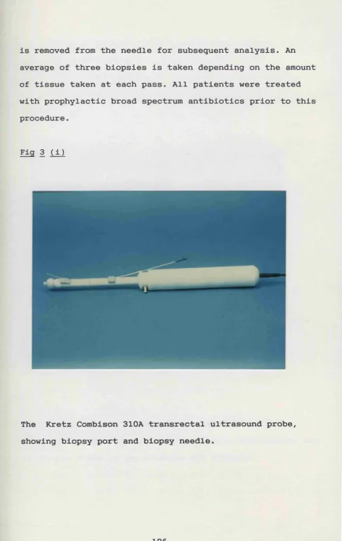
3. (1) The Kretz Combison transrectal ultrasound 106 probe
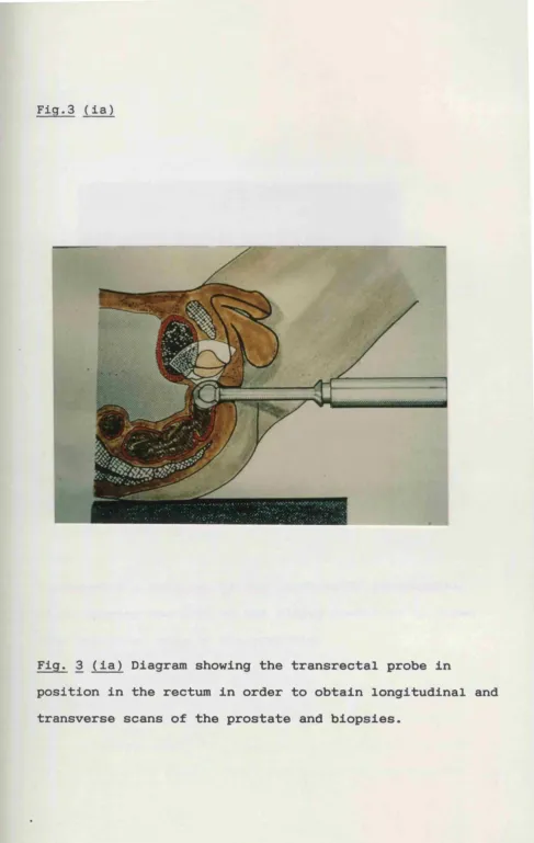
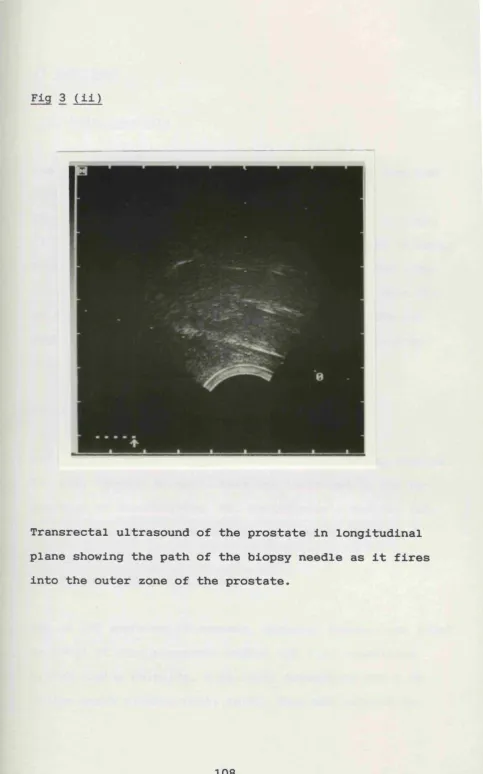
3. (la) The transrectal probe in the rectum 107 3. (ii) Longitudinal transrectal ultrasound of 108
the prostate during biopsy
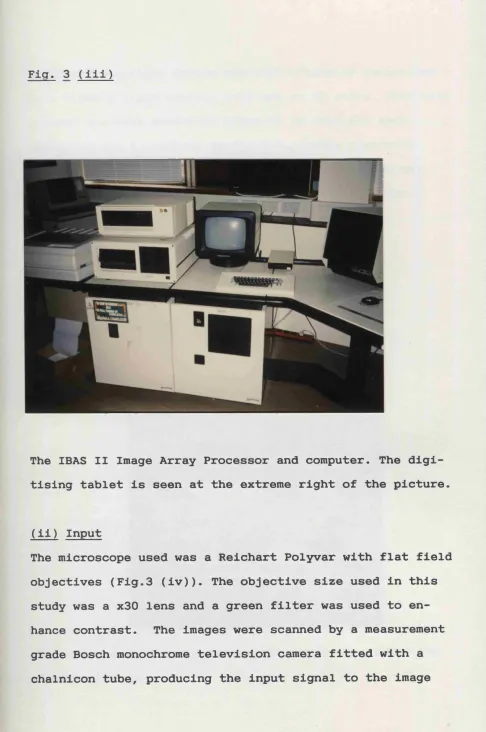
3. (iii) The IBAS image array processor 127 3. (iv) Video camera and microscope 128
3. ( V ) Monochrome Image 130
3. (vi) Binary Image 131
3. (vii) Binary Image inverted and dilated 132
3. (viii) Acinar contours of image 133
3. (ix) Epithelial component of image 133
3. ( X ) Epithelial component in red superimposed
on original image
134
3. (xi) Epithelial component and acinar lumen 135
4. (i) Duration of incubation 140
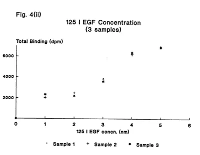
4. (ii) i25i gQp concentration 141
4. (iii) Influence of dilution 142
4. (iv) Comparison SA v SDA oestrogen receptors 147 5. (i) Saturation curve and Scatchard plot of
125i-EGF binding to BPH tissue
155
5. (ii) EGFR SA V SDA in BPH TURP specimens 157 5. (iii) EGFR binding, normal v BPH tissue 159 5. (iv) Immunohistochemistry - BPH - control 163
5. (vi) Immunohistochemistry (HPx320) - BPH - EGF antibody
165
5. (vii) EGFR V ER binding BPH specimens 173 5. (viii) Regional distribution EGFR binding 178 5. (ix) Regional distribution ER binding 179
5. ( X ) Regional distribution PgR binding 181
5. (xi) EGFR V ER binding inner zone 182 5. (xii) EGFR V ER binding outer zone 183 5. (xiii) Steroid hormone and SHBG controls v BPH 185
5. (xiv) T/E2 ratio controls v BPH 187
6. (i) Morphometry Normal v BPH tissue 197 6. (ii) Epithelium % v EGFR binding 202 6. (iii) Glands v EGFR binding (BPH) 202 6. (iv) G/S ratio v EGFR binding (BPH) 203
6. ( V ) % Stroma v ER binding (BPH) 205
6. (vi) G/S ratio v ER binding (BPH) 205 6 . (vii) G/S ratio v T/E2 ratio (BPH) 207 7. (i) Correlation of % increase in flow rate 221
LIST OF TABLES
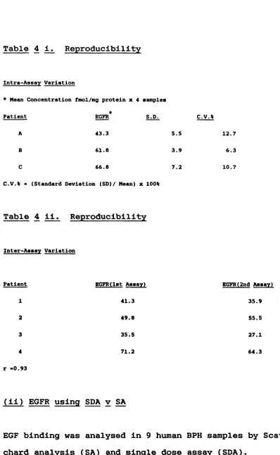
4. i. Reproducibility of EGFR assay - intra assay 144 variation
4. ii. Reproducibility of EGFR assay - inter assay 144 variation
5. i. EGFR assay BPH tissue - SDA 153 5. ii. EGFR SDA v Scatchard analysis (SA) 157 5. iii. EGFR assay normal tissue - SDA 159 5. iv. Immunohistochemistry v SDA - BPH 162
5. V . Immunohistochemistry v SDA - normal 162
5. vi. ER - BPH tissue 168
5. vii. ER - normal tissue 169
5. viii. PgR - BPH tissue 170
5. ix. PgR - Normal tissue 171
5. X . EGFR V Steroid receptors 173
5. xi. Regional distribution EGFR 177 5. xii. Regional distribution ER 179 5. xiii. Regional distribution PgR 180 5. xiv. EGFR and steroid receptor distribution 183
5. X V . Serum hormone levels BPH and controls 184
5. xvi. Serum hormone levels, EGFR and ER binding 186 in BPH
6. ii. Morphometry data - normal post pubertal 194 prostatic tissue
6. iii. Morphometry data - normal prostatic tissue 194 6. iv. Morphometry data - BPH tissue 195 6. V. G/S ratio and "glandularity" 199 6. vi. Morphometry and biochemical analysis - normal 200
prostatic tissue
LIST OF APPENDICES
I Data from validation of methodology 224
II EGFR results raw data 227
III Steroid receptor results raw data 231
IV Regional distribution results 233
CHAPTER ONE
Review of the literature PAGE
I INTRODUCTION 18
II EPIDEMIOLOGY OF BENIGN PROSTATIC HYPERPLASIA 20
1. Demographic factors 20
2. Racial and other risk factors 23 3. Relationship between BPH and malignancy 25
4. Summary 26
III ANATOMY 27
1. Introduction 27
2. Comparative anatomy 28
3. Human foetal and developmental anatomy 30
4. Normal human adult anatomy 32
5. Pathological anatomy 39
6. Ultrasonic anatomy 41
IV ENDOCRINOLOGY / BIOCHEMISTRY 48
1. Historical background 48
2. Steroid hormones in the pathogenesis of BPH 51
3. Mechanisms of hormone action 59
4. Other hormones regulating prostatic growth 70 5. Stromal-epithelial interaction 73
V MORPHOMETRY
1. Introduction 88
2. Morphometric techniques 88
3. Image analysis 91
I INTRODUCTION
Benign prostatic hyperplasia (BPH) is an abnormal, benign growth of the prostate which commonly develops with age in men. Enlargement of the gland gradually occurs and classi cal stromal nodules are formed, together with glandular hyperplasia. As the gland enlarges it often begins to cause mechanical obstruction to the outflow of urine from the bladder during voiding. This leads to the typical
symptoms of poor stream, hesitancy and terminal dribbling. In addition, the obstruction may cause secondary bladder instability which accounts for the irritative symptoms
(frequency, nocturia and urgency) of this condition. Pathological changes frequently begin in the fourth de cade, but significant enlargement with concomitant symp toms does not develop in most cases until after 50 years of age (Swyer 1944, Franks 1954) (Fig.l (i)).
Fig.l (i)
Percentage of age group with BPH
100 r
■ ■ Mieroaeople BPH
I ) Maereaeople BPH
80 rm rm c iim c a i bp h
60
40
20
0 20 40 60 80 100
Host Age (Years)
Epidemiological studies have attempted to define the dimen sions of the clinical problem that surgeons may face In the future, as well as trying to Identify aetlologlcal risk factors. Ageing and the presence of Intact testes are necessary features In the development of BPH, but the
specific biochemical and/or hormonal factors that provoke this condition are unknown.
Better understanding of the biochemistry and endocrino logy of the normal prostate and of the changes which occur In BPH may provide clues to the aetiology and pathogenesis of BPH. This In turn could lead to a more rational ap proach to treatment. Recently the role of Interactions between stroma and epithelium In maintaining physiological homeostasis and In neoplasia has been recognised. There fore morphometric studies make a crucial contribution to Interpreting the biochemical results. Hence the focus of this thesis Is on exploring the relationship between
disturbance of biochemical and hormonal balance In BPH on the one hand and morphometric changes In the prostate gland structure on the other.
II EPIDEMIOLOGY OF BENIGN PROSTATIC HYPERPLASIA
Although benign prostatic hyperplasia (BPH) is the most common benign proliferative abnormality in men, knowledge of the epidemiology of BPH remains fragmentary and re quires more research. This is due mainly to difficulties of collecting reliable epidemiological data and variations in the diagnostic criteria used for BPH (Meyhoff and Hald 1978). Some data are available from the Hospital In-pa tient Enquiry, but these are based on episodes of admis sion and, as an unknown number of in-patient episodes may result from a single case of BPH, they are difficult to interpret (Berry and Jones 1982). Not all BPH is sympto matic and it has been shown that symptoms correlate poorly with prostatic size (Turner-Warwick et al. 1973). This and the lack of standard diagnostic criteria must be borne in mind when considering data from clinical case series.
1. Demographic Factors
Calculation of incidence and prevalence rates depend on the identification of a source population (population at risk) in order to provide a denominator, which is usually difficult to determine. Indeed up to now the only popula tion-based study is that carried out by the Veterans
Administration (Glynn et al. 1985).
diagnostic criteria but, as subjects are not necessarily representative of the general population, they cannot be used to derive prevalence rates. An early Austrian study (Moore 1935) was based on a collection of 675 prostates from every male autopsy conducted in two Vienna hospitals In this study the prevalence of "benign enlargement"
(a term not defined) rose from 0% at age 20-29 years to 75% in men aged 80 and upwards. In Britain, Franks
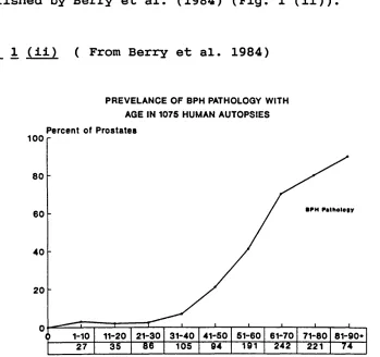
(1954) performed detailed histological studies on autopsy specimens. He also found increased prevalence in each age category from 0% at 20-29 years to 85% in men over 80 years old. Data collated from several autopsy series was published by Berry et al. (1984) (Fig. 1 (ii)).
Fig. 1 (ii) ( From Berry et al. 1984)
PREVELANCE OF BPH PATHOLOGY WITH AGE IN 1075 HUMAN AUTOPSIES Percent of Prostates
100
80
60
40
20
41-50 51-60
21-30 31-40 61-70
11-20 71-60
1-10
94 105
35 221
In a series of nearly 7000 Insurance medicals, also by their nature a selected population, 20% of men in the sixth decade and 43% of men in the eighth decade had
palpable enlargement of the prostate (Lytton et al. 1968). A recent British study (Garraway et al. 1991) suggests that the prevalence of BPH is higher than has been repor ted previously in clinical retrospective and autopsy
studies. They report a prevalence rising to 430 per 1000 men aged 60-69 years using symptom score, flow rate and transrectal ultrasound in 705 men.
All these studies have their limitations, nonetheless it can be concluded that BPH is a very common condition in males over 50 years of age and, in those over 80, histolo gical criteria can be fulfilled in over 80% of subjects. Rotkin (1976) observed that the frequency of symptomatic BPH in American men of all ages, with palpably enlarged glands was 50-70% in most series.
Two studies have attempted to determine time trends in the evolution of BPH. The most recent study by the Veter ans Administration is one of the few that have followed a cohort for a prolonged period (Glynn et al. 1985). Apply ing clear criteria for clinical diagnosis and monitoring surgical treatment they followed 2036 volunteers for up to 25 years. From life-table analysis of these data it was estimated that the lifetime probability of requiring
significant BPH than any clinical assessment of prostatlc size or histological evidence of hyperplasia. This figure however Is higher than that calculated retrospectively by Lytton et al. (1968), who estimated that a 40 year old man had a 10% chance of requiring surgery before he was 80.
2. Racial and other risk factors
There are reports that prevalence of BPH Is higher amongst blacks than In white men and of a relatively low frequency In Orientals. Many of these studies are epldemlologlcally flawed (Rotkin 1983), but the evidence of racial differ ences cannot be Ignored.
Kambal (1977) reported a high prevalence of BPH In North ern Sudan (where Intermarriage with arab migrants occurs) but no case was found In the "pure African" population of Southern Sudan. Significantly higher levels of BPH have been shown In blacks than In whites In New Orleans (Derbes et al. 1937) and Alabama (Morgan and Spruell 1983), but the reports from Africa are largely anecdotal and lack a secure epidemiological basis.
Age is by far the strongest risk factor associated with prevalence and probably the incidence of BPH. Many believe that the age association reflects age-related hormonal changes, and there is now some evidence to support this view (Zumoff et al. 1982).
The Veterans study (Glynn et al. 1985) reported higher rates of BPH among Jewish men and lower rates, particular ly of clinically significant BPH, among smokers. However the former association may reflect socio-economic status and the latter could reflect a reduced likelihood of
surgery among smokers because of their increased anaesthe tic risk or other confounding variables. A link with
hypertension reported by Bourke and Griffin (1966) has not been verified by subsequent studies (Glynn et al. 1985, Morrison 1978).
An association between BPH and diabetes (Bourke and
Griffin 1968) and cirrhosis of the liver (Robson 1964) may also be related to hormonal imbalance. A relative increase in plasma oestrogens may provide the link, due to de
3. Relationship Between BPH and Malignancy
In view of the high prevalence of BPH, its presence in
approximately 20% of all cases of invasive carcinoma does
not necessarily imply that it is a pre-malignant condi
tion. There has been considerable controversy about this
relationship, Armenian et al. (1974) argued on histologi
cal grounds that BPH is an early stage in the development
of prostatic cancer, but Greenwald et al. (1974) found no
evidence to support this conclusion. Relative risks for
development of carcinoma in patients with BPH are quoted
as being between 3 and 5. This may merely reflect the
increased chances of finding one disease if the other is
present and under investigation or treatment (Catalona and
Scott 1978).
At the present time conflicting evidence makes it impos
sible to conclude that BPH is a precursor condition for
the development of prostatic cancer. Very recent ultra
sound evidence (F.Lee, 1990, personal communication)
suggests that these are separate conditions and also that
carcinomas with different malignant potential arise in
different areas of the gland which underlines the hetero
4. Summary
Genetic, hormonal and environmental factors are all apparently associated with the development of BPH. Epide miological studies however, do not reveal any specific risk factors for the development of BPH or its evolution. In particular no factors amenable to preventive interven tion have been identified. It is certain that this condi tion requires more epidemiological investigation.
Ill ANATOMY
1. Introduction
Comparative anatomy, embryonic development and adult
morphology form interlocking bodies of evidence relevant
to human organogenesis and pathogenesis. It is necessary
to consider all of these in order to begin to understand
the function and pathology of the different areas of the
prostate gland.
The heterogeneous composition of the prostate, with
different glandular regions and regions of fibromuscular
tissue tightly fused within the prostatic capsule, make
dissection difficult and this has led to several misinter
pretations of the anatomy. Based on embryological stu
dies, Lowsley (1912) first described the prostatic lobes
(middle, lateral and anterior), but since then several
differing descriptions of these prostatic lobes have been
published (Gil-Vernet 1953, Tisell and Salander 1975).
Whether or not separate morphological "lobes" exist, there
are undoubtedly areas within the normal gland which are
stucturally quite different. For instance the histological
appearances of the central portion of the gland differ
markedly from those of the peripheral part.
The histological distinction between zones is clinically
important because the peripheral zone is almost exclusive
Also the study of the anatomical and histological hetero geneity within the gland, which almost certainly reflects important biological differences, may help an understand ing of the pathogenesis and pathological anatomy of
prostatic neoplasia.
2. Comparative Anatomy
All male mammals possess one or more accessory reproduc tive glands, but the only one found in all orders of
mammals is the prostate (Price and Williams-Ashman 1961). However, there is considerable variation in the compara tive anatomy both between species and within species and it is difficult to identify homologous structures in man. Hunter (1786) identified further difficulties when he
recorded the change in size of the prostate during the rutting season, this observation has since been made in many other species (Price 1963).
The variations in the prostatic anatomy seen in various mammals can be divided into a) a diffuse gland lining the urethra (eg. sheep, goat), b) a discrete "body"
The work of Hill (1953-1961) is invaluable in the study of the comparative anatomy of our phylogenetically closest relations, the primates. The basic pattern of the primate prostate is two anatomically distinct bodies, a cranial gland and a caudal gland, the main bulk of both being dorsal to the urethra. The vas deferens and seminal vesi cle ducts run through a cleft between these two glands, and open separately or as a common ejaculatory duct onto the verumontanum. The caudal prostate tends to be larger and envelops the cranial gland, thus lying more posterior ly. The ducts of the caudal gland enter the urethra below the ejaculatory ducts, whereas the ducts of the cranial gland enter above the verumontanum. In some species there is a suggestion of a "middle" lobe just cranial to the verumontanum, in addition to the main dorsal component of the prostate. There are many variations on this structure but with this basic model we can draw some homologies.
The concept of dual morphology can be supported if we consider that the cranial prostate is homologous to the central zone described by McNeal (1981) which lies cranial to the ejaculatory ducts. This can be extended to include the coagulating gland of the rodents as being homologous to the primate cranial prostate. Van Wagenen (1936) demon strated that the secretion from the cranial lobe of the monkey (Maccaca mulatta) causes coagulation of the fluid
shown to exist in the cranial and caudal prostates of the monkey (Blacklock and Bouskill 1977). Both groups found that the glands of the caudal prostate tend to have smal ler acini and more delicate stroma.
3. Human Foetal and Developmental Anatomy
Androgens trigger the budding of ducts from the urethral lining into the adjacent stroma (Siiteri and Wilson 1974). This process begins in the twelth week of intrauterine life when the embryonic testes begin to produce testoster one. Subsequent duct development was studied in detail by Lowsley (1912) who described five lobes, (posterior,
anterior, middle and two lateral) drained by five separate groups of ducts. However, Franks (1954) and others (Le Duc 1939, McNeal 1972) have since shown that there is little evidence for a lobular structure in later development and they stated that "Lowsleys lobes" do not exist in the adult prostate. McNeal (1968) proposed an alternative embryological basis for the adult structure. He observed that the central zone ducts arose with the ejaculatory ducts and followed their course superiorly through the prostate. He suggested that this implied derivation from the Wolffian ducts rather than from the urogenital sinus.
these ducts blends with the stroma of the ejaculatory ducts. The stroma of the rest of the prostate tends to be finer and more loosely woven. Furthermore the mucosal
pattern of the acini, with epithelial infolding is similar to that of the seminal vesicles and ejaculatory ducts and individual cell characteristics are similar (McNeal 1968).
At birth there is hyperplasia and occasionally squamous metaplasia of the prostatic duct epithelium especially in the zone surrounding the Wolffian ducts; possibly due to maternal oestrogens in the blood (Swyer 1944). These changes subside six to seven weeks after birth and suse-quently there is little change in the prostate up to the age of nine. It is interesting that Blacklock (1982) has demonstrated that sections of pre-pubertal prostates
showing earlier acinar development in the central zone and incomplete differentiation in the peripheral zone. This may be due to greater androgen control of the peripheral zone.
Irregular multiplication of the epithelial Infoldlngs Into the lumen of the follicles.
4. Normal Human Adult Anatomy
Histological studies have shown that In the adult pros tate there Is no lobar pattern, but rather there are two concentric zones of glandular tissue which partially
surround the prostatlc urethra (Le Duc 1939, Franks 1954, Ferguson and Gibson 1956). Differential histology In the mature gland has been described by Moore (1936), but he concentrated on the pleomorphlsm of cells In different acini rather than on the architecture of the gland.
All of these early studies were anatomically Incomplete because they were limited to transverse sections through the middle of the gland and did not Include either the more proximal or distal tissues, nor did they study other planes of section. In contrast, the now classic studies of McNeal (1968) used serial blocks throughout the entire gland In multiple planes of section. From his findings In over 500 adult specimens he was able to construct a three dimensional model of the gland (McNeal 1981).
its midpoint it undergoes a sharp anterior kink of approx imately thirty five degrees where it is joined by the
ejaculatory ducts at the level of the verumontanum.
The central zone ducts arise close to the ejaculatory duct orifices and follow them proximally, branching out later ally towards the base of the prostate. Thus these ducts occupy the majority of the gland in the angle between the ejaculatory ducts and the proximal urethra, and constitute approximately 25% of the glandular prostate. This zone is surrounded by the peripheral zone which also encloses the distal prostatic urethra. The peripheral zone ducts arise separately in the recesses lateral to the base of the verumontanum, and develop laterally into the mesenchyme posterior to the distal urethral segment. Their branches
fan out laterally and proximally to cover a wide area and make up 75% of the glandular tissue.
The ducts of both these zones grow into the same block of mesenchyme, and the two are therefore continuous. The most proximal branches of the peripheral zone lie almost in contact with the most lateral of the central zone, but despite the fusion of the two zones at their borders, an anatomically distinct boundary line still remains visible between them in the normal adult gland (Fig.l (iii)).
Fig.l (iii)
m
10
Fig.l (iii) Transverse slice of normal human prostate age 48 years divided macroscopically to show urethra (U),
verumontanum (V), and central (C ) and peripheral zones of the gland.
The different histological features within the normal prostate have recently been studied in a group of young transplant donors (J.Higgins, personal communication 1991)
;
Fig.l (iv)
Fig.l (iv) Coronal section of whole normal prostate age 19 years (x 2.5). Massons trichrome stain shows capsule and external sphincter.
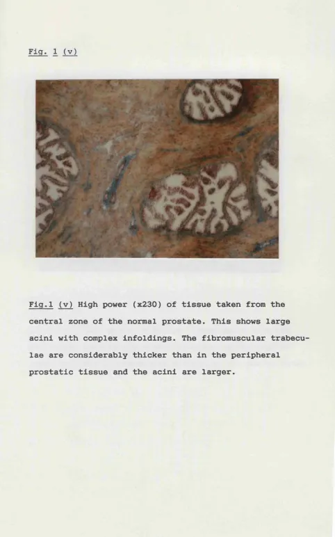
Fig. 1 (v)
%
4
Fig.l (v) High power (x230) of tissue taken from the central zone of the normal prostate. This shows large acini with complex infoldings. The fibromuscular trabecu lae are considerably thicker than in the peripheral
Fig.l (v i )
No major ducts arise from the urethral segment proximal to the verumontanum and this has been called the pre prostatic region. Surrounding this segment, a cylindrical smooth muscle sphincter extends down and is continuous with the bladder neck muscle. Within this cylinder, in the
immediate periurethral stroma, there is a group of glands (the periurethral glands) which appear incompletely deve loped and extend upwards towards the bladder neck. In order to attain full glandular development the periure thral ducts would either have to penetrate the cylindrical barrier or the muscle would have to be incomplete. It is only at the lower end of the prostatic urethra, near the base of the verumontanum where the sphincter is incom plete, that a small group of ducts is found which are able to branch out laterally towards the central zone and thus achieve glandular development. Though there is more duct branching and acinar proliferation than in the other periurethral ducts, it is still only a small area and constitutes only 5% of the glandular prostate. This area is macroscopically indistinguishable from neighbouring zones but is known as the transition zone and is thought to be the site of origin of early benign prostatic hyper plasia (McNeal 1978).
the weight of the gland. It is continuous with the detru sor muscle of the bladder neck and inseparably fused to the gland, producing the anterior convexity.
5. Pathological Anatomy
There is now general support for the concept that benign and malignant diseases of the prostate are independent entities and typically originate in different areas of the gland (McNeal 1969). Morgagni (1769) first observed as early as the 18th century that BPH was due to hyperplasia of the inner group of glands, but the location of these glands was not clearly defined until the end of the nine teenth century. In 1894, Jores described groups of submu cosal glands on the trigone, vesical neck and the urethral walls just below the bladder neck. These enlarged with age and median lobe enlargements arose from these glands. In addition, Albarran and Motz (1902) reported periurethral glands embedded in the inner longitudinal smooth muscle layer of the urethral wall between the verumontanum and bladder neck.
Since these observations there has been controversy concerning the exact site of origin of BPH. Moore (1944) believed that BPH arose in the lateral and middle lobes of the true prostate (central portion) as defined by Lowsley
in and limited to the immediate periurethral tissue, or to
a selected group of neighbouring "inner" prostatic glands
(Franks 1954). It is still not clear whether or not the
hyperplastic glandular tissue involved arises from the
local periurethral ducts (Le Duc 1939 and McNeal 1972) or
whether it is generated from branches of the main
prostatic ducts (Deming and Neumann 1939).
The exact anatomical landmarks and boundaries of this
region were incompletely defined until the work of McNeal
(1977), when he subjected the tissues within and lateral
to the proximal prostatic urethra to systematic examina
tion. Coronal sections were taken through the upper seg
ment of the urethra and, in planes parallel to this,
tracings were made of the anatomical boundaries and nod
ules. As a result he was able to identify the precise area
in which early nodules appeared. He concluded that most
nodules arose in the tissue that lies just proximal to the
verumontanum, close to the muscle of the distal end of the
bladder neck sphincter. Some nodules also seem to develop
in the periurethral glands, but these are small and do not
form the main mass of BPH tissue as described in previous
studies (Le Duc 1939, Pradham and Chandra 1975).
This small area along the long axis of the urethra may
well be the site of origin of BPH, but the further evolu
tion of BPH is unclear and the subject of some specula
tion. Further nodule genesis seems to occur in a focal and
and central zones of the gland, never appearing to spread
out to involve the peripheral zone where the majority of
the glandular tissue is situated. In many men over 70,
enlargement and glandular proliferation takes place within
the nodules already in existence.
Both stromal and epithelial hyperplasia may occur, alone
or together, and it is impossible to say that one occurs
before the other (Franks 1954). Further consideration
will be given to this aspect when discussing
stromal-epithelial interactions and the morphometry of BPH in more
d e t a i l .
The pathological anatomy of BPH highlights the regional
differences within the gland, with early diffuse growth of
the transition zone occurring without change in its archi
tecture, followed by massive enlargement of the nodules to
occupy the centre of the gland.
6. Ultrasonic anatomy
Prostatic ultrasound started with an attempt at
trans-rectal ultrasonography, which is now recognised as the
most suitable method of imaging the prostate routinely. In
1955 Wild and Reid presented their screw type transrectal
scanner, this was planned primarily for diagnosing tumours
of the rectum and large bowel.
The principle was identical with that of modern
of the inferior state of electronic technology at that
time.
Several urologists followed up this idea (Takahashi and
Ouchi 1963, Gotoh and Nishi 1965), reporting on the A-mode
appearances of the prostate via the transrectal route, but
this could not reveal the distribution of tissues in
various planes. The first radial scanning transrectal
probe was developed by Takahashi and Ouchi in 1964 and
although the picture quality was poor they obtained
hori-z onta1 tomograms.
Watanabe et al. (1968 and 1971) were the first to use an
ultrasonic probe with an oscillating disc in clinical
practice. Their images were improved by examining the
patients seated on a chair thus removing interference from
air bubbles. Over the next ten years real time ultrasonic
images steadily improved as a result of electronic
advances and the first linear scanner able to provide
longitudinal scans was introduced by Sekine et al. (1982).
This is still the state of the art scanner, although
additional attachments for needle guidance, allowing
accurate sampling of the prostate have been added (Saitoh
et al. 1981).
It is now possible with transrectal ultrasound (TRUS)
imaging to depict the major divisions and structures of
the prostate. Ultrasonically the organ can be divided into
an anterior component consisting of urethra and its
glandular tissue and ejaculatory apparatus. The centrally placed urethra can be divided into two equal segments as it angles forward at the proximal end of the verumontanum which provides a good reference point.
It is possible to distinguish the central and transition al zones from the peripheral zone both in the normal and the majority of diseased glands. However it is not pos sible to clearly distinguish the transitional and central zones in established BPH and biopsy them separately. In order to clarify the areas which have been biopsied the transitional and central zones, where BPH is confined, are combined and called the inner zone whereas the peripheral gland which is relatively spared is called the outer zone
(Figs.l (vii) and (viii). The peripheral (outer) zone, located posteriorly and laterally makes up most of the apex of the gland in the normal patient. It appears sono-graphically as a homogeneous, "isoechoic" pattern, due to the fine glandular and ductal tissues present here. This is taken as the baseline of isoechogenicity to which other areas are compared.
(inner) zones can be compared to the peripheral (outer)
zone. These sonographic appearances are variable depending
on which tissue component predominates. It m a y be
hypoe-choic with respect to the peripheral zone if the stromal
elements dominate, isoechoic like the peripheral zone, or
relatively hyperechoic if the glandular tissue dominates
(Weiss 1989).
In the majority of patients the boundary between the
peripheral (outer) zone and the remainder of the gland
becomes more clearly demarcated with progress of hyperpla
sia, particularly if the surgical capsule is outlined by a
ring of calcified corpora amylacea. However in other,
often smaller glands, the two zones appear more hypoechoic
and merge imperceptibly into each other.
The ejaculatory ducts are often imaged as paired cystic
structures at the base of the gland, and their associated
muscles may occasionally be seen as a hypoechoic halo
around the ducts. On sagittal scans the ducts are seen
running from the seminal vesicles towards the verumonta
num. The pre-prostatic urethra proximal to the verumonta
num can be seen angling towards the bladder neck, with the
post-prostatic urethra distally. The true prostatic cap
sule is seen sonographically as a bright echo structure
laterally while posteriorly it separates the gland from
the fascia and the rectum. The seminal vesicles lying
cephalad to the prostate are usually hyperechoic relative
Fig.l (vii)
Anterior fibromuscular
/ stroma
Inner zone
Outer zone
Seminal vesicle
F i g
,1
(vii) Diagram of the longitudinal zonal prostaticanatomy (BPH) and the corresponding transrectal ultrasound
image. The peripheral (outer) zone and central (inner)
Fig.l (viii)
A nterio r fib ro m u scu lar
C
strom aIn ner zon e
P Q
Outer zone
Fig.l (viii) Diagram of the tranverse zonal anatomy of the
prostate through line A in Fig.l (vii) and the correspond
The ultrasound appearances of prostate cancer initially were the subject of some controversy. However recent
studies, utilizing newer technology and anatomical infor mation, have shown that prostate cancer, rather than being hyperechoic as originally described, is actually hypoe choic or echopenic. Correlation of pathological abnormali ties of the prostate with sonographic patterns was carried out by Egender et al. (1984) who studied fresh cadaver prostates. They concluded that cancer was echopenic, thus supporting their in vivo experience which had shown that over 70% of patients with prostatic cancer had echopenic
lesions.
Dahnert et al. (1986), showed that BPH had a variable and irregular hypoechoic pattern in the central zone of the gland, whereas prostate cancer showed asymmetry with a tumour bulge posteriorly, and in 76% of cases, malignant lesions were hypoechoic. There was no case where cancer was hyperechoic. In Lee's series (1987) biopsy of relat ively hyperechoic lesions all demonstrated BPH.
Ultrasound has become an increasingly important imaging modality for the visualization of many internal soft
IV ENDOCRINOLOGY / BIOCHEMISTRY
1.Historical Background
(1) Steroid Hormones In growth control
Steroid hormones are Important physiological factors In controlling cell division and proliferation, and they promote long-term responses such as the maintenance of growth and function In their target tissues. John Hunter
(1786) was the first to recognise the Importance of the testes In the maintenance of the growth of the prostate long before the concept of steroid hormones had been postulated. The association of endocrine function with abnormal or neoplastic growth was first recognised by Sir Astley Cooper (1836) In his book "Lectures on the Princi ples and Practice of Surgery". He noticed that there was a correlation between the growth of the human breast, and the phase of the menstrual cycle. In 1896, Sir George Beatson, a Glasgow surgeon, described two patients with Inoperable breast carcinoma, whose tumours significantly regressed following bilateral salplngo-oophorectomy. This may have been the earliest demonstration of an endocrine responsive tumour. However this finding preceded the
Isolation of oestrogen from ovarian tissue and consequent ly the explanation for Beatson*s observation was not
Further advances In understanding of endocrine dependence of tumours were made following the isolation of steroid hormones. Doisy et al. (1929), isolated oestrone from the urine of pregnant women, and MacCorquodale et al. (1936) isolated a crystalline oestrogenic hormone from sow's
(ii) Steroid hormone action on the prostate
John Hunter (1786) contrasted the normal prostate of
animals "large and fleshy", to that of castrated animals
"small, flabby, tough, ligamentous and little secretion".
Although lacking a specific diagnosis. White (1895) trea
ted 111 men with bladder outflow obstruction by castra
tion, and demonstrated rapid atrophy of the enlarged
prostate gland. Cabot (1896) in a study with longer follow
up reported improvement in symptoms in 84 % of men with
BPH following orchidectomy. A Russian sect, the Skoptzs,
who were castrated at 35, were reported to have small
prostates and no symptoms of BPH (Zuckerman 193 6).
That castration or absence of testicular function could
have a preventative effect on the development of BPH was
also demonstrated by Moore (1944). He studied the pros
tates of 28 patients who were older than 45, but were
either eunuchs, eunuchoid or had pituitary infantilism and
had lost their secondary sexual characteristics before the
age of 40. He serially sectioned their prostates but
there was no evidence of BPH in any of this group.
However a further advance in the realisation that sex
hormones could influence the prostate and the growth of
other normal and neoplastic tissues, resulted from the
studies of Huggins and Clark (194 0) . They demonstrated
that orchidectomy or the administration of oestrogens to
reduce circulating androgens in dogs, caused reduction in
also Inhibited the secretion of prostatic fluid. The same year Huggins and Stevens (1940) described 3 men with BPH who were treated by castration. One had a marked reduction in prostatic size and all three were shown to have epithe lial atrophy on subsequent biopsy. This evidence supported the view that the prostatic epithelium at least, was under the control of the testes. They concluded that since the normal target organ is dependent upon hormonal support for growth and metabolic activity then cancer derived from it may be similarly dependent. In a now classic paper (Hug gins and Hodges 1941) it was demonstrated that castration or oestrogen injection caused regression of prostate
cancer and so began a new era in endocrine manipulation of tumours.
2. Studies of Steroid Hormones in the Pathogenesis of BPH (i) Plasma levels of steroid hormones
Many groups have investigated blood hormone levels in BPH patients and normal men, in an attempt to identify differ ences that might lead to conclusions regarding hormonal involvement in the aetiology of BPH. This has yielded very confusing data, and reports of alterations in hormone
lost with age (Bremner et al. 1983) and measurement of 24 hour mean plasma concentrations of testosterone shows a continuous decline with age (Zumoff et al. 1982).
In patients with BPH Hulka et al. (1987) found elevated serum testosterone levels but many others report no
significant difference in comparison to normal controls (Hammond 1978, Bartsch et al. 1979, Drafta et al. 1982, Brochu and Belanger 1987). However several studies
(Vermeulen et al. 1972, Pirke and Doerr 1975, Horton 1976, Rannikko and Aldercreutz 1983) report lower levels of
testosterone in BPH.
Although similar variations have been found for
oestrogens, Zumoff et al. (1982) who measured 24 hour mean oestrone and oestradiol in normal men found no alteration with age. Thus they proposed that a relative rise in
oestrogens may be involved in the pathogenesis of BPH, although two groups have also reported an absolute in crease in oestradiol with age (Rubens et al. 1974, Pirke and Doerr 1975).
fii) Tissue levels of steroid hormones
Several investigators have measured the tissue concentra
tions of various steroid hormones in BPH tissue and
compared these with "normal" prostate tissue. Siiteri and
Wilson (1970) found no significant difference in
testosterone levels but noted a four-fold increase in
dihydrotestosterone (DHT) levels in BPH tissue. Interest
ingly they also found higher DHT levels in the central
periurethral areas (where BPH characteristically
commences), than in the outer regions of the same gland.
This may be important as Farnsworth and Brown (1963) found
that DHT and androstenediol were the principal metabolites
of radioactive testosterone incubated with slices of human
BPH. It therefore appears that testosterone is a prohor
mone, and the active form of the androgen in the prostate
is DHT, produced by the action of the enzyme 5 alpha
reductase (Bruchovsky and Wilson 1968 a,b, Anderson and
Liao 1968). In contrast to most animals high levels of DHT
formation persist in the dog and in man throughout life
(Wilson and Gloyna 1970). Since these are the only two
species to spontaneously develop BPH, this highlights the
likely importance of DHT in prostatic growth control and
speculation on its role in BPH. Subsequently several
groups also found increased local DHT levels in BPH
(Geller et al. 1976, Hammond 1978, Meikle 1978, Kreig et
nediols and androsterone in parallel with an increase in DHT. Moore et al. (1979) showed increased prostatic DHT concentrations in androgen induced prostatic hyperplasia in the dog.
Walsh et al. 1983, have questioned these findings, since they found a similar DHT content in normal peripheral and BPH tissue obtained at open surgical procedures. A review of the literature reporting an elevated local DHT content in BPH reveals that in all series normal tissue was taken from autopsy specimens. Incubation of fresh prostatic tissue at 37°C even for a few hours, results in consider ably reduced DHT levels, and therefore in all these series the normal DHT levels were probably highly underestimated.
This casts considerable doubt on the role of DHT as a major causative factor in the pathogenesis of BPH. In fact experiments in age-matched beagles, demonstrated no dif ference in DHT content between dogs with histologically normal prostates and those with spontaneous BPH (Ewing et al. 1983). This has led to the search for other factors which may sensitise this tissue and accelerate growth.
(iii) Androgen Metabolism
meta-bollseâ further by 3-alpha or 3-beta hydroxysteroid dehy drogenase into the corresponding androstanediols and this step is known to be reversible. However the irreversible conversion of 3-beta androstanediol to the triol compound may play a role in the elimination of androgenic activity. The enzyme 17-hydroxy steroid dehydrogenase reversibly catalyses the interconversion between testosterone and an-drostenedione and DHT and androstanedione. Any change in any of these enzyme activities might cause a shift in the concentrations of several metabolites.
In view of the now controversial reports of a rise in DHT in BPH tissue, studies have tended to concentrate on 5-alpha reductase (DHT formation) and 3-5-alpha/beta dehydro genase (DHT removal). Most of these studies indicate an increased activity of 5-alpha reductase (Morfin et al. 1978, Kreig et al. 1979, Bruchovsky and Lievovsky 1979, Isaacs et al. 1983), with two of these (Bruchovsky and Lievovsky 1979 and Isaacs et al. 1983) also reporting a decrease in 3-alpha/beta hydroxysteroid dehydrogenase and or 17-hydroxysteroid dehydrogenase. Such changes would lead to an increase in intracellular DHT.
However these results must be treated with caution as they may be dependent on tissue heterogeneity and regional
concentrations in the stromal than in the epithelial
fraction, although Habib et al. (1983) did not support
this, and found that the capacity to metabolise testoster
one was evenly distributed between stroma and epithelium.
These studies are influenced by the techniques used for
the separation of tissue fractions and this may account
for some of the differences. If there is an uneven distri
bution of some of the enzymes between tissue fractions,
then the tissue composition (i.e. epithelial/stromal
ratio) could influence the metabolic capacity of the tis
sue. In a large biopsy series (Schweikert et al. 1985), no
correlation between 5-alpha reductase activity and epithe
lial content could be found.
( i v ) Role of Oestrogens in the Pathogenesis of BPH
Many researchers have speculated that oestrogens may have
a stimulatory role in the development of BPH. Walsh and
Wilson (1976) demonstrated that oestrogens were essential
for the development of BPH in the dog. Castrated dogs
treated with androgens plus oestrogens developed large
prostates, whereas those treated with androgen alone did
not. De Klerk et al. (1979) showed that BPH could be
experimentally induced with a combination of oestradiol
and DHT. Many other authors (Thompson et al. 1979, Murphy
et al. 1980 and Krieg et al. 1981) have also reported
BPH, and their activity in the development of hyperplasia. Only one study has indicated that BPH may be a result of an absolute increase in oestradiol in ageing men (Rannikko and Aldercreutz 1983). However an increase in the ratio of oestrogen to androgen, due to a fall in testosterone
(Zumoff et al. 1982) or a rise in sex hormone binding
globulin (SHBG) which reduces available androgen is a more likely stimulus for the development of BPH. Oestrogens may be produced locally in the prostate or peripherally
(adipose/muscle tissue) by aromatisation of androstene-dione (forming oestrone) or testosterone (forming oestra diol ). This conversion is catalysed by the cytochrome P450 dependent enzyme complex, aromatase. Certainly the experi ments of Schweikert (1979) using cultured BPH fibroblasts, and Kaburagi et al. (1987) and Stone et al. (1986), using whole tissue homogenates provide some evidence for local oestrogen production. In contrast other studies (Bartsch et al. 1987) have failed to show any endogenous aromatase activity in BPH tissue, by two different aromatase assays (oestrone formation and tritium release). This has recent ly been confirmed by J.Davies (1991 personal communica tion), who was also unable to find any increase in speci fic mRNA.
adrenal androgens. Tunn et al. (1985), studied the effects of treating patients with BPH with testolactone, a weak aromatase inhibitor and although this was not a randomised double blind study and the numbers were small they noted improvement in 60% of patients (7/13). More recently the same group (Schweikert and Tunn 1987) showed that
prostatic size was significantly reduced (15-26%) in 50% of patients taking testolactone and that compared to a control group, symptoms of outflow obstruction were im proved. However these results must be treated with caution as the observed effect may be due to inhibition of 17-beta hydroxysteroid dehydrogenase rather than of aromatase. This would result in a decrease in the formation of oe stradiol from oestrone and of testosterone from androste-nedione. Coffey (1988) recently confirmed that inhibition of endogenous aromatase activity was not an effective treatment for spontaneous BPH in the dog.
Further studies using the specific cDNA probe for the aromatase mRNA would enable determination of any gene overexpression either due to increased transcription or gene amplification. This would help clarify whether or not aromatase inhibition was a worthwhile therapeutic man oeuvre in BPH.
3) Mechanisms of steroid hormone action
Steroid hormones circulating in the blood are bound
predominantly to plasma proteins such as albumin, specific steroid binding globulins and alpha-feto protein; each of these carrier systems has a characteristic affinity and capacity. Testosterone, the major androgen in the circula tion, and oestrogens are transported largely bound to sex hormone binding globulin (SHBG) while progesterone circu lates predominantly bound to corticosteroid binding globu lin or transcortin. The delivery of hormone to specific target tissue is potentially a controlling mechanism and variation in the concentration of SHBG could be one of the factors that influence the biological effects of androgens on the prostatic cell. No control mechanism at the cell membrane has been found and testosterone and oestrogen are
Jensen and Jacobsen in 1962 at the University of Chicago synthesised tritiated oestradiol with a high specific activity and demonstrated its selective uptake and reten tion in target organs (pituitary, uterus and vagina) of female rats. The difference between target organ and non target organ is the ability to retain steroid (Peck et al. 1973).
( i ) Receptors
Toft and Gorski (1966) identified an oestrogen binding protein in the cytoplasm of rat uterine cells and referred to this as an oestrogen receptor. In 1968 Gorski et al. and Jensen et al. independently suggested a two step model for the mechanism of interaction of a steroid hormone with its target cell (Fig.l (ix)). In this model the steroid enters the cytoplasmic compartment of the target cell, and binds to its specific receptor protein. These receptors are characterised by their specificity, their high affin ity and low binding capacity (Giannoupolous and Gorski 1971). They have also been demonstrated to be heat and pH labile (Toft and Gorski 1966, Myatt et al. 1979). The cytoplasmic steroid-receptor complex has to undergo acti vation which is a temperature sensitive process by which
where it binds to specific nuclear sites, "acceptor" sites on the chromatin (Puca et al. 1974). The interaction of the activated cytoplasmic steroid-receptor complex with the nuclear acceptor sites results in the activation of RNA polymerases and an increased rate of transcription of specific DNA segments. This includes specific messenger RNA (mRNA) (Borthwick and Smellie 1975) which is transpor ted to the cytoplasm and results in synthesis of new
cellular proteins (Edwards et al. 1980) and often in cell growth and division. Following translocation the depleted cytoplasmic receptor pool is replenished possibly by new synthesis (Dix and Jordan 1980), although receptor recyc ling probably also occurs (Clark and Peck 1976).
F i g . l ( i x )
SHBG
lECEPTOR COMPLEX
JT ACTIVAT!
▲
FREE LIGAND
FREE CYTOPLASMIC RECEPTOR
TRANSLOCATION — )LACCEFTOF
SITE
CELL DIVISION DNA
SYNTHESIS RNA
PROTEIN SYNTHESIS
The mechanism for androgen action based on the two step
model for steroid receptors was reviewed extensively in a
monograph by Mainwaring (1977), and was accepted as the
mechanism by which androgens influenced prostatic cell
biochemistry until recently. In the light of further
studies the two step model for steroid receptor action has
been reconsidered. King and Greene (1984) and Welshons et
al. (1984) using immunocytochemical and biochemical
techniques have been unable to identify a cytoplasmic
receptor, and have proposed that the only intracellular
receptor is located within the nucleus (one-step model)
(Fig.l.(x). Welshons et al. (1984) have suggested that the
cytoplasmic receptor may be an artefact of tissue prepara
tion.
In support of the one step model are the observations on
the interaction of androgen with intact nuclear envelopes
prepared from rat ventral prostate tissue (LePebvre et al.
1985) and the exclusive nuclear localisation of 5-alpha
reductase in the human prostate (Houston et al. 1985,
Habib and Chisholm 1989). However there is no doubt that
unoccupied receptor which is only weakly nucleus associa
ted, is readily identified in cytosolic preparations.
There has now been a return of support for a cytoplasmic
steroid hormone receptor and it is thought that the anti
bodies used by Greene and co-workers may fail to recognise
the 8s oligomeric complex of ER (Pratt et al. 1989, Jensen
Fig.l (x)
SHBG
d)
TESTOSTERONE
s a REDUCTASE
RECEPTOR
CELL DIVISION m R N A DNA
SYNTHESIS
PROTEIN SYNTHESIS
DIAGRAM OF T H E MECHANISM OF ANDROGEN ACTION BASED ON THE ’ ONE S T E P ’ MODEL
(11) Androgen receptors In BPH