C A S E R E P O R T
Open Access
Riedel
’
s thyroiditis - a case report with genes
’
expression studies
Katarzyna Wojciechowska-Durczy
ń
ska
1, Adam Durczy
ń
ski
2, Stanis
ł
aw Sporny
3, Janusz Strzelczyk
2and
Andrzej Lewi
ń
ski
1*Abstract
Background:Genetic background of Riedel’s thyroiditis remains unknown. Herein, we describe our results of studies on genes expression levels in Riedel’s thyroiditis.
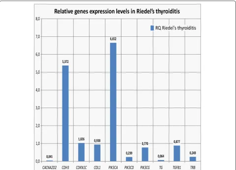
Case report and genetic findings:We report the case of 48-year old woman with Riedel’s thyroiditis who has presented unusual course of disease with non-specific cervical discomfort, though as with no pain and/or no compression symptoms. After surgery, thyroid specimens were quantitatively evaluated, regardingPIK3CA, PIK3CD, PIK3CG, Tg, TGFB1, THRB, COL1, CDKN1C, CDH3andCACNA2D2genes expression levels, by real-time PCR in the ABI PRISM® 7500 Sequence Detection System. Out of 10 above genes, in 2 cases the expression was higher than in respective Controls of unchanged thyroid tissue. In the remaining 8 cases, expression in question became comparable or lower as in Controls.
Discussion:The association between increased expression levels ofPIK3CAandCDH3genes and Riedel’s thyroiditis is not well-defined. However, the increased expression ofPIK3CAandCDH3genes in our case report and in previous studies of other authors on various malignancies may suggest possible molecular relation between Riedel’s thyroiditis and certain neoplastic processes, the relation of which requires further genetic evaluation. It is to be stressed that gene expression studies in Riedel’s thyroiditis are difficult to perform, mainly due to fibrosis, resulting in scarce thyroid specimens and - in consequence - small amount of genetic material.
Keywords:Riedel?’?s thyroiditis, Genes?’? expression, Real-time PCR
Background
Riedel’s thyroiditis is a rare fibrosclerotic infiltrative thyroid disorder of unclear etiology, during which normal thyroid tissues are replaced by fibrous tissue, often expanding out-side the thyroid capsule. The diagnosis can be difficult to establish prior to surgical removal of the thyroid and to sub-sequent histopathological examination of the resected gland [1-3]. Reports on genetic findings in Riedel’s thyroiditis are scarce in medical literature. Herein, we describe our results of studies on genes expression levels in Riedel’s thyroiditis.
Case report
We report the case of 48-year old women, diagnosed originally as a goiter with eminently increased
consistency on palpation, presented with non-specific cervical discomfort, though with no pain and/or no compression symptoms. Clinically, the thyroid was enlarged, painless, however very firm. Serum thyrotropin (TSH) and free thyroid hormones (FT3 and FT4) con-centrations were in normal ranges. Prior to surgery, the result of chest X-ray examination showed no pathologi-cal changes and all biochemipathologi-cal laboratory tests were normal. Ultrasound examination of the thyroid gland revealed few normoechoic foci in both lobes. Based on benign cytology of the nodules (normal thyroid follicular cells and colloid - Bethesda II) and the absence of com-pression symptoms, a subtotal thyroidectomy was per-formed. During surgery, a stony hard, white thyroid was disclosed. No complications after thyroid surgery were observed. Histopathological findings led to the diagnosis of Riedel’s thyroiditis, based on disappearance of thyroid follicles, which were replaced by the fibrous tissue and * Correspondence: [email protected]
1Department of Endocrinology and Metabolic Diseases, Polish Mother’s
Memorial Hospital Research Institute, Medical University of Lodz, Rzgowska St. No. 281/289, 93-338 Lodz, Poland
Full list of author information is available at the end of the article
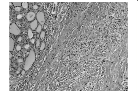
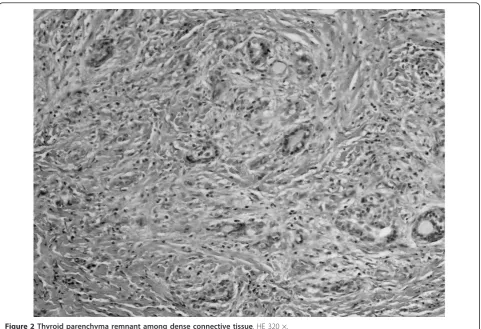
lymphocytes infiltrations, and features of focal endoar-teritis (Figures 1, 2, 3, 4). Postoperatively, the patient was treated with levothyroxine, 100 μg per day. At 4-months follow up, results TSH, FT3, FT4, PTH, cal-cium-phosphorus homeostasis and autoantibodies against thyroid peroxidase and TSH receptor were in normal ranges; autoantibodies against thyroglobulin (anti-Tg) were elevated. Ultrasound examination of the neck did not showed features of disease recurrence.
Genetic findings
After surgery, following conventional histopathological examination, thyroid specimen, received from patient with Riedel’s thyroiditis, was quantitatively evaluated regarding PIK3CA, PIK3CD, PIK3CG, Tg, TGFB1,
THRB, COL1, CDKN1C, CDH3 and CACNA2D2genes
expression levels by real-time PCR in the ABI PRISM
®7500 Sequence Detection System. Macroscopically
unchanged thyroid tissue, surgically removed from the patient with non-toxic nodular goitre (NTG), served as a control forreal-timePCR experiment.
Total RNA from tissue was extracted by use of an RNeasy Midi Kit (Qiagen, Hilden, Germany) based on modified Chomczyński and Sacchi’s according to
manufacturers’ recommendations. The purity of total RNA was assessed by NanoDrop® ND-100 spectropho-tometr and next the RNA was used in the first strand cDNA synthesis with TaqMan® Reverse Transcripton Reagents (Applied Biosystem) according to maufac-turers’ instruction. Real-time PCR was performed by using TaqMan® Universal PCR Master Mix (Applied Biosystem) and TaqMan® Gene Expression Assays probe and primer mix (Applied Biosystem) according to the manufacturer’ specification. Thermal cycler condi-tions were as follows: hold for 10 min. at 95°C, follow by two-step PCR for 50 cycles of 95°C for 15 s followed by 60°C for 1 min. Amplification reactions, in triplicate for each sample, were performed and the results were normalized to theACTBgene expression level.
An analysis of relative gene expression data was per-formed, using the 2-ΔΔCT method on an ABI PRISM® 7500 Sequence Detection System Software. The fold change in studied gene expression, normalised to endo-genous control, was calculated using: RQ = 2-ΔΔCT.
Out of 10 above genes, in 2 cases [PIK3CA- responsi-ble for coding alpha catalytic subunit of class I PI3K (phosphoinositide 3-kinase) andCDH3- responsible for coding P-cadherin] the expression was higher than in
Figure 1Thyroid parenchyma peripherally to dense connective tissue hyperplasia. HE 320 ×.
Wojciechowska-Durczyńskaet al.Thyroid Research2012,5:2 http://www.thyroidresearchjournal.com/content/5/1/2
respective Controls of unchanged thyroid tissue. In the remaining 8 cases, expression in question became com-parable or lower as in Controls (Figure 5).
Discussion
Riedel’s throiditis is a rare disease that most commonly occurs in females in their fifties and usually demon-strates as a goitre without clinical evidence of thyroid nodules. On palpation, thyroid demonstrates firm or stony consistency ("iron-hard thyroiditis”). Symptoms of regional compression might progressively appear. How-ever, the clinical course of this disease is mostly variable and may include cases with no thyroid-related symp-toms, as we have noted in our patient [3]. Before sur-gery, Riedel’s thyroiditis is often difficult to differentiate from aggressive types of thyroid cancers (i.e., undifferen-tiated thyroid carcinoma), nevertheless, a long-term fol-low-up shows no disease-specific mortality [4]. Likewise in presented patient, in vast majority of cases, only his-topathological, postoperative examination leads to diag-nosis of Riedel’s thyroiditis, due to no specific symptoms. Cytological evaluation is also of limited use in Riedel’s thyroiditis, due to firm thyroid tissue consis-tency and scanty amount of cellular material thus often
non-diagnostic thyroid specimens [5]. According to our opinion, in the case described by us, unusual course of the Riedel’s thyroiditis with the absence of compressive symptoms may be related to early-stage of the disease.
A number of hypotheses have been proposed regard-ing etiology and pathogenesis of Riedel’s thyroiditis. The disease may be associated with subacute and chronic thyroiditis, thus some authors proposed the, so called, “intrathyroidal hypothesis”. According to that hypoth-esis, the entity may represent the late stage (i.e., the pro-gression to fibrotic stage) of chronic inflammatory disorder [6-8]. On the contrary, the “pharmacological hypothesis” suggests that some particular medications may trigger Riedel’s thyroiditis development [8]. Although not proven, inherited susceptibility and devel-opment was hypothesized, as well [8]. Nowadays, the “systemic autoimmune hypothesis” seems to have the most support and it basically views Riedel’s thyroiditis as a manifestation of disordered fibroplastic proliferation because of hypersensivity reaction and consequent release of stimulating growth factors [9]. This theory considers a fibroblast not only as a final cell responsible for fibrosis, but also as a main target for well-defined systemic autoimmune attack [8]. In our case, the
Figure 3Arterial vessel among fibrosing thyroid parenchyma. HE 320 ×.
Figure 4Thyroid parenchyma peripherally to dense connective tissue hyperplasia. HE 320 ×.
Wojciechowska-Durczyńskaet al.Thyroid Research2012,5:2 http://www.thyroidresearchjournal.com/content/5/1/2
“systemic autoimmune hypothesis”of Riedel’s thyroiditis is the most plausible, it is supported by laboratory find-ings of autoimmune thyroid disease (anti-Tg), which have persisted after surgery.
PI3K/Akt (phosphoinositide 3-kinase/serine-threonine protein kinase) pathway participates in cellular signaling in response to various growth factors including FGF (fibro-blast growth factor). Activating mutations and amplifica-tion ofPIK3CAgene have been associated with enhancing PIK3CA kinase activity and Akt phosphorylation. Increased PI3K/Akt expression has been observed in both Hashimoto thyroiditis and well-differentiated thyroid can-cer [10,11]. Still, little is known about the precise role of
PIK3CAin fibrosclerotic disorders, however, it has been
proved that hepatic Akt expression correlated with advanced liver fibrosis in chronic hepatitis C patient [12].
Our results demonstrate that CDH3gene expression may increase in Riedel’s thyroiditis, as well. Cadherins, such as CDH3, are integral membrane glycoproteins responsible for calcium-dependent cell-cell adhesion. Our contemporary knowledge about the role of
P-cadherin in fibrosis and autoimmune processes is scanty and, so far, it has been only demonstrated that aberrant expression of E-cadherin/b-catenin complex is asso-ciated with a wide variety of human malignancies and fibrosis [13].
The association between increased expression levels of
PIK3CAandCDH3genes and Riedel’s thyroiditis is not
well-defined. However, the increased expression of
PIK3CAandCDH3genes in our case report and in
pre-vious studies of other authors on various malignancies may suggest possible molecular relation between Riedel’s thyroiditis and certain neoplastic processes, the relation which requires further genetic evaluation.
Low expression levels of the remaining studied genes could be associated with destruction of glandular thyr-oid tissue. The levels of TGFB1, COL1 and CDKN1C
genes expression were comparable in Riedel’s thyroiditis and in macroscopically unchanged tissue. It is to be emphasized that high expression levels of TGFB1and of
COL1A1 genes in chronic thyroiditis and its correlation
with thyroid fibrosis has previously been observed [14].
These results call into question the “intrathyroidal hypothesis”of Riedel’s disease pathogenesis. One should remind once again that gene expression studies in Rie-del’s thyroiditis are difficult to perform, mainly due to fibrosis, resulting in scarce thyroid specimens and - in consequence - small amount of genetic material.
Consent
Written informed consent was obtained from the patient for publication of this report and any accompanying images.
Author details
1Department of Endocrinology and Metabolic Diseases, Polish Mother’s
Memorial Hospital Research Institute, Medical University of Lodz, Rzgowska St. No. 281/289, 93-338 Lodz, Poland.2Department of General and Transplant Surgery, Medical University of Lodz, Barlicki University Hospital, Lodz, Poland.
3
Department of Dental Pathology, Medical University of Lodz, The Clinical-Didactic Centre, Lodz, Poland.
Authors’contributions
KW-D designed and coordinated the study, carried out the molecular genetic studies, writing a manuscript. AD, SS and JS participated in coordination of the study. AL senior supervision, writing a manuscript. All authors have read and approved the final manuscript.
Competing interests
The authors declare that they have no competing interests.
Received: 17 January 2012 Accepted: 7 March 2012 Published: 7 March 2012
References
1. Schwaegerle SM, Bauer TW, Esselstyn CB Jr:Riedel’s thyroiditis.Am J Clin Pathol1988,6:715-722, Review.
2. Papi G, Corrado S, Cesinaro AM, Novelli L, Smerieri A, Carapezzi C:Riedel’s thyroiditis: clinical, pathological and imaging features.Int J Clin Pract
2002,1:65-67.
3. Fontaine S, Gaches F, Lamant L, Uzan M, Bennet A, Caron P:An unusual form of Riedel’s thyroiditis: a case report and review of the literature. Thyroid2005,1:85-88.
4. Fatourechi MM, Hay ID, McIver B, Sebo TJ, Fatourechi V:Invasive fibrous thyroiditis (Riedel thyroiditis): the Mayo Clinic experience, 1976-2008. Thyroid2011,7:765-772.
5. Junik R, Juraniec O, Pypkowski J, Krymer A, Marszałek A:A difficult diagnosis: a case report of combined Riedel’s disease and fibrosing Hashimoto’s thyroiditis.Endokrynol Pol2011,62(4):351-356. 6. Zelmanovitz F, Zelmanovitz T, Beck M, Cerski CT, Schmid H,
Czepielewski MA:Riedel’s thyroiditis associated with high titers of antimicrosomal and antithyroglobulin antibodies and hypothyroidism.J Endocrinol Invest1994,9:733-737.
7. Papi G, Corrado S, Carapezzi C, De-Gaetani C, Carani C:Riedel’s thyroiditis and fibrous variant of Hashimoto’s thyroiditis: a clinicopathological and immunohistochemical study.J Endocrinol Invest2003,5:444-449. 8. Papi G, LiVolsi VA:Current concepts on Riedel thyroiditis.Am J Clin Pathol
2004,121:50-63.
9. Biddinger PW:Thyroiditis.InDiagnostic Pathology and molecular Genetics of the Thyroid.Edited by: Nikiforov YE. Lippincott Williams 2009:39-59. 10. Larson SD, Jackson LN, Riall TS, Uchida T, Thomas RP, Qiu S, Evers BM:
Increased incidence of well-differentiated thyroid cancer associated with Hashimoto thyroiditis and the role of the PI3k/Akt pathway.J Am Coll Surg2007,5:764-773.
11. Wojciechowska-Durczyńska K, Krawczyk-Rusiecka K, Cyniak-Magierska A, Zygmunt A, Gałecka E, Lewiński A:Relative quantification ofPIK3Cgene expression level in fine-needle aspiration biopsy thyroid specimens
collected from patients with papillary thyroid carcinoma and non-toxic goitre by real-time RT-PCR.Thyroid Res2010,3:5.
12. Huang JF, Chuang YH, Dai CY, Yu ML, Huang CF, Hsiao PJ, Hsieh MY, Huang CI, Yeh ML, Yang JF, Lin ZY, Chen SC, Chuang WL:Hepatic Akt expression correlates with advanced fibrosis in patients with chronic hepatitis C infection.Hepatol Res2011,5:430-436.
13. Tian X, Liu Z, Niu B, Zhang J, Tan TK, Lee SR, Zhao Y, Harris DC, Zheng G: E-cadherin/β-catenin complex and the epithelial barrier.J Biomed Biotechnol2011,2011:567305.
14. Cyniak-Magierska A, Januszkiewicz-Caulier J, Brzeziańska E, Lewiński A:
Analysis of correlation between the process of thyroid fibrosis and TGFB1 gene expression level in fine-needle aspiration biopsy (FNAB) thyroid specimens collected from patients with Hashimoto’s thyroiditis and non-toxic goitre.Exp Clin Endocrinol Diabetes2010,7:420-426.
doi:10.1186/1756-6614-5-2
Cite this article as:Wojciechowska-Durczyńskaet al.:Riedel’s thyroiditis
-a c-ase report with genes’expression studies.Thyroid Research20125:2.
Submit your next manuscript to BioMed Central and take full advantage of:
• Convenient online submission
• Thorough peer review
• No space constraints or color figure charges
• Immediate publication on acceptance
• Inclusion in PubMed, CAS, Scopus and Google Scholar
• Research which is freely available for redistribution
Submit your manuscript at www.biomedcentral.com/submit
Wojciechowska-Durczyńskaet al.Thyroid Research2012,5:2 http://www.thyroidresearchjournal.com/content/5/1/2