R E S E A R C H
Open Access
Aging impacts isolated lymphoid follicle
development and function
Keely G McDonald
1†, Matthew R Leach
1†, Conway Huang
2†, Caihong Wang
1, Rodney D Newberry
1*Abstract
Background:Immunosenescence is the age-related decline and dysfunction of protective immunity leading to a marked increase in the risk of infections, autoimmune disease, and cancer. The majority of studies have focused on immunosenescence in the systemic immune system; information concerning the effect of aging on intestinal immunity is limited. Isolated lymphoid follicles (ILFs) are newly appreciated dynamic intestinal lymphoid structures that arise from nascent lymphoid tissues, or cryptopatches (CP), in response to local inflammatory stimuli. ILFs promote“homeostatic” responses including the production of antigen-specific IgA, thus playing a key role in mucosal immune protection. ILF dysfunction with aging could contribute to immunosenescence of the mucosal system, and accordingly we examined phenotypic and functional aspects of ILFs from young (2 month old) and aged (2 year old) mice.
Results:We observed that aged mice have increased numbers of ILFs and increased numbers of structures corresponding to an early stage of CPs transforming into ILFs. The cellular composition of ILFs in aged mice is altered with a smaller B-lymphocyte population and an increased T-lymphocyte population. The ILF T-lymphocyte population is notable by the presence of CD4+ CD8aa+ T-lymphocytes, which are absent from the systemic compartment. The smaller B-lymphocyte population in ILFs from aged mice is directly correlated with decreased mRNA and protein expression of CCL20 and CXCL13, two chemokines that play crucial roles in recruiting B-lymphocytes into ILFs. Aged mice had elevated levels of serum and fecal immunoglobulins and despite the decreased B-lymphocyte population, ILFs from aged mice displayed increased IgA production. The immunoglobulin repertoire was skewed in aged mice, and ILFs demonstrated a repertoire usage similar to that of the systemic pool in both young and aged mice.
Conclusions:Here we observed that ILF development, cellular composition, and immunoglobulin production are altered with aging suggesting that ILF dysfunction contributes to mucosal immunosenescence.
Background
Immunosenescence is the age-related decline and dys-function in protective immunity with serious clinical consequences [1-4]. With aging, bacterial and viral infections in the lungs, skin, and urinary tract become more common [5-7]. Compounding this susceptibility to infection, the rates of seroconversion after prophylactic vaccination decrease proportionally with advancing age [8,9]. Related to the decreased ability to mount effective immune responses to pathogens, immunosenescence
also leads to a decline in effective immune surveillance potentiating an increased incidence of malignancy [10]. Finally, immunosenescence is not only associated with declining host immune competence, but also with immune dysregulation manifested by an increased inci-dence in autoimmune and chronic inflammatory disor-ders with increasing age [11].
Despite the earlier thoughts that the mucosal immune compartment was largely unaffected by aging, the muco-sal immune response is now believed to be compro-mised in old animals and elderly humans [12-14]. The impact of mucosal immunosenescence is highlighted by epidemiological studies demonstrating a marked increase in mortality due to gastrointestinal infections in the elderly in comparison to young adults [13]. Likewise, * Correspondence: [email protected]
†Contributed equally
1
Department of Internal Medicine, Washington University School of Medicine, St. Louis, Missouri 63110, USA
Full list of author information is available at the end of the article
age is also an important risk factor for colon cancer, the third most deadly cancer in the United States. Coinci-dent with this decline is an increased incidence of indi-viduals diagnosed with inflammatory bowel disease in their seventh decade of life, thus demonstrating a ten-dency toward the development of inappropriate mucosal immune responses with aging [15,16].
The mucosal immune system is a complex network generating immune responses that both protect the host and mitigate potential damage due to uncontrolled inflammation [17,18]. In the gastrointestinal tract this system includes diffuse effector sites, such as the intest-inal lamina propria (LP) and the intraepithelial lympho-cyte (IEL) compartment, as well as organized lymphoid structures that are collectively referred to as the gastroin-testinal-associated lymphoid tissue (GALT). Isolated lym-phoid follicles have recently become appreciated as distinct members of the GALT. ILFs resemble Peyer’s patches (PPs), the most widely studied lymphoid struc-ture in the small intestine, in architecstruc-ture and cellular composition. Like PPs, ILFs can possess germinal centers and an overlying follicle-associated epithelium (FAE) containing M cells [19]. In contrast to PP, whose forma-tion is developmentally driven, with early vital events occurring only during embryogenesis, ILFs develop after birth and arise from nascent lymphoid tissues, or crypto-patches (CPs), in response to luminal stimuli including alterations in the intestinal microbiota [19,20]. The adult murine intestine contains ~1000 CP, and thus in compar-ison to the relatively small (~10) fixed number of PP, there is potentially a much greater number of the ILFs that can contribute to mucosal immune responses. The function of CP and ILFs are incompletely understood. ILFs, but not CP, are known to act as sites for the induc-tion of adaptive immune responses [21], and studies of young animals demonstrate that ILFs function in a com-pensatory manner, promoting‘homeostatic’responses to local inflammatory stimuli including the production of antigen specific IgA [22]. Therefore a dysfunction in ILF development or function with aging has the potential to contribute to the immunosenescence of the intestinal immune system in a substantial way.
To gain insight into the role the ILFs play in intestinal immunosenescence, we examined the phenotypic and functional aspects of ILFs from aged and young mice. To our surprise, we observed an increase in the number of structures corresponding to transitioning CP and ILFs with aging. However, consistent with immune dysfunc-tion with aging, we found that ILFs from aged mice had a smaller B-lymphocyte population and an increased T-lymphocyte population when compared with their young counterparts. This finding correlated with decreased expression of the chemokines CXCL13 and CCL20, known to be important in recruiting B-lymphocytes into
ILFs, and decreased expression of their corresponding ligands, CXCR5 and CCR6, in ILFs of aged mice. Further analysis of the ILF T-lymphocyte population in aged mice revealed additional abnormalities characterized by the presence of a unique CD4+ CD8aa+ T-lymphocyte population. In agreement with prior observations, we found that the serum immunoglobulin levels in the aged mice were significantly increased and that fecal IgA levels were elevated with aging. Consistent with ILFs contribut-ing to dysfunction of the mucosal immune system with aging, we observed that ILFs from aged animals had ele-vated production of IgA. Evaluation of the B-lymphocyte immunoglobulin repertoire revealed that the ILF B-lym-phocyte population was reflective of the systemic B-cell population and developed a skewed VH usage in aged mice. These observations indicate that ILFs are altered with aging and that ILF dysfunction contributes to muco-sal immunosenescence.
Results
ILF formation is augmented at an early stage with aging
ILFs and CP are components of a spectrum of lymphoid aggregates that have been termed solitary intestinal phoid tissue (SILT) [20]. CPs are clusters of unique lym-phoid tissue inducer like cells lacking expression of surface markers defining mature bone marrow derived lineages. These clusters act as nascent lymphoid tissues giving rise to ILFs in response to inflammatory stimuli. As CP progress to become ILFs they become infiltrated with a substantial population of dendritic cells [23], and accordingly the presence of CD11c+ clusters can be used to identify stages of SILT ranging from CP pro-gressing to immature ILFs to fully developed mature ILFs. In comparison to CP, ILFs are larger, more com-plex cellular aggregates containing mature B- and T-lymphocytes. ILFs can range from lymphocyte con-taining cellular clusters encompassing the crypt and extending up the villi to more organized aggregates hav-ing a follicular appearance possesshav-ing an overlyhav-ing folli-cle associated epithelium (FAE). ILFs in all stages of development can be identified as clusters of B220+ cells and the more developed or mature ILFs can be identi-fied by their nodular appearance and FAE identiidenti-fied withUlex europaeus(UEA-I) staining.
to young mice (Figure 1b). We also observed an increased number of CD11c+ clusters identifying com-ponents of the SILT spectrum encompassing CP transi-tioning to iILFs and mILFs (Figure 1c). Prior reports indicate that the numbers of CP are relatively static, and we observed no difference in the numbers of CP between aged and young animals (not shown). Studies in young animals indicate that the influx of CD11c+ cells in response to inflammatory stimuli is an early event in the transformation of CPs into ILFs [23]. In conjunction with our findings of augmented ILF devel-opment with aging, this suggests that during aging the intestine is subject to chronic inflammation.
The cellular composition of the ILFs is altered with aging
The cellular composition of ILFs in young mice resembles that of PP and lymph nodes, and includes B-lymphocytes, T-lymphocytes (CD4 or CD8 single positive), and dendri-tic cells [19-21]. To gain insight into the function of ILFs in aging, we examined the cellular compositions of ILFs in aged and young mice using flow cytometry. We found that in comparison to young mice, the aged mice had a smaller population of B-lymphocytes (CD19+ cells) and an increased population of T-lymphocytes (TCRb+ cells) (Figure 2a, 2b). We observed no significant differences in
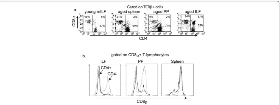
the antigen presenting cell population (CD19-, MHC II+) from the young and aged mice (Figure 2c). Further analy-sis of the increased T-lymphocyte population from aged mice revealed the presence of a unique CD4+CD8a+ (double positive) TCRb+ population (Figure 3a). These double positive T lymphocytes were rare in spleen and the PPs of the aged mice. The double positive cells repre-sented 2.33 +/- 0.145% and 19.05 +/- 3.55% (mean +/- SD) of the total ILF cellular population falling within the lym-phocyte gate for the young and aged mice, respectively. CD8 is a transmembrane glycoprotein that binds to class I MHC proteins and serves as a co-receptor for TCR. It forms a dimer, most commonly consisting of CD8aand CD8b(CD8ab+ heterodimer). Less commonly, homodi-mers of two CD8achains are also expressed. Further ana-lysis of the CD4+CD8a+ cells from ILFs of the aged mice revealed that these cells did not express CD8b, and hence are CD4+ CD8aa+ TCRb+ T-cells (Figure 3b). Likewise the minor population of CD4+ CD8a+ T-cells from the PP of aged mice were CD8b-, while the spleen of aged mice did not contain a CD8a+ CD8b- population (Figure 3b). The ILFs and PP of aged mice also contained CD8aa+ and CD8ab+ populations, while CD4-CD8aa+ populations were largely absent from the spleen (Figure 3b).
aged young * 150
100
50
0
mature ILFs
aged young *
150 300
0
immature ILFs
aged young *
400
200
0 600
CD1
1c+ clusters
a. b. c.
Figure 1ILF formation is augmented at an early stage with aging. To evaluate if the process of ILF development was altered with aging, whole mount techniques were used to identify well developed SILT containing a follicle associated epithelium indicative of mature ILFs (panel a), poorly developed or immature ILFs comprising a loose cluster of B-lymphocytes (panel b), and nascent lymphoid tissues transitioning into ILFs as identified by CD11c+ clusters (panel c) in young and aged mice. Aged mice had significantly increased numbers of mature ILFs (panel a), immature ILFs (panel b), and CD11c+ clusters (panel c), indicating that all stages of CP transitioning into ILFs are augmented with aging. n = 3 or more mice in each group. * = p < 0.05.
% APCs
(CD19- MHCII+)
15
10
5
0 c. 60
40
20
0
% B-lymphocytes
(CD19+)
a.
* 45
30
15
0
% T
-lymphocytes (TCR
β
+)
b. *
aged young aged young aged young
ILFs from aged mice have altered expression of chemokines and chemokine receptors required for ILF development
We previously reported that CC chemokine receptor-6 (CCR6) and its ligand CCL20 are highly expressed within ILFs and that B-lymphocytes are the largest CCR6-expressing population within ILFs [18]. Further-more, CCR6-sufficient B-lymphocytes are essential for the formation of ILFs [18]. Prior studies have demon-strated that CXC chemokine receptor-5 (CXCR5) and its ligand, CXCL13 or B-lymphocyte chemoattractant (BLC), are required for the migration of B lymphocytes to organized lymphoid follicles and the development of PP and SILT [23-25]. To assess the roles of these che-mokines and their receptors in the age-related augmen-tation of SILT formation and aberrant ILF cellular populations, we examined the mRNA expression of CCR6, CCL20, CXCR5, and CXCL13 in ILFs using real-time polymerase chain reaction. We noted that the mRNA expression of chemokines CCL20, CXCL13, and their receptors CCR6 and CXCR5 were significantly lower in ILFs of the aged mice than in those of the young mice, which correlated with decreased expression of CCL20 and CXCL13 protein in the intestine of aged mice (Figure 4). These factors are essential for B-lym-phocyte homing to lymphoid follicles, and their decreased expression is in agreement with the smaller B-lymphocyte population in the ILFs of aged mice.
Aged mice produce higher levels of systemic and mucosal immunoglobulins
The literature on immunoglobulin production in aged animals and humans is limited and somewhat conflict-ing. Some studies have reported increases or no change in the serum IgA levels in old animals and humans [26-29]. A few studies also reported the absence of age-related differences in the amounts of nonspecific immu-noglobulins secreted into the intestinal lumenin vivo or into the medium by cultured duodenal biopsies [28-31], while other studies observed that the production of anti-gen-specific IgA is diminished with aging [14,32,33].
We observed significantly elevated levels of IgA, IgG and IgM in the serum of the aged mice (Figure 5a-c), and increased levels of IgA in the feces of aged mice (Figure 5d). ILFs preferentially induce B-lymphocytes to become IgA producing plasma cells [22]. To evaluate the contribution of the ILFs to the increase in fecal IgA with aging, we isolated and cultured ILF cellular popula-tions and evaluated the IgA levels in the culture super-natant. Consistent with ILFs contributing to the increase in fecal IgA with aging, we found that ILFs from aged mice had elevated IgA production (Figure 5e).
ILF immunoglobulin repertoire reflects the systemic compartment and becomes skewed with aging
The development of ILFs is not completely understood, with some studies suggesting that ILFs develop from the
Figure 3ILFs from aged mice contain a population of CD4+ CD8aa+ T-lymphocytes. ILF, PP, and splenocyte populations from young and
aged mice were isolated and evaluated by multi-color flow cytometry for the presence of CD4+ and CD8a+ T-lymphocytes (panel a). ILFs from
aged mice contained a significant population of CD4+ CD8a+ T-cells, which was largely absent from the spleen and PP of aged mice and from
the ILFs of young mice (panel a). Further analysis of the CD4+ CD8a+ population within ILFs from aged mice revealed that this population was
CD8b-, and therefore CD8aa+ (black line histogram panel b). This CD4+ CD8aa+ T-lymphocyte population was also present in PP from aged
mice, but absent from the splenocyte population of aged mice (black line histogram panel b). Within the ILF and PP population from aged mice
the CD4- CD8a+ T-lymphocytes were both CD8b+ and CDb-, while in the spleen this population was only CD8b+ (gray line histograms panel
recruitment of peripheral B-lymphocytes and others sug-gesting that B-lymphocytes within ILFs expandin situto generate follicles. In support of the former possibility we previously reported that individual ILFs from young mice contain a population of polyclonal B-2 B lymphocytes that was reflective of the systemic pool of B-lymphocytes [22]. Consistent with the second possibility, others reported that the predominant immunoglobulin VHgene used within individual ILFs in aged activation-induced cytidine deaminase (AID)-deficient mice varied, suggest-ing that different B-lymphocytes expand within individual ILFs skewing the immunoglobulin repertoire usage such
that it was not reflective of the systemic compartment [34]. While these two possibilities are not mutually exclu-sive, they could suggest different pathways by which the number of ILFs would expand during aging; with the for-mer implying that the increased number of ILFs occurs by recruitment of B-lymphocytes representative of the systemic pool and the latter that increased ILF develop-ment results from expansion of B-lymphocytes within the ILFs, possibly reflective of a B-lymphocyte intrinsic pro-cess. To address these possibilities, we analyzed the immunoglobulin heavy chain repertoire usage within the spleen, PP, and individual ILFs from young and aged
aged young
0 1 2 3 4 5
6 CCL20
CCR6 CXCL13 CXCR5
fold change
0 25 50 75 100 125
CCL20 pg/mg
aged young
*
CXCL13 pg/mg
aged young 0
10 20 30 40
*
a. b.
Figure 4ILFs from aged mice express lower levels of chemokines and chemokine receptors demonstrated to play a role recruiting
B-lymphocytes into ILFs. RNA was isolated from pooled ILFs from aged and young mice. The mRNA expression of CCL20, CXCL13, CCR6, and
CXCR5 was evaluated using real time PCR (panel a), and the protein expression for intestinal CCL20 and CXCL13 was measured by ELISA (panel b). ILFs from aged mice showed decreased mRNA expression of chemokines CCL20 (~3 fold), CXCL13 (~4 fold) and their respective receptors CCR6 (~3.5 fold), CXCR5 (~6 fold) (panel a), and significantly decreased intestinal protein expression of CCL20 and CXCL13 (panel b). * = p < 0.05.
0.0 0.1 0.2 0.3 0.4 0.5 0.6
0 1 2 3 4 5 6 7 8 9
0 1 2 3
IgA
mg/ml
IgG mg/ml IgM mg/ml
a. b. c.
0 100 200 300 400 500
IgA
µg/ml
0 2500 5000 7500
IgA
ng/ml
d. e.
* * *
*
aged young aged young aged young
aged young aged young
serum
feces
ILF culture supernatant
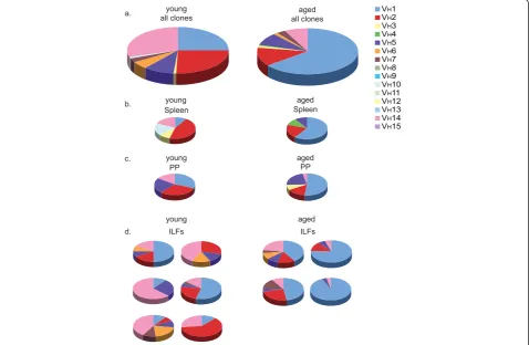
mice. We observed that the VH usage within ILFs from young mice was diverse, indicative of a polyclonal popu-lation, paralleling that of the spleen, which is representa-tive of the systemic pool (Figure 6a-d). Likewise, in the aged mouse the VHusage within all the ILFs examined was highly reflective of that seen in the spleen, and thus reflective of VHusage within the systemic compartment (Figure 6a-d). While both young and old mice had a simi-lar usage of VHfamilies, with VH1, VH2, and VH14 repre-senting greater than 80% of the VHfamilies used by both groups, the usage of an individual VH family became more skewed with aging as the VH1 family became the predominant family used in each tissue, representing over 60% of the VHfamily usage overall.
Discussion
Immunosenescence is a state of dysregulated immune function contributing to the increased susceptibility of
the elderly to infection, autoimmune diseases, chronic inflammatory diseases, and cancer [1-4]. The majority of studies on immunosenescence have examined the sys-temic immune system and support that syssys-temic immu-nity is affected with aging. Studies of the impact of aging on the mucosal immune system are less numer-ous, however emerging data more directly suggests an affect of aging on the mucosal immune system. Age greater than 65 has been identified as an independent risk factor for the development of C. difficlecolitis [35], an enteric infection whose recurrence and clinical course are related to the production of anti-toxin speci-fic IgA in the intestinal lumen [36]. Aging is associated with a decreased ability to produce antigen specific immunoglobulins, including IgA, following mucosal immunization [32,37-39], implying a direct impact of aging on the mucosal immune system, with a resultant increased risk of enteric infection. While it is becoming
VH1 VH2 VH3 VH4 VH5 VH6 VH7 VH8 VH9 VH10 VH11 VH12 VH13 VH14 VH15
young all clones
aged all clones
Spleen Spleen
PP PP
ILFs ILFs
a.
b. young
young
aged
aged
c. young aged
d.
Figure 6ILFs from young and aged mice contain a B-lymphocyte repertoire that is reflective of the systemic B lymphocyte pool; the
B-lymphocyte repertoire becomes skewed with aging. RNA isolated from the spleen, PP, and individual ILFs from a young and aged mouse
was transcribed into cDNA and immunoglobulin genes amplified using a universal VHprimer and a constant IgM region primer. Gel-purified
products were ligated into vectors and individual clones containing the appropriate size insert were sequenced and assigned to a VHfamily.
Analysis of all clones identified from the young (left) and aged mice (right) revealed a similar preferential usage of VHfamilies with VH1, VH2, and
VH14 representing greater than 80% of the VH families used by either mouse (panel a). With aging, the Ig repertoire became more skewed with
dominant usage of the VH1 family (panel a right). Comparison of the VHusage within the spleen (panel b), reflective of the systemic pool, to the
PP (panel c) and individual ILFs (panel d) revealed similar VHusage between these tissues within aged and young mice and revealed that the Ig
more clear that the mucosal immune system is affected by aging, the components of the mucosal immune sys-tem that are affected by aging and the functional conse-quences of these changes are still largely unexplored.
Organized lymphoid tissues play a crucial role in immunity by providing sites for the efficient interactions of antigens, antigen presenting cells, and lymphocytes. These structures are essential for the initiation of pri-mary immune responses, and the regulated environment they provide is believed to be necessary to prevent inap-propriate immune responses. Traditionally the organized lymphoid tissues of the intestine were thought to be limited to secondary lymphoid structures, or Peyer’s patches (PP). The foundations for PP formation only occur during embryogenesis, and therefore the number and positioning of PP are fixed throughout life [40-43]. Recent studies have shown that the adult intestine con-tains an additional population of organized lymphoid tissues, or solitary intestinal lymphoid tissues (SILT). SILT are a continuum of lymphoid tissues that have a unique plasticity allowing for the development and regression of a fully functional lymphoid tissue that can mediate homeostatic responses [17,19-21]. Within this continuum, CP act as nascent lymphoid tissues that sequentially become invested with dendritic cells and B-lymphocytes, to become ILFs [23]. Poorly developed, or immature ILFs (iILFs), can subsequently expand and develop a dome like structure with a follicle associated epithelium (FAE) to become mature ILFs (mILFs), which have the potential to initiate controlled adaptive immune responses to luminal antigens [21]. The murine intestine contains ~1000 SILT, and therefore in contrast to PP which are fixed and more limited in number (~10), SILT have a greater potential to act as sites for the induction of intestinal immune responses and con-versely to be affected by immunosenescence.
There is precedence for newly developed lymphoid tis-sues to contribute to chronic inflammatory and autoim-mune pathologies. Inducible lymphoid tissues, are markedly increased in conditions with inappropriate immune responses, such as those seen in inflammatory bowel disease (IBD) [20]. These aberrantly formed or tertiary lymphoid tissues have been proposed to initiate or propagate the inappropriate responses seen in auto-immune and chronic inflammatory conditions and har-bor the earliest manifestations of IBD [44,45]. Thus it is possible that ILFs formed aberrantly as a result of immunosenescence might subsequently contribute to immune dysfunction and participate in the increased incidence of IBD seen in the seventh decade of life.
Here we demonstrate that ILF formation is augmented with aging. We observed that the numbers of CPs con-taining DCs, iILFs, and mILFs were significantly increased in the aged mice. Aged mice were on average
25% larger than their gender and strain matched coun-terparts; average of 28.4 gm vs. 22.6 gm for 17 month old vs. 11 week old female Balb/c mice (information from the National Institute on Aging rodent colony). While this increase in body size could translate to a pro-portional increase in the number of ILFs, alone it is not sufficient to explain the three-fold increase in ILFs we observed. Furthermore, the cellular compositions of ILFs from the aged mouse are abnormal with a decreased B-lymphocyte population and the appearance of CD4 +CD8+ double positive T-lymphocytes. To assess the factors contributing to the decreased B lymphocyte population in ILFs with aging, we examined the expres-sion of the chemokines CCL20 and CXCL13, which have been demonstrated to play a role in recruiting B-lymphocytes into SILT [18,23,25]. Similar to the process of ILF formation, the production of CCL20 is induced by inflammatory conditions, and CCR6, the only identi-fied receptor of CCL20, is expressed by B-lymphocytes [46-49]. CCR6 is required for the development of ILFs and CCR6 deficient B-lymphocytes have reduced ability to localize to PPs and ILFs [18]. In a similar fashion, CXCR5 expression by B-lymphocytes is required for the development of ILFs, and ILF dendritic cells were iden-tified as sources for CXCL13 production to support ILF development [23,25]. CXCL13 not only recruits CXCR5 + B-lymphocytes, but also induces the expression of lymphotoxin (LT) by antigen-naïve B-lymphocytes, which further induces the production of CXCL13 by lymphotoxin beta receptor (LTbR) expressing stromal cells [24]. In addition, LT expression by B-lymphocytes is required for the development of SILT with large folli-cles, or mature ILFs [17]. Since CCR6+ B lymphocytes are also CXCR5+ [50], CCL20 and CXCL13 may act synergistically to recruit antigen naïve B-lymphocytes into ILFs and promote the development of mature folli-cles via LT/LTbR pathways. We observed a significant decrease in the expression of CCL20, CXCL13, and their respective receptors CCR6 and CXCR5 in the ILFs of aged mice when compared with those of the young mice. The decreased expression of CCL20 and CXCL13 and resultant decreased B-lymphocyte population in the ILFs of the aged mice is consistent with these previously observed functions of CCL20 and CXCL13. The combi-nation of a decreased expression of these chemokines and a decreased ILF B-lymphocyte population with a paradoxical increase in the number of ILFs suggests that alternative pathways may be emphasized in ILF develop-ment during aging.
specific differences (helper vs. cytolytic function respec-tively) as well as differences in major histocompatibility complex (MHC) restriction (MHC II vs. MHC I respec-tively). An exception to this generalization in which mature peripheral T-lymphocytes express both CD4 and CD8 co-receptors has been noted across many species [51-54]. The double positive cells can be grouped on the basis of CD8bexpression, i.e. CD4+CD8aa+ and CD4 +CD8ab+. There is evidence that the CD4 and CD8 co-receptors are functional in these double positive cells [55-57]. Regulating these double positive cells would be complex, as they could be more prone to generating inappropriate responses due to the multiplicity of recep-tors. The origin and function of the double positive cells is unclear. They have been suggested to be 1) recent thy-mic emigrants that are precursors of single positive cells, 2) single positive cells that have upregulated expression of the reciprocal coreceptor upon activation, 3) extrathy-mic T lymphocytes generated within the intestine, at the sites of CP, in response to the involution of the thymus with aging, or 4) T lymphocytes that were aberrantly selected in the thymus [53,54,56,58,59]. The double posi-tive cell were less common in PP and rare in the spleen of aged animals, supporting the preferential localization of this rare T-lymphocyte population to the intestinal mucosa. Double positive T-lymphocytes have been noted to accumulate in the peripheral blood and intraepithelial lymphocyte compartment with aging [54,55,59,60] and in target organs affected in autoimmune and chronic inflammatory conditions such as atopic dermatitis, sys-temic sclerosis, and autoimmune thyroid disease, sug-gesting that these cells may be responsible for the pathologic immune responses seen in these conditions [59]. Coupled with our observations, this suggests that with aging ILFs become sites of dysregulated immune responses potentially contributing to the increased inci-dence of IBD with aging.
A characteristic feature of the mucosal immune sys-tem is its ability to produce and secrete IgA. Some stu-dies reported the absence of age-related differences in the amounts of nonspecific immunoglobulins secreted into the intestinal lumen in vivoor into the medium by cultured duodenal biopsies, while others reported that the level of IgA increased in the intestinal juice from aged mice [31,61]. The sources contributing to lumenal IgA are complex and include both B1 and B2 lympho-cytes arising from the systemic immune system, mucosal immune system, and the peritoneal cavity. Mature ILFs are sites for the generation of IgA producing plasma cells from B2 B-lymphocyte precursors in response to lumenal antigens [21,22]. Therefore alterations in ILF development and/or function with aging could impact IgA production and mucosal protection. We observed that with aging IgA was significantly elevated in the
serum (~ 2 fold) and further elevated in the feces (~ 4 fold), implying a mucosal source that contributes to these elevated fecal IgA levels. While the relative contri-bution of each of the above mentioned sources to the elevated mucosal IgA levels can not be determined, the increased number of mature ILFs (~3 fold) seen with aging and the increased production of IgA by ILF cellu-lar populations (~ 5 fold) with aging are consistent with ILFs as contributors to the increased lumenal IgA and suggest that they become a more significant contributor to lumenal IgA with increasing age. The increased IgA seen in the aged ILFs occurs despite the decreased population of ILF B-lymphocytes. In concert with the increased number of ILFs, this further suggests the mucosal immune system becomes dysregulated with aging.
B-lymphocytes are a prominent hematopoietic cellular population within ILFs. The sources of B-lymphocytes that contribute to the emergence of the developing SILT are not completely understood, but they may originate from two sources that are not mutually exclusive and include B-lymphocytes that are recruited from the sys-temic pool or B-lymphocytes that expandin situ to gen-erate ILFs. To better understand how ILFs are gengen-erated during aging we evaluated the immunoglobulin reper-toire usage within ILFs and compared it to the systemic pool as represented by the immunoglobulin repertoire usage within the spleen. We observed that in both young and aged mice the ILF immunoglobulin reper-toire was reflective of that seen in the spleen. Like prior studies we also observed that B-lymphocyte repertoire usage becomes less diverse with aging [62]. However, this skewed repertoire usage was consistent throughout the tissues evaluated in the aged mice further supporting that ILF B-lymphocytes are recruited from the systemic pool. Multiple factors could contribute to the skewing of the B-lymphocyte repertoire, including diminished B-lymphocyte precursors and expansion of oligoclonal lymphocyte populations [63]. Skewing of the B-lymphocyte repertoire with aging is associated with poor health [62], and the observation that this also occurs within the mucosal immune compartment is further evi-dence that the mucosal immune system is impacted by aging and adversely affected by immunosenescence.
Conclusions
demonstrate that like the components of the systemic immune system, aging impacts these recently recognized contributors to mucosal immunity.
Methods
Mice
Female BALB/c mice of indicated age were purchased from National Institute on Aging (NIA) and were housed in a specific pathogen free facility and fed rou-tine chow diet. Young mice were 8-12 weeks old, and old mice were 21-23 months old. Mice were housed for at least 2 weeks prior to experiments to allow the muco-sal immune system to accommodate to changes in bac-terial flora. No differences in the measured parameters were observed with longer periods of housing. Animal procedures and protocols were carried out in accor-dance with the institutional review board at Washington University School of Medicine.
Whole mounts of small intestine
Small intestines were removed intact, flushed with cold PBS, and opened along the mesenteric border. Intestines were mounted, lumen facing up and fixed with cold 10% buffered formalin phosphate (Fisher Scientific) for 1 h at 4°C. Intestines were washed three times in cold PBS, incubated in a solution of 20 mM DTT, 150 mM Tris, and 20% ethanol at room temperature for 45 min, washed three times in cold PBS, and incubated in a solu-tion of 1% H2O2 for 15 min at room temperature to block endogenous peroxidases. Intestines were washed three times in PBS, followed by incubation in PBS con-taining 1% BSA and 0.3% Triton X-100 for 30 min. Intes-tines were incubated with HRP-conjugated lectin from Ulex europaeus(UEA-I) (Sigma-Aldrich, St. Louis, MO) in PBS containing BSA and Triton X-100 overnight at 4° C to facilitate the identification of PP and mature ILF (mILF). The following day, intestines were washed three times in PBS, incubated in DAB metal peroxide substrate (Pierce, Rockford, IL) for 15 min, rinsed twice in distilled water, and returned to PBS for further analysis. Under low-power microscopy (25-65X) the following criteria were used to determine the presence of mILF: 1) pre-sence of a nodular structure with size equal to or greater than the width of one villus, 2) nodular structure posses-sing an overlying dome resembling the FAE of PP, and 3) nodular structures occurring singly or in groups of two (three or more nodules of approximately the same size were considered to be PP).
For anti-B220 and anti-CD11c staining of whole mounts to determine the numbers of immature ILFs (iILFs) and CD11c+ clusters, identifying CP transitioning into ILFs and ILFs, intestines were removed intact, flushed with PBS, opened along the mesenteric border, and mounted as above. Intestines were then incubated
three times in HBSS (BioWhittaker, Walkersville, MD) containing 5 mM EDTA at 37°C with shaking for 10 minutes to remove epithelial cells. Intestines were then fixed in 10% buffered formalin phosphate and trea-ted with 1% H2O2 for 15 minutes at room temperature as above. Intestines were incubated in a solution of 50 mM Tris pH 7.2, 150 mM NaCl, 0.6% Triton-X 100, and 0.1% BSA for one hour at 4°C to block non-specific antibody binding and then incubated with rat mouse B220 antibody or biotin conjugated hamster anti-mouse CD11c (both from BDbiosciences, San Diego, CA) diluted in the above solution overnight at 4°C. Intestines were washed three times in the above solution and incubated with a horseradish peroxidase conjugated goat anti-rat IgG antibody or streptavidin conjugate horseradish peroxidase (Jackson ImmunoResearch Laboratories, West Grove, PA) diluted in the above solution at room temperature for one hour. Intestines were washed three times and incubated in DAB metal peroxide substrate as above. Intestine whole mounts were examined under a dissecting microscope at 25-65X. Immature ILFs (iILFs) were counted as clusters of B220+ cells occurring at the base of villi and not con-taining an overlying dome. Dendritic cell clusters were counted as clusters of CD11c+ cells occurring at the base of villi [17,20].
Isolation of cellular populations from spleen, PP, and ILFs
Spleens and PP were removed from BALB/c mice and disrupted by mechanical dissociation. Intestines were flushed with cold PBS, opened along the mesenteric border, and mounted with the lumen facing up in cold PBS, as described above. PP were identified, cut out, and disrupted using mechanical disassociation. Using a dis-secting microscope and a blunt-end 26-gauge needle and syringe, the contents of multiple mILFs were aspi-rated and placed in cold PBS. RBC were lysed from each cellular suspension and then used for flow-cytometric analysis as described below. Average yield of viable mononuclear ILFs cells ranged from 3-7 × 10⁵ cells/ intestine [17].
ELISAs for immunoglobulins, CCL20, and CXCL13
lysates obtained from sections of intestine from aged and young mice using commercially available assays (R and D systems, Minneapolis, MN) per the manufac-turers recommendations.
Flow Cytometric Analysis
Single cell suspensions, obtained as above were used for flow cytometric analysis. Briefly, single cell suspension were resuspended in PBS with 1% BSA (Fisher Scienti-fic) and 1 mg/ml human IgG (Sandoz Pharmaceuticals, East Hanover, NJ) at 2 × 10⁷ cells/ml. Cells were incu-bated with antibodies for 30 min on ice in the dark. Cells were washed in PBS and fixed with 1% paraformal-dehyde in PBS. Events were analyzed using a FACScan cytometer (BDbiosciences) retrofitted with a second laser. Data acquisition was performed using Cellquest (BDbiosciences) and Rainbow (Cytek, Fremont, CA) software. Dead cells were excluded based on forward and side light scatter. Gates for positive staining were defined such that 1% of the analyzed population stained positive with the appropriate isotype control antibodies (all from BD Biosciences).
RNA isolation and real-time polymerase chain reaction
Mature ILFs were isolated from both young and aged mice using a dissection microscope and 26-gauge blunt needle as described above. The aspirates containing the overlying FAE, stromal elements, and mononuclear cells from the same mouse were pooled together. RBCs were lysed from each cellular suspension. RNA was isolated using the Arc-turus PicoPure RNA isolation kit (Applied Biosystems, Foster City, CA), and treated with DNase I (Ambion, Aus-tin, TX) to remove contaminating DNA. cDNA was synthesized from 2μg of total RNA using Superscript II RNase H- reverse transcriptase (Invitrogen, Carlsbad, CA). Expression of targets was detected by real time PCR using ABI prism 7700 sequence detection system and SYBR Green PCR master mix (Applied Biosystems). The follow-ing primers were used for detection of the targets, forward primers are listed first followed by reverse primers: 18S 5’ -CGGCTACCACATCCAAGGAA-3’and 5’ -GCTGGAAT-TACCGCGGCT-3’,b-actin 5’-GCTTCTTTGCAGCT CCTTCGT-3’and 5’ -ATATCGTCATCCATGGCGAAC-3’, CCL20 5’-TGATGCTTTTTTGGGATGGAA-3’and 5’-AGCCTTCAACCCCAGCTGT-3’, CCR6 5’-TGTTC TGCTATCTGTTCATTATCAAGA-3’and 5’-CACGA CTCGGATGGCTCTGT-3’, CXCR5 5’-GGGCCCCTG TCTGTTTCTGT-3’and 5’ -GCCCAAGCTCGAGTTG-GAT-3’, and CXCL13 5’ -CAGAATGAGGCTCAGCA-CAGC-3’ and 5’-CAGAATACCGTGGCCTGGAG-3’. Samples were measured in triplicate. Relative quantitation of target expression using 18S orb-actin as a housekeep-ing gene was determined ushousekeep-ing the comparative CT
method as described in the ABI Prism 7700 sequence detection system user bulletin.
Immunoglobulin heavy chain repertoire analysis
Single cell populations from spleen, PP, and individual ILFs were isolated as described above and the RNA was isolated using the Arcturus PicoPure RNA isolation kit. cDNA was synthesized using the isolated RNA as tem-plate as described previously. The immunoglobulin genes were amplified with PCR using a universal VH primer 5’ -AGGTSMARCTGCAGSAGTCWGG-3’in combination with an IgM constant region primer 5’- CCCTGGAT-GACTTCAGTGTTG - 3’[65]. Amplified products were ligated into the pCR-BluntII-TOPO vector (Intivtrogen Corporation, Carlsbad, CA) and plasmids were isolated from individual clones and sequenced. Sequences were analyzed using The International Immunogenetics Infor-mation System (IMGT/V-Quest). Four hundred and forty seven individual clones were sequenced of which 298 clones could be definitively assigned to a VHfamily.
Statistical analysis
Data analysis using Student’s t test and one-way ANOVA followed by Tukey’s multiple comparisons post-test was performed using GraphPad Prism (Graph-Pad Software). A value of p < 0.05 was used as a cutoff for statistical significance.
List of abbreviations
CCL20: Chemokine (C-C motif) ligand 20; CCR6: C-C chemokine receptor 6; CP: cryptopatch; CXCL13: Chemokine (C-X-C motif) ligand 13; CXCR5: C-X-C chemokine receptor 5; IBD: inflammatory bowel disease; ILF: isolated
lymphoid follicle; iILF: immature ILF; LT: lymphotoxin; LTβR: lymphotoxin
beta receptor; mILF: mature ILF; PP: Peyer’s patch.
Acknowledgements
This work was supported in part by grants DK64798-RDN and AG028309-RDN. We thank the Alvin J. Siteman Cancer Center at Washington University School of Medicine and Barnes-Jewish Hospital in St. Louis, MO, for the use of the High Speed Cell Sorter Core, which provided the flow cytometry service. The Siteman Cancer Center is supported in part by an NCI Cancer Center Support Grant #P30 CA91842.
Author details
1
Department of Internal Medicine, Washington University School of
Medicine, St. Louis, Missouri 63110, USA.2University of Texas Southwestern
Medical School, Austin Texas, 78701, USA.
Authors’contributions
KM performed whole mount analysis of intestines, flow cytometry of cellular populations, isolation and in vitro culture of ILF cellular populations, and ELISAs. ML performed experiments related to the analysis of the immunoglobulin repertoire and ELISAs for CCL20 and CXCL13. CH and CW performed ELISAs for immunoglobulins and gene expression analysis. CH helped draft the manuscript. RN conceived of the study, participated in the design and analysis of the experiments, and drafted the manuscript. All authors read and approved the final manuscript.
Competing interests
Received: 30 August 2010 Accepted: 7 January 2011 Published: 7 January 2011
References
1. Castle SC, Uyemura K, Rafi A, Akande O, Makinodan T:Comorbidity is a
better predictor of impaired immunity than chronological age in older adults.J Am Geriatr Soc2005,53(9):1565-1569.
2. Castle SC, Uyemura K, Fulop T, Makinodan T:Host resistance and immune
responses in advanced age.Clin Geriatr Med2007,23(3):463-479.
3. Castle SC:Impact of age-related immune dysfunction on risk of
infections.Z Gerontol Geriatr2000,33(5):341-349.
4. Pawelec G:Immunosenescence: impact in the young as well as the old?
Mech Ageing Dev1999,108(1):1-7.
5. Hakim FT, Gress RE:Immunosenescence: deficits in adaptive immunity in
the elderly.Tissue Antigens2007,70(3):179-189.
6. Janssens JP, Krause KH:Pneumonia in the very old.Lancet Infect Dis2004,
4(2):112-124.
7. Castle SC:Clinical relevance of age-related immune dysfunction.Clin
Infect Dis2000,31(2):578-585.
8. Bouree P:Immunity and immunization in elderly.Pathol Biol (Paris)2003,
51(10):581-585.
9. Kang I, Hong MS, Nolasco H, Park SH, Dan JM, Choi JY, Craft J:
Age-associated change in the frequency of memory CD4+ T cells impairs long term CD4+ T cell responses to influenza vaccine.J Immunol2004,
173(1):673-681.
10. Zhang HG, Grizzle WE:Aging, immunity, and tumor susceptibility.
Immunol Allergy Clin North Am2003,23(1):83-102.
11. Johnson SA, Cambier JC:Ageing, autoimmunity and arthritis: senescence
of the B cell compartment - implications for humoral immunity.Arthritis Res Ther2004,6(4):131-139.
12. Schmucker DL, Heyworth MF, Owen RL, Daniels CK:Impact of aging on
gastrointestinal mucosal immunity.Dig Dis Sci1996,41(6):1183-1193.
13. Schmucker DL, Owen RL, Outenreath R, Thoreux K:Basis for the
age-related decline in intestinal mucosal immunity.Clin Dev Immunol2003,
10(2-4):167-172.
14. Fujihashi K, McGhee JR:Mucosal immunity and tolerance in the elderly.
Mech Ageing Dev2004,125(12):889-898.
15. Lindner AE:Inflammatory bowel disease in the elderly.Clin Geriatr Med
1999,15(3):487-497.
16. Grimm IS, Friedman LS:Inflammatory bowel disease in the elderly.
Gastroenterol Clin North Am1990,19(2):361-389.
17. McDonald KG, McDonough JS, Newberry RD:Adaptive immune responses
are dispensable for isolated lymphoid follicle formation: antigen-naive, lymphotoxin-sufficient B lymphocytes drive the formation of mature isolated lymphoid follicles.J Immunol2005,174(9):5720-5728.
18. McDonald KG, McDonough JS, Wang C, Kucharzik T, Williams IR,
Newberry RD:CC chemokine receptor 6 expression by B lymphocytes is
essential for the development of isolated lymphoid follicles.Am J Pathol
2007,170(4):1229-1240.
19. Hamada H, Hiroi T, Nishiyama Y, Takahashi H, Masunaga Y, Hachimura S,
Kaminogawa S, Takahashi-Iwanaga H, Iwanaga T, Kiyono H,et al:
Identification of multiple isolated lymphoid follicles on the antimesenteric wall of the mouse small intestine.J Immunol2002,
168(1):57-64.
20. Lorenz RG, Chaplin DD, McDonald KG, McDonough JS, Newberry RD:
Isolated Lymphoid Follicle Formation Is Inducible and Dependent Upon Lymphotoxin-Sufficient B Lymphocytes, Lymphotoxin beta Receptor, and TNF Receptor I Function.J Immunol2003,170(11):5475-5482.
21. Lorenz RG, Newberry RD:Isolated Lymphoid Follicles Can Function as
Sites for Induction of Mucosal Immune Responses.Ann N Y Acad Sci
2004,1029:44-57.
22. Wang C, McDonald KG, McDonough JS, Newberry RD:Murine isolated
lymphoid follicles contain follicular B-lymphocytes with a mucosal phenotype.Am J Physiol Gastrointest Liver Physiol2006,291(4):G595-604, Epub 2006 Jun 15.
23. McDonald KG, McDonough JS, Dieckgraefe BK, Newberry RD:Dendritic
cells produce CXCL13 and participate in the development of murine small intestine lymphoid tissues.Am J Pathol176(5):2367-2377.
24. Ansel KM, Ngo VN, Hyman PL, Luther SA, Forster R, Sedgwick JD,
Browning JL, Lipp M, Cyster JG:A chemokine-driven positive feedback
loop organizes lymphoid follicles.Nature2000,406(6793):309-314.
25. Velaga S, Herbrand H, Friedrichsen M, Jiong T, Dorsch M, Hoffmann MW,
Forster R, Pabst O:Chemokine receptor CXCR5 supports solitary intestinal
lymphoid tissue formation, B cell homing, and induction of intestinal IgA responses.J Immunol2009,182(5):2610-2619.
26. Schmucker DL, Thoreux K, Owen RL:Aging impairs intestinal immunity.
Mech Ageing Dev2001,122(13):1397-1411.
27. Finkelstein MS, Tanner M, Freedman ML:Salivary and serum IgA levels in
a geriatric outpatient population.J Clin Immunol1984,4(2):85-91.
28. Arranz E, O’Mahony S, Barton JR, Ferguson A:Immunosenescence and mucosal
immunity: significant effects of old age on secretory IgA concentrations and intraepithelial lymphocyte counts.Gut1992,33(7):882-886.
29. Koga T, McGhee JR, Kato H, Kato R, Kiyono H, Fujihashi K:Evidence for
early aging in the mucosal immune system.J Immunol2000,
165(9):5352-5359.
30. Penn ND, Purkins L, Kelleher J, Heatley RV, Mascie-Taylor BH:Ageing and
duodenal mucosal immunity.Age Ageing1991,20(1):33-36.
31. Senda S, Cheng E, Kawanishi H:Aging-associated changes in murine
intestinal immunoglobulin A and M secretions.Scand J Immunol1988,
27(2):157-164.
32. Thoreux K, Owen RL, Schmucker DL:Intestinal lymphocyte number,
migration and antibody secretion in young and old rats.Immunology
2000,101(1):161-167.
33. Schmucker DL:Intestinal mucosal immunosenescence in rats.Exp
Gerontol2002,37(2-3):197-203.
34. Fagarasan S, Muramatsu M, Suzuki K, Nagaoka H, Hiai H, Honjo T:Critical
roles of activation-induced cytidine deaminase in the homeostasis of gut flora.Science2002,298(5597):1424-1427.
35. McDonald LC, Owings M, Jernigan DB:Clostridium difficile infection in
patients discharged from US short-stay hospitals, 1996-2003.Emerg Infect
Dis2006,12(3):409-415.
36. Warny M, Vaerman JP, Avesani V, Delmee M:Human antibody response to
Clostridium difficile toxin A in relation to clinical course of infection.
Infect Immun1994,62(2):384-389.
37. Schmucker DL, Daniels CK, Wang RK, Smith K:Mucosal immune response
to cholera toxin in ageing rats. I. Antibody and antibody-containing cell response.Immunology1988,64(4):691-695.
38. Taylor LD, Daniels CK, Schmucker DL:Ageing compromises
gastrointestinal mucosal immune response in the rhesus monkey.
Immunology1992,75(4):614-618.
39. Enioutina EY, Visic VD, Daynes RA:Enhancement of common mucosal
immunity in aged mice following their supplementation with various antioxidants.Vaccine2000,18(22):2381-2393.
40. Chaplin DD, Fu Y:Cytokine regulation of secondary lymphoid organ
development.Curr Opin Immunol1998,10(3):289-297.
41. De Togni P, Goellner J, Ruddle NH, Streeter PR, Fick A, Mariathasan S,
Smith SC, Carlson R, Shornick LP, Strauss-Schoenberger J,et al:Abnormal
development of peripheral lymphoid organs in mice deficient in lymphotoxin.Science1994,264(5159):703-707.
42. Rennert PD, Browning JL, Hochman PS:Selective disruption of
lymphotoxin ligands reveals a novel set of mucosal lymph nodes and unique effects on lymph node cellular organization.Int Immunol1997,
9(11):1627-1639.
43. Rennert PD, Browning JL, Mebius R, Mackay F, Hochman PS:Surface
lymphotoxin alpha/beta complex is required for the development of peripheral lymphoid organs.J Exp Med1996,184(5):1999-2006.
44. Fujimura Y, Kamoi R, Iida M:Pathogenesis of aphthoid ulcers in Crohn’s
disease: correlative findings by magnifying colonoscopy, electron microscopy, and immunohistochemistry.Gut1996,38(5):724-732.
45. Lockhart-Mummery HE, Morson BC:Crohn’s disease (regional enteritis) of
the large intestine and its distinction from ulcerative colitis.Gut1960,
1:87-105.
46. Tanaka Y, Imai T, Baba M, Ishikawa I, Uehira M, Nomiyama H, Yoshie O:
Selective expression of liver and activation-regulated chemokine (LARC) in intestinal epithelium in mice and humans.Eur J Immunol1999,
29(2):633-642.
47. Izadpanah A, Dwinell MB, Eckmann L, Varki NM, Kagnoff MF:Regulated
MIP-3alpha/CCL20 production by human intestinal epithelium: mechanism for modulating mucosal immunity.Am J Physiol Gastrointest Liver Physiol2001,280(4):G710-719.
48. Kaser A, Ludwiczek O, Holzmann S, Moschen AR, Weiss G, Enrich B,
expression of CCL20 in human inflammatory bowel disease.J Clin Immunol2004,24(1):74-85.
49. Kwon JH, Keates S, Bassani L, Mayer LF, Keates AC:Colonic epithelial cells
are a major site of macrophage inflammatory protein 3alpha (MIP-3alpha) production in normal colon and inflammatory bowel disease.
Gut2002,51(6):818-826.
50. Bowman EP, Campbell JJ, Soler D, Dong Z, Manlongat N, Picarella D,
Hardy RR, Butcher EC:Developmental switches in chemokine response
profiles during B cell differentiation and maturation.J Exp Med2000,
191(8):1303-1318.
51. Ishimoto Y, Tomiyama-Miyaji C, Watanabe H, Yokoyama H, Ebe K, Tsubata S,
Aoyagi Y, Abo T:Age-dependent variation in the proportion and number
of intestinal lymphocyte subsets, especially natural killer T cells, double-positive CD4+ CD8+ cells and B220+ T cells, in mice.Immunology2004,
113(3):371-377.
52. Yamada K, Kimura Y, Nishimura H, Namii Y, Murase M, Yoshikai Y:
Characterization of CD4+ CD8alphaalpha+ and CD4-CD8alphaalpha+ intestinal intraepithelial lymphocytes in rats.Int Immunol1999,
11(1):21-28.
53. Jimenez E, Sacedon R, Vicente A, Hernandez-Lopez C, Zapata AG, Varas A:
Rat peripheral CD4+CD8+ T lymphocytes are partially
immunocompetent thymus-derived cells that undergo post-thymic maturation to become functionally mature CD4+ T lymphocytes.J Immunol2002,168(10):5005-5013.
54. Colombatti A, Doliana R, Schiappacassi M, Argentini C, Tonutti E, Feruglio C,
Sala P:Age-related persistent clonal expansions of CD28(-) cells:
phenotypic and molecular TCR analysis reveals both CD4(+) and CD4(+) CD8(+) cells with identical CDR3 sequences.Clin Immunol Immunopathol
1998,89(1):61-70.
55. Laux I, Khoshnan A, Tindell C, Bae D, Zhu X, June CH, Effros RB, Nel A:
Response differences between human CD4(+) and CD8(+) T-cells during CD28 costimulation: implications for immune cell-based therapies and studies related to the expansion of double-positive T-cells during aging.
Clin Immunol2000,96(3):187-197.
56. Kenny E, Mason D, Saoudi A, Pombo A, Ramirez F:CD8 alpha is an
activation marker for a subset of peripheral CD4 T cells.Eur J Immunol
2004,34(5):1262-1271.
57. Hori T, Paliard X, de Waal Malefijt R, Ranes M, Spits H:Comparative analysis
of CD8 expressed on mature CD4+ CD8+ T cell clones cultured with IL-4 and that on CD8+ T cell clones: implication for functional significance of CD8 beta.Int Immunol1991,3(7):737-741.
58. Akari H, Terao K, Murayama Y, Nam KH, Yoshikawa Y:Peripheral blood CD4
+CD8+ lymphocytes in cynomolgus monkeys are of resting memory T lineage.Int Immunol1997,9(4):591-597.
59. Parel Y, Chizzolini C:CD4+ CD8+ double positive (DP) T cells in health
and disease.Autoimmun Rev2004,3(3):215-220.
60. Lee WW, Nam KH, Terao K, Akari H, Yoshikawa Y:Age-related increase of
peripheral CD4+ CD8+ double-positive T lymphocytes in cynomolgus monkeys: longitudinal study in relation to thymic involution.Immunology
2003,109(2):217-225.
61. Ebersole JL, Smith DJ, Taubman MA:Secretory immune responses in
ageing rats. I. Immunoglobulin levels.Immunology1985,56(2):345-350.
62. Gibson KL, Wu YC, Barnett Y, Duggan O, Vaughan R, Kondeatis E,
Nilsson BO, Wikby A, Kipling D, Dunn-Walters DK:B-cell diversity decreases
in old age and is correlated with poor health status.Aging Cell2009,
8(1):18-25.
63. Cancro MP, Hao Y, Scholz JL, Riley RL, Frasca D, Dunn-Walters DK,
Blomberg BB:B cells and aging: molecules and mechanisms.Trends
Immunol2009,30(7):313-318.
64. Tsuji M, Suzuki K, Kitamura H, Maruya M, Kinoshita K, Ivanov II, Itoh K,
Littman DR, Fagarasan S:Requirement for lymphoid tissue-inducer cells in
isolated follicle formation and T cell-independent immunoglobulin A generation in the gut.Immunity2008,29(2):261-271.
65. Stoel M, Jiang HQ, van Diemen CC, Bun JC, Dammers PM, Thurnheer MC,
Kroese FG, Cebra JJ, Bos NA:Restricted IgA repertoire in both B-1 and B-2
cell-derived gut plasmablasts.J Immunol2005,174(2):1046-1054.
doi:10.1186/1742-4933-8-1
Cite this article as:McDonaldet al.:Aging impacts isolated lymphoid
follicle development and function.Immunity & Ageing20118:1.
Submit your next manuscript to BioMed Central and take full advantage of:
• Convenient online submission
• Thorough peer review
• No space constraints or color figure charges
• Immediate publication on acceptance
• Inclusion in PubMed, CAS, Scopus and Google Scholar
• Research which is freely available for redistribution