Structural bioinformatics
Self-consistency test reveals systematic bias
in programs for prediction change of stability
upon mutation
Dinara R. Usmanova
1, Natalya S. Bogatyreva
2,3,4, Joan Ari
no Bernad
~
5,
Aleksandra A. Eremina
6, Anastasiya A. Gorshkova
7,
German M. Kanevskiy
8, Lyubov R. Lonishin
9, Alexander V. Meister
10,
Alisa G. Yakupova
7, Fyodor A. Kondrashov
11and Dmitry N. Ivankov
4,11,*
1Department of Systems Biology, Columbia University Medical Center, New York, NY 10032, USA,
2Bioinformatics
and Genomics Programme, Centre for Genomic Regulation (CRG), Barcelona 08003, Spain,
3Universitat Pompeu
Fabra (UPF), Barcelona 08003, Spain,
4Laboratory of Protein Physics, Institute of Protein Research of the Russian
Academy of Sciences, Pushchino, Moscow Region 142290, Russia,
5Centre de Formacio´ Interdisciplina`ria
Superior, Universitat Polite`cnica de Catalunya, Barcelona 08028, Spain,
6School of Biological Sciences, College of
Science and Engineering, University of Edinburgh, Edinburgh EH9 3BF, UK,
7Biological Faculty, Lomonosov
Moscow State University, Moscow 119991, Russia,
8Higher Chemical College of the Russian Academy of Sciences,
Moscow
125047,
Russia,
9Faculty
of
Technical
Physics,
Institute
of
Physics,
Nanotechnology
and
Telecommunications, Peter the Great Saint-Petersburg Polytechnic University, Saint-Petersburg 195251, Russia,
10Department of Medicine, Novosibirsk State University, Novosibirsk 630090, Russia and
11Institute of Science and
Technology, Klosterneuburg 3400, Austria
*To whom correspondence should be addressed. Associate Editor: Alfonso ValenciaReceived on October 5, 2017; revised on March 15, 2018; editorial decision on April 23, 2018; accepted on April 30, 2018
Abstract
Motivation:
Computational prediction of the effect of mutations on protein stability is used by
researchers in many fields. The utility of the prediction methods is affected by their accuracy and
bias. Bias, a systematic shift of the predicted change of stability, has been noted as an issue for
several methods, but has not been investigated systematically. Presence of the bias may lead to
misleading results especially when exploring the effects of combination of different mutations.
Results:
Here we use a protocol to measure the bias as a function of the number of introduced
muta-tions. It is based on a self-consistency test of the reciprocity the effect of a mutation. An advantage of
the used approach is that it relies solely on crystal structures without experimentally measured
stability values. We applied the protocol to four popular algorithms predicting change of protein
stability upon mutation, FoldX, Eris, Rosetta and I-Mutant, and found an inherent bias. For one program,
FoldX, we manage to substantially reduce the bias using additional relaxation by Modeller. Authors
using algorithms for predicting effects of mutations should be aware of the bias described here.
Availability and implementation:
All calculations were implemented by in-house PERL scripts.
Contact:
Supplementary information:
Supplementary data
are available at
Bioinformatics
online.
Note:
The article 10.1093/bioinformatics/bty348, published alongside this paper, also addresses the
problem of biases in protein stability change predictions.
VCThe Author(s) 2018. Published by Oxford University Press. 3653
This is an Open Access article distributed under the terms of the Creative Commons Attribution Non-Commercial License (http://creativecommons.org/licenses/by-nc/4.0/), which permits non-commercial re-use, distribution, and reproduction in any medium, provided the original work is properly cited. For commercial re-use, please contact [email protected]
doi: 10.1093/bioinformatics/bty340 Advance Access Publication Date: 2 May 2018 Original Paper
1 Introduction
Protein stability, a feature largely defined by protein sequence (Anfinsenet al., 1961;Tanford, 1968), is one of the most important factors that defines the function of globular proteins (Tanford, 1968). Experimental measurements of change of protein stability caused by mutations are laborious and feasible only for proteins that can be purified (Stevens, 2000). Therefore, the computational pre-diction of the effect of amino acid changes on protein structure and stability has become vital to many fields, including medical applica-tions (Kiel and Serrano, 2014), protein design (Goldenzweiget al., 2016) and evolutionary biology (Shahet al., 2015;Tokurikiet al., 2007) to name a few key fields.
Several computational methods for prediction of the effect of amino acid changes on protein stability are available, which differ in processing time and accuracy (Benedixet al., 2009;Capriottiet al., 2005b;Gilis and Rooman, 2000;Gueroiset al., 2002;Rohlet al., 2004;Seeliger and de Groot, 2010;Yinet al., 2007). The molecular dynamics protocol that uses alchemical free energy simulations is the most time-consuming method that shows the highest correlation with experimental data, up to r¼0.86 (Seeliger and de Groot, 2010). Programs such as FoldX (Gueroiset al., 2002;Schymkowitz et al., 2005), Eris (Yinet al., 2007) and Rosetta (Rohlet al., 2004) manipulate structures more quickly, resulting in correlations with experimental change in free energy in independent tests ofr¼0.50 for FoldX andr¼0.26 for Rosetta (Eris was not assessed) (Potapov et al., 2009). Machine-learning methods such as I-Mutant (Capriotti et al., 2005a,b) work even faster; I-Mutant achieves correlation of r¼0.54 (Potapovet al., 2009) based solely on the original protein structure or sequence, without requiring construction of the mutant protein structure.
Accuracy is usually used as the main and only descriptor of a method’s utility. Developers and testers of the programs for predic-tion of the change in protein stability attempt to maximize and quantify accuracy (Potapov et al., 2009). Although it has been reported that some methods are biased, the datasets used to detect the bias were small (Capriottiet al., 2008;Christensen and Kepp, 2012;Frappieret al., 2015;Thiltgen and Goldstein, 2012), and it has not been systematically investigated.
A straightforward approach to detect the bias in the measure-ments of protein stability is to rely on the principle of symmetry, a common feature in several areas of physics. Specifically, for a state function the values of the function for forward and reverse changes sum up to zero. This idea was used by several authors to detect the bias in different programs predicting the effect of protein substitu-tions. First,Capriottiet al.(2008)noticed that the methods could suffer from the bias without quantifying the effect.Frappieret al. (2015)found the bias after application of the prediction methods to a dataset containing 303 stabilizing and destabilizing mutations. To estimate the bias quantitatively, Christensen and Kepp (Christensen and Kepp, 2012) measured the deviation of the reverse change of stability from that expected from the forward ones, where both wild-type and mutant structures were produced by homology mod-eling. Next, Thiltgen and Goldstein measured the bias on the small dataset of 65 pairs (Thiltgen and Goldstein, 2012). Finally, Fariselli et al. when developing the INPS method (Fariselli et al., 2015), avoided the bias by adding symmetrical mutations to the training dataset to make it ideally balanced.
Here we measure systematically and accurately the bias inherent to methods predicting change of protein stability as a function of the number of introduced mutations. Compared to previous realiza-tions, our approach has one or more of the following advantages.
First, it does not use any experimental data on protein stabilities or change of protein stability. Second, it uses protein crystal structures and the computational method without requiring additional struc-ture predictions or manipulations. Our dataset contains thousands of protein structure pairs. Finally, it is independent of protein struc-tures being wild-type because processing forward and reverse substi-tutions are identical in terms of computational procedures. We explored the presence of the bias for four of the most popular pre-diction algorithms, FoldX (Gueroiset al., 2002;Schymkowitzet al., 2005), Eris (Yin et al., 2007), Rosetta (Rohl et al., 2004) and I-Mutant (Capriottiet al., 2005a,b). We found that all four algo-rithms have an inherent bias, whereby for many instances the effect of the forward and the reverse substitution was predicted to be sub-stantially different in magnitude. The value of the bias increases with the number of introduced amino acid substitutions.
2 Materials and methods
2.1 Dataset
We created a high-quality dataset of the protein structures differing by few amino acid residues (from one to ten). For that, we retained from protein data bank (PDB, www.rcsb.org) (Bermanet al., 2000): 1. X-ray determined structures with resolution lower than 2.5 A˚ ; 2. monomeric structures (according to the REMARK 350 of PDB
header). If several chains were presented in the PDB file, we selected the first one;
3. PDB structures without unresolved backbone atoms coordinates; 4. PDB structures without non-standard residues.
In the dataset built from the sequences of the selected PDB files we found pairs of structures differing by one to ten amino acids (the number of found pairs is given in theSupplementary Table S1) using stand-alone version of BLAST (Altschul, 1997). From every set of pairs differing by a given number of mutations, we selected all pairs if their number was lower than 1000; otherwise, we chose 1000 pairs at random to minimize the computation time, the list is given in theSupplementary Table S2.
2.2 FoldX
We used FoldX (Gueroiset al., 2002;Schymkowitzet al., 2005) 4.0 version (http://foldxsuite.crg.eu/products), predictions are given in Supplementary Table S2. At the moment, FoldX does not have a web-server version.
During a single run of ‘BuildModel’ procedure, FoldX samples different rotamers of the new amino acid residue from the rotamer library to achieve a lower free energy. The recommendation is to make one run; to check the convergence, multiple runs can be done (FoldX manual, http://foldxsuite.crg.eu/command/BuildModel). We found that the bias itself is not changed (both for the default proto-col and for the modified one, see Section 3.4), while the standard de-viation of the bias decreases with the number of models. So, to increase the reliability of the presented results we used ten-run modeling.
2.3 Eris
We used the stand-alone version v1.0 of Eris (Yinet al., 2007) with default parameters. We renumbered amino acid residues in PDB files starting with 1 and retaining only the first protein chain with small molecules belonging to that chain. The web-version can be found at http://redshift.med.unc.edu/eris/login.php.
2.4 Rosetta
We used Rosetta (Rohlet al., 2004) version 32.58837. We renumbered amino acid residues in PDB files starting with 1 and retaining only the first protein chain with small molecules belonging to that chain. Then we made five relaxed structures by ‘relax’ procedure of Rosetta pack-age with parameters ‘-relax: dualspace true; -ex1; -ex2; -use_input_sc; -flip_HNQ; -no_optH false; -relax: min_type lbfgs_armijo_nonmono-tone; -ignore_unrecognized_res; -database $ROSETTA/main/database; -nstruct 5; -nonideal’. Then we chose the structure with the lowest free energy. In that structure we calculated change of stability using the ‘ddg_monomer’ procedure of Rosetta package with parameters ‘ddg::iterations 3; ddg::dump_pdbs false; ignore_unrecognized_res; -ddg::local_opt_only false; -ddg::suppress_checkpointing true; -in::file::-fullatom; -ddg: min_cst true; -ddg: mean false; -ddg: min true; -ddg: sc_min_only false; ddg: ramp_repulsive true; ddg: opt_radius 12.0; -score: fa_max_dis 9.0; -ddg::output_silent true’.
2.5 I-Mutant
I-Mutant (Capriottiet al., 2005a,b) is a machine-learning method trained to estimate free energy change of single mutations in two modes: based on protein structure or protein sequence alone. The algorithm was trained to rely on the character of the mutation and the environment of the mutated position. For structural prediction the neighboring residues in physical space are used (or their absence in case of solvent-accessible position), and for sequence-based prediction a sequence window of 69 residues is used (Capriotti et al., 2005a,b). The web-version can be found at http://folding.bio fold.org/cgi-bin/i-mutant2.0.cgi. We used stand-alone version of I-Mutant 3.0 in PDB mode (i.e. predicting change of stability using the protein crystal structure) with default options.
2.6 Relaxation of structure by Modeller
We used Modeller (Webb and Sali, 2014) version 9v4 following the basic procedure for modelling a sequence with high identity to tem-plate. For each protein structure in question we generated with Modeller ten models of wild-type sequence and ten models of mu-tant sequence. Then for each Modeller model we ran ten rounds of FoldX RepairPDB and took final stability. Finally, we calculated the difference between average stability of ten mutant models and ten wild type models.
3 Results
3.1 Measurement of the bias
Using pairs of homologous proteins with known structure, we used the following protocol for accurate measurement of the bias in prediction of protein change of stability after mutation, which can be described as follows [see also (Christensen and Kepp, 2012; Frappieret al., 2015;Thiltgen and Goldstein, 2012)].
Suppose, we have protein structures A and B having free energy change of foldingDGAandDGBdiffering by one amino acid in the pos-ition X: structure A has residue XA, while structure B has residue XB (Fig. 1). LetDDGAB¼DGB–DGAbe the free energy change of structure A due to mutation XA ->XB, where DGA and DGB are folding free energies of the structures A and B, respectively. Similarly,
DDGBA¼DGA–DGBis the free energy change of structure B due to mutation XB->XA. From the definition ofDDGABandDDGBA:
DDGAB¼–DDGBA; or
DDGABþDDGBA¼0:
However, calculations are not ideal: to obey this equation, programs should generate the lowest energy structure B from the structure A andvice versa, which may be hard, considering internal specific fea-tures of the programs. FoldX, for instance, does not move backbone chain upon mutation; it also does not move sidechains of all residues but neighbors. As a result, we expect the modeled structure B to be less stable than the crystal structure B by some valuedAB. Similarly, modeled structure A is expected to be less stable than the crystal structure A by some valuedBA:
DDGABþDDGBA¼dABþdBA; or DDGAB¼–DDGBAþðdABþdBAÞ:
In this way, we can measure sum of the two delta values (dABþdBA) and calculate the average bias per mutation as <(dABþdBA)/ 2>¼<(DDGABþDDGBA)/2>considering all available protein pairs A and B.
The used approach can be extended to pairs of structures that differ by more than one amino acid substitution. The advantage of the approach is that it does not depend in any way on the experi-mental determination of the free energy of the protein structure. Furthermore, it does not depend on the knowledge of which of the two sequences, if any, is the wild type variant.
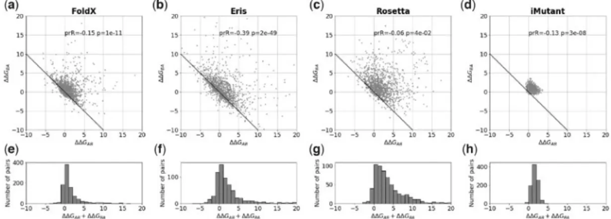
3.2 The bias for single and multiple substitutions
For single substitutions the average bias (DDGBAþDDGAB)/2 signifi-cantly deviates from zero (Table 1 and Fig. 2), ranging from 0.7460.05 kcal/mol for FoldX to 2.0860.12 kcal/mol for Rosetta. The clouds inFigure 2a–dconsist of pairs near the non-biased lineDDGBA¼–DDGABthat do not contribute to the bias and a dispersed group of points that comprises the bias. We investigated influence of different factors for one of the program, FoldX. We found that mutations in more buried positions and mutations with more dramatic change in the amino acid size tend to give larger bias (Supplementary Fig. S3). The change in hydrophobicity, change in charge of the mutated residues, and other factors have little or no in-fluence on the bias (Supplementary Fig. S3).
We also investigated the bias for multiple mutants (Fig. 3) for FoldX, Eris and Rosetta (I-Mutant was not studied because it does not allow input of multiple mutations). The bias increases with the Fig. 1.Design of the protocol. Two example protein structures, A and B, differ-ing by one amino acid residue are shown on the left. If we measure free en-ergy change for the forward and reverse mutations, their sum must be zero:
DDGABþDDGBA¼0 (arrows on the left). For predicted destabilizations (shown
on the right), their sum may not be zero due to errors. Here, the case is given, whenDDGABþDDGBA>0
number of introduced mutations (from two to ten); however, the in-crement becomes less pronounced with additional mutations.
3.3 Influence of additional parameters on the bias for
FoldX
We took one program, FoldX, as an example, and introduced some modifications in its default protocol to reduce or eliminate the bias. To reduce the running time of the modeling, we sampled 100 random pairs from the original pool of pairs differing by the one to ten substitutions. For reference, the bias for the default protocol of FoldX for 100-pairs subsample for single substitution was 0.8060.16 kcal/mol.
3.3.1 Initial relaxation of protein structure
Most programs manipulating protein structures as the first step of the algorithm prepare the protein structure to avoid artifacts coupled with the initial unrelaxed structure. In FoldX this procedure is called ‘RepairPDB’. First, it recovers all absent atoms and residues in the protein, then flips Asp, Gln and His side chains to avoid incor-rect 180-degree rotation. Then, for amino acid residues with high free energy it tests different rotamers from the rotamer library to ob-tain a better free energy estimation. The recommendation of the FoldX manual is to use RepairPDB once (http://foldxsuite.crg.eu/ command/RepairPDB), as we have done to obtain the data shown in the previous section.
In a computational experiment, one may want to consider changes of free energy of completely different proteins in one pool.
To avoid possible artifacts coupled with unequal relaxation of dif-ferent proteins, one may require the full relaxation of protein struc-tures, i.e. the free energy after the relaxation should reach a plateau (within a reasonable threshold, we chose here 0.1 kcal/mol). We found that FoldX reaches a plateau only after 7–10 rounds of RepairPDB (Supplementary Fig. S4).
Thus, a possible concern for the default FoldX procedure is that an incomplete initial relaxation influences the predicted values of stability change and the bias. To test this possibility, we made ten rounds of RepairPDB instead of the default single round. We found that, on average, change of stability (r¼0.99, slope¼1.01 when intercept fixed at zero) and the bias (r¼0.99, slope¼0.97 when intercept fixed at zero) were the same (Supplementary Fig. S5). So, full initial relaxation of the structures, although being physically rea-sonable, does not reduce the bias.
3.3.2 Additional relaxation after introducing mutations
The residues are mutated in FoldX by the procedure called ‘BuildModel’. In the mutated position, BuildModel removes the ori-ginal residue and assigns different rotamers for the new residue from the rotamer library. Simultaneously, BuildModel reconsiders the side-chain of the residues neighboring the mutated residue in physic-al space since the new amino acid residue may bump into its
Fig. 2.The bias for single substitutions for FoldX, Eris, Rosetta and I-Mutant. (a–d) The relationship between predicted changes of stabilityDDGABandDDGBAfor
the forward and reverse mutations, where the structures A and B differ by a single substitution. The ‘ideal’ relationshipDDGABþDDGBA¼0 is shown as a solid line.
Because of symmetry of the protocol, every pair of structures A and B is plotted as (A; B) and (B; A), so the plot is symmetric relative to the y¼x line. ‘prR’ and ‘p’ are the Pearson correlation coefficient and the associatedP-value. (e–h) The histograms of the sum of two changes of stabilityDDGABþDDGBAfor the forward and
reverse mutations for FoldX, Eris, Rosetta and I-Mutant, respectively Table 1.Bias for single substitutions
Program Bias, kcal/mol r(P-value) Binary fraction of errors FoldX 0.7460.05 0.15 (1011) 0.35
Eris 1.2560.11 0.39 (21049) 0.27
Rosetta 2.0860.12 0.06 (0.04) 0.51 I-Mutant 0.8060.01 0.13 (3108) 0.74
Note: Bias is given for an individual substitution, i.e. bias¼(DDGABþDDGBA)/
2, with the standard error of mean.r, Pearson correlation coefficient with the asso-ciatedP-value. Binary fraction of errors is the fraction of errors in binary classifica-tion. A pair was considered as correctly classified if the signs of forward and reverse change of stability were opposite.
Fig. 3.The bias for multiple mutations for FoldX, Eris and Rosetta. The indi-vidual value of the bias depending on the number of amino acid substitutions separating protein variants A and B in pair of structures. The error bars repre-sent 3 standard errors of mean
neighbors. The final prediction of the free energy change is calculated as the difference in free energy between the mutant structure and the reference wild-type structure. For better prediction, BuildModel moves ‘the same neighbours in the WT and in the mutant producing for each mutant PDB a corresponding PDB for its WT’ (FoldX manual, http://foldxsuite.crg.eu/command/ BuildModel).
FoldX changes only the mutated position and several neighbors keeping the rest of the protein structure the same, which may seem like a biologically unrealistic requirement. We checked if the inde-pendent relaxation of the rest of the structure of mutant and the cor-responding wild-type protein can decrease the bias. The additional relaxation indeed helps, and ten rounds are needed again to achieve full relaxation (Supplementary Fig. S6) decreasing the bias to 0.6160.13 kcal/mol for single mutations (Fig. 4, gray bars), or 25% lower than before. For multiple mutants, the reduction was even more significant (Fig. 4, white and gray bars).
3.4 Using Modeller to decrease the bias of FoldX
FoldX does not move the backbone upon mutation, which may be the reason for the bias found here. To test this hypothesis, after mutation we used Modeller (Webb and Sali, 2014) to relax the structure including the backbone. After that, we applied ten-fold re-laxation by FoldX, because the Modeller force-field could not be op-timal for FoldX force-field.We found that using Modeller for single mutants of FoldX removes the bias (Fig. 4, black bars) but at a cost of increasing the noise of the predictions (Supplementary Fig. S7). For multiple mutants using Modeller did not eliminate the bias; nevertheless, it was reduced significantly for structures differing by as many as eight substitutions. For sequences with eight to ten mutations Modeller did not reduce the observed bias (Fig. 4).
All physically reasonable modifications to the default protocol suggested here require additional computations; however, for more careful analysis one might prefer a more accurate but slower proto-col, with the necessary computations, which can be performed in a feasible timeframe on a computational cluster.
4 Discussion
We accurately and systematically measured the bias for programs predicting the effect of substitutions on protein stability, for one to ten substitutions. The design of the protocol allows its application without knowing any experimental data on protein free energy change (Christensen and Kepp, 2012;Frappieret al., 2015;Thiltgen and Goldstein, 2012). The protocol uses two protein structures dif-fering by one or several mutations. A program predicts protein free energy change upon mutations in the forward and reverse directions, and the bias is detected if the sum of free energy changes deviates, on average, from zero. We used the protocol on four representative programs, FoldX (Gueroiset al., 2002;Schymkowitzet al., 2005), Eris (Yinet al., 2007), Rosetta (Rohlet al., 2004) and I-Mutant (Capriottiet al., 2005a,b), and showed that they have an inherent bias in the prediction of the mutation effect. For single mutants, the bias was 0.7460.05 kcal/mol for FoldX, 1.2560.11 kcal/mol for Eris, 2.0860.12 kcal/mol for Rosetta and 0.8060.01 kcal/mol for I-Mutant (Table 1).
The bias was noticed before. For example,Christensen and Kepp (2012) in their investigation of beta-lactamase mutants estimated the bias for FoldX equal to0.5 kcal/mol. It is close to our results for single mutants; however, their estimate characterizes both single
and multiple mutants together, making it impossible to differentiate between the influence of single mutations and their interactions. Moreover, to estimate the bias they used protein models obtained by homology modeling. These manipulations lead to additional uncer-tainty about the nature of the observed bias. In the work (Thiltgen and Goldstein, 2012) the authors found the values for the bias, which agrees to our results, but only for single mutations and using only 65 protein pairs.
The exact reasons for the observed bias and reduced accuracy on a balanced dataset remain largely obscure. One of the reasons, gen-eral to all the programs, could be that programs are trained on ex-perimental datasets which have much more destabilizing mutations than stabilizing ones, as discussed in (Capriotti et al., 2008). Therefore, if such programs are given a balanced dataset (as we use here), then the algorithm will predict more destabilizing mutations, reflecting the tendencies of the training dataset.
The reason specific to FoldX could be that FoldX fixes the bone when making the mutant structure. Obviously, the fixed back-bone is optimal for the starting structure, not the mutated one, which is expected to estimate the prediction to be more destabiliz-ing. When applying FoldX to a balanced dataset of 84 mutations (42 forward and 42 reverse) in the original paper (Gueroiset al., 2002) the authors used additional relaxation by WHATIF program (Vriend, 1990) for the mutation increasing the sidechain. In that way, they were able to obtain unbiased results [seeFig. 3inGuerois et al.(2002)]. Using WHATIF program was similar to our usage of Modeller in the present work.
For I-Mutant, the specific factor could be that it considers the se-quence/structure context of the mutated position. The context con-tribution is the same both for forward and reverse mutations. Being trained on a dataset containing more destabilizing mutations it may erroneously predict that some context on average creates more destabilized predictions.
The identification of the bias does not immediately lead to a bet-ter prediction of experimental mutation effects because it is not clear if the bias results from misestimating the impact of the forward or the reverse mutation. For example, in independent tests I-Mutant showed the strongest correlation between experimental and predicted effects of mutations (Potapov et al., 2009); however, I-Mutant was also the noisiest method in our test (Fig. 2d). This sug-gests, that in our work we tested for different parameters of quality of prediction programs than are explored by test for agreement with experimental data. Hopefully, the bias explored here may be addressed in the course of development of new approaches [as in Fig. 4.The bias for different modifications of the default protocol of FoldX. The error bars represent standard error of mean
INPS (Fariselliet al., 2015)], or modification of the existing methods for prediction of free energy change (Christensen and Kepp, 2013, 2012). For example, the protocol used here can be utilized as an in-dependent training part of new programs, when a program requires the forward and reverse mutations to have opposite effects.
There are situations when the bias does not influence the inter-pretation of the results. One of them is when we compare protein variants that have the same number of mutations and are generated from the same template structure. In this case the bias is, on average, the same for all mutants (Sarkisyanet al., 2016; Tokurikiet al., 2007). Otherwise, mutant structures obtained from the same refer-ence structure but with different number of substitutions will have different bias, which will make them hard to compare.
To summarize, we measured accurately and systematically the bias in prediction of change in protein structure stability, both for single and multiple substitutions, utilizing the protocol based on self-consistency. The users might evaluate if the bias can influence the interpretation of their results. The developers could reduce or re-move the bias from the predictions by using artificially completed balanced datasets or by requiring the method to predict the same but opposite in sign effects for forward and reverse mutations. Our findings have important applications in the studies involving the protein structure destabilization predictions.
Acknowledgements
We thank Rita Casadio and Emidio Capriotti for providing us with stand-alone version of I-Mutant 3.0 and Nikolay V. Dokholyan for providing us with stand-alone version of Eris. We thank Alexander S. Mishin for technical support.
Funding
This work was supported by the HHMI International Early Career Scientist Program [55007424], the MINECO [BFU2015-68723-P], Spanish Ministry of Economy and Competitiveness Centro de Excelencia Severo Ochoa 2013-2017 [grant SEV-2012-0208], Secretaria d’Universitats i Recerca del Departament d’Economia i Coneixement de la Generalitat’s AGAUR [pro-gram 2014 SGR 0974], the European Research Council under the European Union’s Seventh Framework Programme [FP7/2007-2013, ERC grant agree-ment 335980_EinME] and Russian Scientific Foundation (RSF #14-24-00157, the part about I-Mutant calculations). The work was started at the School of Molecular and Theoretical Biology supported by the Dynasty Foundation.
Conflict of Interest: none declared.
References
Altschul,S.F. (1997) Gapped BLAST and PSI-BLAST: a new generation of pro-tein database search programs.Nucleic Acids Res.,25, 3389–3402. Anfinsen,C.B.et al. (1961) The kinetics of formation of native ribonuclease
during oxidation of the reduced polypeptide chain.Proc. Natl. Acad. Sci. USA,47, 1309–1314.
Benedix,A.et al. (2009) Predicting free energy changes using structural ensem-bles.Nat. Methods,6, 3–4.
Berman,H.M.et al. (2000) The Protein Data Bank.Nucleic Acids Res.,28, 235–242.
Capriotti,E.et al. (2008) A three-state prediction of single point mutations on protein stability changes.BMC Bioinformatics,9, S6.
Capriotti,E.et al. (2005a) I-Mutant2.0: predicting stability changes upon mu-tation from the protein sequence or structure. Nucleic Acids Res.,33, W306–W310.
Capriotti,E.et al. (2005b) Predicting protein stability changes from sequences using support vector machines.Bioinformatics,21, ii54–ii58.
Christensen,N.J. and Kepp,K.P. (2012) Accurate stabilities of laccase mutants predicted with a modified FoldX protocol. J. Chem. Inf. Model., 52, 3028–3042.
Christensen,N.J. and Kepp,K.P. (2013) Stability mechanisms of laccase iso-forms using a modified FoldX protocol applicable to widely different pro-teins.J. Chem. Theory Comput.,9, 3210–3223.
Fariselli,P.et al. (2015) INPS: predicting the impact of non-synonymous varia-tions on protein stability from sequence.Bioinformatics,31, 2816–2821. Frappier,V.et al. (2015) ENCoM server: exploring protein conformational
space and the effect of mutations on protein function and stability.Nucleic Acids Res.,43, W395–W400.
Gilis,D. and Rooman,M. (2000) PoPMuSiC, an algorithm for predicting pro-tein mutant stability changes: application to prion propro-teins.Protein Eng., 13, 849–856.
Goldenzweig,A.et al. (2016) Automated structure- and sequence-based design of proteins for high bacterial expression and stability. Mol. Cell, 63, 337–346.
Guerois,R.et al. (2002) Predicting changes in the stability of proteins and pro-tein complexes: a study of more than 1000 mutations.J. Mol. Biol.,320, 369–387.
Kiel,C. and Serrano,L. (2014) Structure-energy-based predictions and network modelling of RASopathy and cancer missense mutations.Mol. Syst. Biol., 10, 727.
Potapov,V.et al. (2009) Assessing computational methods for predicting pro-tein stability upon mutation: good on average but not in the details.Protein Eng. Des. Sel.,22, 553–560.
Rohl,C.A.et al. (2004) Protein structure prediction using Rosetta.Methods Enzymol.,383, 66–93.
Sarkisyan,K.S.et al. (2016) Local fitness landscape of the green fluorescent protein.Nature,533, 397–401.
Schymkowitz,J.et al. (2005) The FoldX web server: an online force field. Nucleic Acids Res.,33, W382–W388.
Seeliger,D. and de Groot,B.L. (2010) Protein thermostability calculations using alchemical free energy simulations.Biophys. J.,98, 2309–2316. Shah,P.et al. (2015) Contingency and entrenchment in protein evolution
under purifying selection.Proc. Natl. Acad. Sci. USA,112, E3226–E3235. Stevens,R.C. (2000) High-throughput protein crystallization.Curr. Opin.
Struct. Biol.,10, 558–563.
Tanford,C. (1968) Protein denaturation.Adv. Protein Chem.,23, 121–282. Tokuriki,N.et al. (2007) The stability effects of protein mutations appear to
be universally distributed.J. Mol. Biol.,369, 1318–1332.
Thiltgen,G. and Goldstein,R.A. (2012) Assessing predictors of changes in pro-tein stability upon mutation using self-consistency.PLoS ONE,7, e46084. Vriend,G. (1990) WHAT IF: a molecular modeling and drug design program.
J. Mol. Graph.,8, 52–56, 29.
Webb,B. and Sali,A. (2014) Comparative protein structure modeling using Modeller.Curr. Protoc. Bioinf.,47, 5.6.1–5.6.32.
Yin,S.et al. (2007) Eris: an automated estimator of protein stability.Nat. Methods,4, 466–467.