Received: 13 July 2018 Revised: 31 August 2018 Accepted: 04 September 2018
Version of Record published: 04 October 2018
Review Article
Measuring microtubule dynamics
Alexander James Zwetsloot
1,2, Gokhan Tut
1,2and Anne Straube
1,31Centre for Mechanochemical Cell Biology, University of Warwick, Coventry, CV4 7AL, U.K.;2MRC Doctoral Training Partnership, University of Warwick, Coventry, CV4 7AL, U.K.;
3Division of Biomedical Sciences, Warwick Medical School, Coventry, CV4 7AL, U.K.
Correspondence:Anne Straube ([email protected])
Microtubules are key players in cellular self-organization, acting as structural scaffolds, cel-lular highways, force generators and signalling platforms. Microtubules are polar filaments that undergo dynamic instability, i.e. transition between phases of growth and shrinkage. This allows microtubules to explore the inner space of the cell, generate pushing and pulling forces and remodel themselves into arrays with different geometry and function such as the mitotic spindle. To do this, eukaryotic cells employ an arsenal of regulatory proteins to control microtubule dynamics spatially and temporally. Plants and microorganisms have developed secondary metabolites that perturb microtubule dynamics, many of which are in active use as cancer chemotherapeutics and anti-inflammatory drugs. Here, we summa-rize the methods used to visualize microtubules and to measure the parameters of dynamic instability to study both microtubule regulatory proteins and the action of small molecules interfering with microtubule assembly and/or disassembly.
Why measure microtubule dynamics?
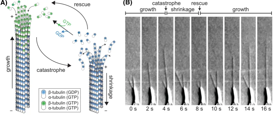
One of the most striking properties of microtubules is theirdynamic instability (Figure1). Extensive phases of microtubule growth are followedby rapid disassembly andregrowth [1].Microtubules grow by the addition ofGTP-boundαβ-tubulin heterodimers to their ends.Incorporation into the microtubule facilitatesGTP hydrolysis, resulting in the growing tip being cappedbyGTP-tubulin while the micro
-tubulelattice consists primarily ofGDP-tubulin. ExposingGDP-tubulin at the tip results in catastrophe, the switch from growth to shrinkage.Microtubuledynamics, therefore, aredriven by thedelicate balance ofGTP-tubulin incorporation andrate ofGTP hydrolysis [2].
Dynamic instability enables the formation oflong filaments that can be reorganizedon the timescale of minutes. For example, at the onset of mitosis, interphase microtubules aredisassembledandnew mi
-crotubules form the bipolar mitotic spindle, capture, align andthen separate chromosomes by making
dynamiclinkages to the kinetochore [3,4]. Also in non-dividing cells, microtubules perform important functions. For example, neuronalcells maintain polarizedmicrotubule bundles in axons and dendrites for theduration of an animal’slife, thereby allowing thedirectionaltransport of cargoes by kinesins and dynein [5,6]. Similarly, muscle cells arrange microtubules paraxially to provide structuralintegrity and
resistance to contractile forces [7,8].In motile cells, microtubules control directionality of cell locomo
-tion in a crosstalk with the actin cytoskeleton andby mediating the turnover of adhesion sites [9]. Thus, proper microtubule arrangement andthe spatiotemporalcontrolof microtubuledynamics are integrally important to cellmorphology, function andtheir ability to faithfully proliferate.Due to their ability to in
-terfere with microtubuledynamics andthus inhibit cellproliferation, microtubule-targeting agents such as taxanes andvinca alkaloids are widely usedin the clinic as cancer chemotherapeutics [10,11].
To understandhow the cellular machinery regulates microtubuledynamics andto revealthe mecha
Figure 1.Microtubule dynamic instability
(A) Microtubules are tubular filaments assembled from αβ-tubulin heterodimers arranged head-to-tail in 13 helically ar-ranged protofilaments. Microtubules grow by the addition of GTP-tubulin (green) to their ends. GTP hydrolysis occurs when
β-tubulin is buried in the lattice. Catastrophe, i.e. transition to shrinkage occurs when the GTP cap is lost and GDP-tubu-lin is exposed at the microtubule end. The transition from shrinkage to growth is called rescue. Note that α-tubulin con-tains a non-exchangeable GTP trapped at the intradimer interface. (B) DIC images from a time series of microtubules grow-ing from an axoneme (large object at bottom of image). Imaggrow-ing data courtesy of Douglas Drummond and Rob Cross. Abbreviation: DIC, differential interference contrast.
behaviour of individualmicrotubules andthedirect effect of any addedprotein or chemical.In vitroexperiments usually permit measurement of allfour parameters of dynamic instability (growth, shrinkage speeds, catastrophe andrescue frequencies). Additionalinformation on nucleation frequency, flexuralrigidity or tubulin turnover in the microtubule shaft are also accessible in such reconstitution experiments. Alternatively, or additionally,dynamic microtubules can be studiedinside living cells.In many cellular systems, the highdensity of microtubules poses challenges to observing individualmicrotubules andthus to measuring allparameters of microtubule instability. Therefore, a number of complementary approaches andmarkers might needto be usedto glean information about thedynamic state of microtubules.Here, we review the current approaches in the fieldto investigate microtubule
dynamics both in reconstitution experiments as wellas in cells.
Measuring microtubule dynamics
in vitro
A simple way to measure microtubule assembly is to measure the turbidity of a solution of soluble tubulin upon the addition ofGTP as the forming microtubules scatter thelight roughly proportionally to their mass [12,13]. Such bulk measurements allow some insight into whether a compoundhas a stabilizing ordestabilizing effect on microtubules, andcan be performedat high throughput.Without any addition oflabels, individualmicrotubules can be visualized
usingdarkfieldordifferentialinterference contrast (DIC) microscopy (Figure1B) andtheir growth andshrinkage speeds as wellas transition rates measuredfrom timelapse images [14,15].DarkfieldorDICmicroscopy are stillthe methods of choice if working withlimiting sources of tubulin such as single isoform tubulin preparations [16,17], or to exclude effects from fluorescentlabels [18].However, the availability of both commercialimaging systems and
fluorescentlylabelledtubulin, andthe ability to study thedynamic relocalization of the protein-of-interest at the same time makes totalinternalreflection fluorescence (TIRF) microscopy currently the most popular methodfor studying microtubuledynamicsin vitro.
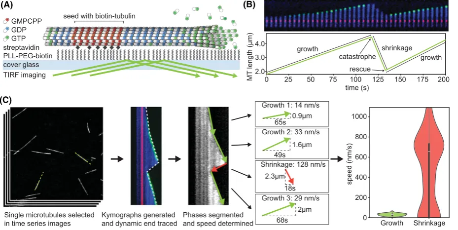
Typically,dynamic microtubules are assembledfrom a template (Figure 2A). This can either be purifiedcentro
-somes that are a source ofγ-tubulin ring complexes from which new microtubule growth may be nucleatedor ax
Figure 2.Microtubule dynamics measurementin vitro
(A) Typical TIRF-based assay to reconstitute and measure microtubule dynamicsin vitroin which a GMPCPP-stabilized microtubule seed is immobilized on a passivated glass surface that nucleates a microtubule. The seed and free tubulin typically contain 5–20% tubulin that is labelled with different fluorophores. Microtubule-binding proteins or small molecules can be added and their binding as well as effect on microtubule dynamics measured in this assay. (B) Montage and life history plot of a dynamic microtubule with seed (red), dynamic microtubule lattice (blue) and microtubule tip labelled with EB3-GFP (green). Phases are labelled as growth and shrinkage, transitions marked as catastrophe and rescue. (C) Workflow to measure microtubule dynamics parameters including the generation of kymographs (space–time plots), automatic or manual detection of the edge and extraction of speed and transition rates. The final violin plots are vertical histograms that show the full distribution of growth and shrinkage speeds that make up the median, which is also depicted by an inset boxplot.
in close proximity to, the surface thereby allowing TIRF microscopy [21].Growing microtubules can be observed
either through the addition of a few percent fluorescentlylabelledtubulin within the reaction mixture or by fluores
-centlylabelledmicrotubule-associatedproteins (MAPs) such as EB3 [22].Imaging over a periodof severalminutes allows the observation of growth andshrinkage of microtubules (Figure 2B). To extract the parameters ofdynamic instability, the microtubule ends can be tracked directly in the timelapse movie, but it is more common to gener
-ate kymographs andtrace the path of the microtubule endeither manually or using automatic image analysis tools (Figure 2C). For example, the automatic extraction of microtubuledynamics parameters from kymographs ofDIC
images, is possible if using optimizedfiltering andedgedetection algorithms [23].Gaussian models have been fitted directly to fluorescence images of microtubule todetect microtubule motility, but alsodepolymerization speeds with nanometre precision [24].However, todetermine the endposition in microtubuledynamics assays with immobilized
seeds, thedirect fitting of a 2D Gauss error function to the microtubule endis sufficient todetectlength changes at subpixelresolution. The variance of theGauss error function can also revealthe extent of taper at the microtubule end, i.e. thedifference between theleading and lagging protofilaments [25]. All direct fitting approachesdependon high signal–noise ratio (SNR) [26]andwillonly result in meaningful data on the microtubule tip structure if alarge pro
-portion (>25%) of fluorescentlylabelledtubulin is used(Fitton&Straube, unpublished data). Finally, automatically or manuallyderivedtrackingdata are usedto calculate speeds andtransition rates (Figure 2C). For that purpose, the catastrophe rate isdefinedas the number of growth-to-shrinkage transitionsdividedby the totaltime microtubules spent growing andinversely, the rescue frequency is calculatedas the number of events when a shrinking microtubule resumes growth before reaching the seed dividedby the totaltime spent shrinking.
size couldbe measuredinstead. The only caveat to this approach is that EB1itself is changing theGTP cap andmight accelerateGTP hydrolysis [29].
Finally, microtubules experience resisting forces when assembling inside cells. This can be mimickedin vitroby growing microtubules into barriers. Such experimentsdemonstrate that microtubule polymerization speedandcatas
-trophe frequency are strongly forcedependent [30].It is important to study the forcedependence as these effects couldbe further exacerbatedin the presence of microtubule regulators that accelerate growth by assembling elab
-orate tip structures, which arelikely to be unstable under force.Measuring microtubuledynamics under force also enables very precise measurements of nanoscalelength changes. Todo this, the force is kept constantlylow andthe
displacements of a beadattachedto a microtubule seedor axoneme are usedtoderive nanoscale microtubulelength measurements [31,32]. Such nanoscale measurements can revealchanges in the fluctuationsduring a growth phase providing insights into the mechanism of microtubule regulators.
While reconstitution experiments are powerfulto revealthedirect effect of a protein or smallmolecule on mi
-crotubule assembly and disassembly, the complex interaction oflattice andtip binding microtubule regulators and
spatialconstraints that together bring about spatiotemporally regulatedmicrotubuledynamics in cells isdifficult to reconstitute. A bridge between the reductionism ofin vitroreconstitution andcomplexity of in cellexperiments can be achievedwith the use of cellextracts.Xenopus egg extracts have been usedextensively over the years to study microtubuledynamics, nucleation andspindle formation.Microtubules are visualizedby addition of fluorescently
labelledtubulin to the extracts, while extraneous nucleation can be inducedby the addition of purifiedcentrosomes [33].Individualproteins can bedepletedfrom the extract using specific antibodies and labelledcomponents can be titratedinto the extract forlocalization or to study concentration-dependence [34-36].
To gain a more holistic view of how adrug or protein affects microtubuledynamics, mechanistic understandings gainedfromin vitroexperimentation must be contextualizedinside cells, andconversely, phenotypes observedin cells must be understoodmechanisticallyin vitro. For this reason, many people complementin vitroresearch with cellular studies.
Observing microtubule dynamics in cells
Measuring microtubuledynamic instability in cells requires being able to visualize individualmicrotubules.While it is possible to see microtubules without anylabels in thin sections of the cellusing video-enhanced differentialinter
-ference contrast microscopy [37,38], fluorescent techniques either using the injection of chemicallylabelledtubulin [39,40], cellpermeabledyes [41]orDNA-encodedfluorescent protein fusions allow, in principle, to image every mi
-crotubule in the cell.However,due to thedensity of microtubule arrays in many celltypes andin particular within the mitotic spindle, it is often not possible to observe individualmicrotubules for their entirelife history andthus ob
-tain allparameters ofdynamic instability from a single experiment as it is possible in the reconstitution experiments
describedabove. Thusdifferentlabelling techniques are usedto obtain insights intodistinct aspects of microtubule
dynamics.
Directlabelling of microtubules can generally be achievedby fusing a fluorescent protein (FP) to the N-terminus ofα-tubulin or theC-terminus ofβ-tubulin (Figure 3A,B) [42-44].While it is not possible to replace allendogenous tubulin with FP fusionsdue to steric hindrance, endogenous tagging of tubulin has been successfully achievedin mammalian cells [45,46]as these express severalisoforms of bothα-andβ-tubulin andthus only a subset of the
dimers in the microtubule willcarry alabel. An alternative is the use of smallorganic fluorophores. These can be conjugatedto purifiedtubulin andthen introducedby microinjection [39,40]ordirectlylabelledinliving cells using genetic code expansion andcellpermeable fluorescentdyes such as silicon rhodamine [47]. Thelatter is an especially powerfulapproach as thedye attachment site can be chosen freely andthus avoid locations for protein–protein inter
-action andpost-translationalmodifications.Due to the relatively high cytoplasmic poolof tubulin, an increasedSNR can often be obtainedwhenlabelling aMAP or smallmolecule rather than tubulindirectly. Lattice-decoratingMAPs such asMAP4and MAP7/ensconsin are commonly usedto achieve this (Figure 3C) [48-52]. As many fluorescent fusions ofMAPs retain fullfunction, endogenous tagging avoids perturbing microtubuledynamics as may occur with tubulin orMAP overexpression [53]. Thedocetaxel-basedcell-permeabledye SiR-tubulin is a popular alternative for hard-to-transfect cells. At concentrations up to100nM, it has been shown to havelittle effect on cellproliferation andit’s binding on the inside of the microtubule makes it particularly suitable for superresolution techniques [41].
A popular alternative tolabelling the entire microtubule is to use tip tracking proteins (+TIPs) such as EB1, EB3 orCLIP-170that track the growing microtubule end(Figure 3D) [54,55]. EBs allow the reliable measurement of microtubule assembly even withindense microtubule structures andare the marker of choice to measure micro
Figure 3.Microtubule dynamics measurement in cells
(A) Widefield image of a human hTERT RPE1 cell expressing GFP-tubulin. Individual microtubules can be observed, especially close to the cell cortex (inset). (B) Widefield image ofUstilago maydiscell expressing YFP-tubulin (grey) and histone4-CFP (blue). Individual microtubules are visible throughout the cell, enabling measurement of four parameters of dynamics instability. Diamond graphs show growth speed (right), shrinkage speed (left), rescue frequency (top) and catastrophe frequency (bottom). Data are plotted for a temperature-sensitive dynein mutant Dyn2ts and the respective control strain at permissive (22◦C) and restrictive temperature (29◦C) [89]. (C) Microtubule labelling using the ensconsin microtubule domain fused to three tandem copies of GFP [49] results in higher signal noise than direct tubulin labelling in the same cell line as shown in (A). Measurement of microtubule dynamics by recording the length at different time points is indicated in the zoomed section for one growing microtubule. (D) Example of an hTERT RPE1 cell expressing EB3-tdTomato that has been analysed using plusTipTracker [79]. Tracks are presented colour-coded indicating average speed, and according to directionality relative to long axis of the cell [90].
3D) [7,56-61].However,+TIPs only provide insights into microtubule assembly andit remains challenging to mea
-sure other aspects of microtubuledynamics indense arrays such as the mitotic spindle or in neurites.One approach to measure microtubule turnover is to use photoactivatable or convertible FP fusions to tubulin andthen measure thedissipation of the activatedproteins from a region of interest [62]. Alternatively, the incorporation time of sin
-gle tubulin subunits can bedirectly measuredin sparselylabelledsamples [63].In addition, both methods provide information on thedisplacement of microtubules.
In most cells, microtubules grow at an average rate of0.2–0.4μm/s andshrink substantially faster. Thus to not miss rapidshortening events, imaging at1frame per secondis required.Using widefieldor confocalmicroscopy, usually only one imaging plane can be acquiredat this rate without causing photodamage that alters both microtubule
dynamics as wellas cellmorphology. For adherent cells, TIRF microscopy offers an increase in SNR andreduced
this rate for many minutes without photodamage [65]. LLSMhas already been successfully usedto quantify EB tracks throughout the entire mitotic spindle [61]andislikely to become the state-of-the art.
Determining the four parameters ofdynamic instability in cells is relatively straightforwardin cellular systems with very few or spacedout microtubules [43,66-69](Figure 3B).However, a complication todetermining parameters of
dynamic instability in cells is the spatialregulation of microtubuledynamics, thus reporting globalparameters might not always make sense.In many cells, microtubules grow relatively unperturbeduntilthey reach the cellboundaries andcatastrophes andrescues occur primarily near the cellcortex [69,70].Microtubule assembly–or atleast the progress of EB comets–slowsdown in the proximity of the cellcortex [71], possiblydue to increasedresistance in the actin-denselamella andretrograde actin flow [72]. Also, microtubuledynamics changeduring the cellcycle and
some parameters ofdynamic instability change up to4-foldif comparing interphase andmitotic microtubules [73]. Therefore, to obtain insights into the function of a microtubule regulator, it is important to compare similar regions of cells with similar morphology in the same cellcycle state.
Data analysis and presentation
The most common andmost reliable way to extract microtubulelife historydata is to track the position of the micro
-tubule endor to measure thelength of a microtubule manually at every time point [74,75]. To account for changes in the curvature of the microtubule, a curve can be tracedor fittedto the entirelength of the microtubule [69].If the entire microtubule is not visible, this can bedone from an arbitrary point on the microtubulelattice (Figure 3C).If microtubulesdo not undergo significant sideways movements, generating kymographs as forin vitrodata is a faster alternative as itdoes not require measuring every timepoint individually. An elegant way to visualize microtubule growth andshortening events is sequentialsubtraction analysis [76]. By subtracting images with a time shift of one or severalframes from each other, both regions with polymer gain as wellas regions with polymerloss can be ca
l-culatedandtracked.In cells, in whichlateralmovement of microtubules is slow in comparison withlength changes, this technique allows tracking growth andshrinkage even indense networks [76,77].
If growing microtubules have beenlabelledwith+TIPs, automatic tracking andanalysis of microtubule assembly parameters is a possibility.Using a pattern recognition approach in Labview, EB comets were trackedinC. elegans
embryos to conduct a screen for factors requiredfor microtubule growth andnucleation [57]. To study microtubule polarity inDrosophila oocytes,limitations oflow SNR mainlydue to the autofluorescence from yolk were overcome by a combination of probabilistic foregroundextraction andadaptive mean filtering to allowedautomatic segmentation andtracking of EB foci [78]. PlusTipTracker is aMATLAB-basedpackage for tracking andanalysing EB comets or any bright spots basedon the knowledge that microtubule trajectories are almost straight [71,79](Figure 3D). The software package also infers shrinkage rates andrescue frequencies from comets reappearing on the previous trajectory of a comet.Indense microtubule networks, the inferred data are probably not informative. Further, if the
data are from a single plane, comets arelost andgained due to growing out or into the focalplane, further complicating any analysis beyondmicrotubule assembly speed.However, EB tracking performedon timelapsedata from the entire cellvolume as available fromlatticelight sheet imaging allows extracting information from birth to catastrophe.In pioneering work, 3Dtracking of EB comets was performedusingImaris followedby post-analysis inMATLAB to
determine microtubule assembly properties indifferent stages of human mitosis [61].
It is common practice to present microtubuledynamics parameters as mean+−SDin a table. A visualalternative are
diamondgraphs that show the four parameters ofdynamic instability andallow easy comparison betweendifferent treatments [80](Figure 3B).However, growth andshrinkage velocities are not normallydistributedandit is feasible to expect that some microtubule regulators speedup velocity in the cellbody but result in reducedassembly near the cellcortex or vice versa. Such changes to thedistribution wouldbe ignoredifjust reporting meandata for the entire cell.Violin plots (Figure 2C) are a space-saving way to show andcomparedistributions betweendifferent conditions, while speedmaps are usefulto visualize their spatial distribution (Figure 3C).
Measuring microtubule nucleation and self-renewal
It is becoming recognizedat one of the most important events in thelife of a microtubule is its birth. Thus regulating where andwhen microtubules are nucleatedis a key aspect in organizing microtubule arrays. To measure microtubule nucleation ratesin vitro, counting microtubules formed 15min after warming the sample or adding free tubulin to a template is a simple andeffective approach that wouldusually be performedfor a range of tubulin concentrations [15,81]. Nucleating activity wouldthen bedetectedby a shift of the curve tolower tubulin concentrations. Todeter
or incubation on ice, before their regrowth is observedfromdifferentlocations in the cell[59,82]. The experiment works on the assumption that nucleation templates stay in position when microtubules are removed. Newly appearing EB comets can indicate either a microtubule birth or rescue–aslong as out of focus entry can be excluded. Especially if comparing nucleation fromdifferent subcellular structures such as the centrosome andtheGolgi, counting ema
-nating EB tracks is a robust methodtodetermine relative nucleation efficiency [58].Corroborating evidence can be providedbylocalizing known microtubule nucleators such as centrosome components, minus endstabilizers of the
CAMSAP family, or minus-end directedkinesin fusions such as Nod-KHC[82-84]. An elegant approach todeter
-mine the position of microtubule minus ends in the mitotic spindle is to cut microtubules with alaser and determine the extent of microtubuledepolymerization [85].
Another emerging topic in the measurement of microtubuledynamics is the exchange of tubulin not at the tip of the microtubule, but along thelattice. Tubulin exchange occurs at sites of microtubuledefects that either occur spontaneouslyduring microtubule assembly andcorrelate with the speedof microtubule polymerization or can be introducedby mechanicalstress on the microtubule such as repeatedbending [86,87]. Tubulin exchange can bede
-tectedby changing the colour of the free tubulin pool.In vitro, microfluidics allows the gentle exchange of the so -lution with tubulin containing adifferentlabel[87].In cells, photoconversion of mEos2-tubulin has been usedto the same effect [88]. Spots in the‘new’tubulin colour appear at sites of repair after severalminutes of incubation. These findings suggest that microtubule repair couldresult in a significant turnover of tubulin subunits inlong-lived
microtubules away from the microtubule tips and lattice bindingMAPs might have a role in regulating this tubulin exchange mechanism.
Conclusions
We expect that our knowledge in microtubule regulation willincreasedramatically over the nextdecadedue to sev
-eralrecent breakthroughs:(1) Advances inlive cellimaging allow 3Dtimelapse imaging with unprecedentedtempo
-ralresolution, (2) the cryoEMrevolution enables uncovering the structuralchanges in tubulin following nucleotide hydrolysis andthe interactions of microtubule regulators with tubulin and(3) the purification of recombinant hu
-man tubulin enables studying point mutations, modifications andisoforms. Therefore, establishing robust assays to measure microtubuledynamics bothin vitroandinliving cells willbe important to push the boundary of our un -derstanding. The biggest challenges forin vitroexperiments are the use of buffers that might not be physiologically relevant andalack of standardin tubulin preparations across bothlaboratories andprep-to-prep variability.In vivo, it is increasingly apparent thatdifferent celltypes expressdifferent compositions of tubulin isoforms, andregulate mi
-crotubuledynamicslocally andtemporally;thus, microtubuledynamics is contextdependent. To truly understand
how simple tubulindimers can buildthe beautifulandcomplex microtubule network, we must gain context-specific understanding of how tubulin isoforms, tubulin modifications and MAPs guide the fascinating phenomenon ofdy
-namic instability.
Summary
• The proper regulation of microtubule dynamics is essential for faithful chromosome segregation, cellular morphology and motility.
• Microtubules are long polymers undergoing phases of continuous growth and shrinkage.
• The four parameters of dynamic instability typically measured are growth speed, shrinkage speed, catastrophe rate and rescue rate.
• Microtubule dynamics can be reconstituted in vitro, which is useful to measure the direct effect of individual proteins or small molecules.
• A number of strategies are available to label microtubules in cells to observe and measure their dynamics in their physiological context.
Acknowledgements
Funding
A.J.Z. and G.T are funded by the MRC Doctoral Training Partnership [grant references MR/J003964/1 and MR/N014294/1]. A.S. is a Prize Fellow of the Lister Institute of Preventive Medicine and a Wellcome Trust Investigator [200870/Z/16/Z]. Work in her lab is also supported by the Leverhulme Trust [RPG-2016-260].
Competing interests
The authors declare that there are no competing interests associated with the manuscript.
Abbreviations
CFP, cyan fluorescent protein; DIC, differential interference contrast; EB, end-binding protein; FP, fluorescent protein; GFP, green fluorescent protein; GMPCPP, Guanosine-5’-[(α,β)-methyleno]triphosphate; LLSM, lattice light sheet microscopy; MAP, microtubule-associated protein; RPE, retinal pigment epithelium; SiR, silicon rhodamine; SNR, signal–noise ratio; +TIP, tip track-ing protein; TIRF, total internal reflection fluorescence; YFP, yellow fluorescent protein.
References
1 Mitchison, T. and Kirschner, M. (1984) Dynamic instability of microtubule growth.Nature312, 237–242, https://doi.org/10.1038/312237a0
2 Hyman, A.A., Salser, S., Drechsel, D.N., Unwin, N. and Mitchison, T.J. (1992) Role ofGTP hydrolysis in microtubule dynamics: information from a slowly hydrolyzable analogue,GMPCPP.Mol. Biol. Cell3, 1155–1167,https://doi.org/10.1091/mbc.3.10.1155
3 McIntosh, J.R. and Landis, S.C. (1971) The distribution of spindle microtubules during mitosis in cultured human cells.J. Cell Biol.49, 468–497,
https://doi.org/10.1083/jcb.49.2.468
4 Auckland, P. and McAinsh, A.D. (2015) Building an integrated model of chromosome congression.J. Cell Sci.128, 3363–3374,
https://doi.org/10.1242/jcs.169367
5 Tas, R.P., Chazeau, A., Cloin, B.M.C., Lambers, M.L.A., Hoogenraad, C.C. and Kapitein, L.C. (2017) Differentiation between oppositely oriented microtubules controls polarized neuronal transport.Neuron96, 1264e5–1271e5, https://doi.org/10.1016/j.neuron.2017.11.018
6 Burton, P.R. and Paige, J.L. (1981) Polarity of axoplasmic microtubules in the olfactory nerve of the frog.Proc. Natl. Acad. Sci. U.S.A.78, 3269–3273,
https://doi.org/10.1073/pnas.78.5.3269
7 Mogessie, B., Roth, D., Rahil, Z. and Straube, A. (2015) A novel isoform of MAP4 organises the paraxial microtubule array required for muscle cell differentiation.Elife4, e05697,https://doi.org/10.7554/eLife.05697
8 Robison, P., Caporizzo, M.A., Ahmadzadeh, H., Bogush, A.I., Chen, C.Y., Margulies, K.B. et al. (2016) Detyrosinated microtubules buckle and bear load in contracting cardiomyocytes.Science352, aaf0659,https://doi.org/10.1126/science.aaf0659
9 Kaverina, I. and Straube, A. (2011) Regulation of cell migration by dynamic microtubules.Semin. Cell Dev. Biol.22,968–974,
https://doi.org/10.1016/j.semcdb.2011.09.017
10 Dumontet, C. and Jordan, M.A. (2010) Microtubule-binding agents: a dynamic field of cancer therapeutics.Nat. Rev. Drug Discov.9, 790–803,
https://doi.org/10.1038/nrd3253
11 Stanton, R.A.,Gernert, K.M., Nettles, J.H. and Aneja, R. (2011) Drugs that target dynamic microtubules: a new molecular perspective.Med. Res. Rev.
31, 443–481, https://doi.org/10.1002/med.20242
12 Borisy,G.G., Olmsted, J.B. and Klugman, R.A. (1972) In vitro aggregation of cytoplasmic microtubule subunits.Proc. Natl. Acad. Sci. U.S.A.69, 2890–2894, https://doi.org/10.1073/pnas.69.10.2890
13 Mirigian, M., Mukherjee, K., Bane, S.L. and Sackett, D.L. (2013) Measurement of in vitro microtubule polymerization by turbidity and fluorescence.
Methods Cell Biol.115, 215–229, https://doi.org/10.1016/B978-0-12-407757-7.00014-1
14 Horio, T. and Hotani, H. (1986) Visualization of the dynamic instability of individual microtubules by dark-field microscopy.Nature321,605–607,
https://doi.org/10.1038/321605a0
15 Walker, R.A., O’Brien, E.T., Pryer, N.K., Soboeiro, M.F., Voter, W.A., Erickson, H.P. et al. (1988) Dynamic instability of individual microtubules analyzed by video light microscopy: rate constants and transition frequencies.J. Cell Biol.107, 1437–1448, https://doi.org/10.1083/jcb.107.4.1437
16Vemu, A., Atherton, J., Spector, J.O., Szyk, A., Moores, C.A. and Roll-Mecak, A. (2016) Structure and dynamics of single-isoform recombinant neuronal human tubulin.J. Biol. Chem.291, 12907–12915, https://doi.org/10.1074/jbc.C116.731133
17 Hussmann, F., Drummond, D.R., Peet, D.R., Martin, D.S. and Cross, R.A. (2016) Alp7/TACC-Alp14/TOGgenerates long-lived, fast-growing MTs by an unconventional mechanism.Sci. Rep.6, 20653, https://doi.org/10.1038/srep20653
18 Komarova, Y., DeGroot, C.O.,Grigoriev, I.,Gouveia, S.M., Munteanu, E.L., Schober, J.M. et al. (2009) Mammalian end binding proteins control persistent microtubule growth.J. Cell Biol.184,691–706, https://doi.org/10.1083/jcb.200807179
19Reber, S. (2011) Isolation of centrosomes from cultured cells.Methods Mol. Biol.777, 107–116,https://doi.org/10.1007/978-1-61779-252-6˙8
20 Waterman-Storer, C.M. (2001) Microtubule/organelle motility assays.Curr. Protoc. Cell Biol., https://doi.org/10.1002/0471143030.cb1301s00
21 Telley, I.A., Bieling, P. and Surrey, T. (2011) Reconstitution and quantification of dynamic microtubule end tracking in vitro using TIRF microscopy.
Methods Mol. Biol.777, 127–145, https://doi.org/10.1007/978-1-61779-252-6˙10
22 MontenegroGouveia, S., Leslie, K., Kapitein, L.C., Buey, R.M.,Grigoriev, I., Wagenbach, M. et al. (2010) In vitro reconstitution of the functional interplay between MCAK and EB3 at microtubule plus ends.Curr. Biol.20, 1717–1722, https://doi.org/10.1016/j.cub.2010.08.020
24 Ruhnow, F., Zwicker, D. and Diez, S. (2011) Tracking single particles and elongated filaments with nanometer precision.Biophys. J.100, 2820–2828,
https://doi.org/10.1016/j.bpj.2011.04.023
25 Demchouk, A.O.,Gardner, M.K. and Odde, D.J. (2011) Microtubule tip tracking and tip structures at the nanometer scale using digital fluorescence microscopy.Cell. Mol. Bioeng.4, 192–204, https://doi.org/10.1007/s12195-010-0155-6
26Bohner,G.,Gustafsson, N., Cade, N.I., Maurer, S.P.,Griffin, L.D. and Surrey, T. (2016) Important factors determining the nanoscale tracking precision of dynamic microtubule ends.J. Microsc.261,67–78, https://doi.org/10.1111/jmi.12316
27 Walker, R.A., Pryer, N.K. and Salmon, E.D. (1991) Dilution of individual microtubules observed in real timein vitro: evidence that cap size is small and independent of elongation rate.J. Cell Biol.114, 73–81, https://doi.org/10.1083/jcb.114.1.73
28 Duellberg, C., Cade, N.I., Holmes, D. and Surrey, T. (2016) The size of the EB cap determines instantaneous microtubule stability.Elife5,
https://doi.org/10.7554/eLife.13470
29Maurer, S.P., Cade, N.I., Bohner,G.,Gustafsson, N., Boutant, E. and Surrey, T. (2014) EB1 accelerates two conformational transitions important for microtubule maturation and dynamics.Curr. Biol.24, 372–384, https://doi.org/10.1016/j.cub.2013.12.042
30 Janson, M.E., de Dood, M.E. and Dogterom, M. (2003) Dynamic instability of microtubules is regulated by force.J. Cell Biol.161, 1029–1034,
https://doi.org/10.1083/jcb.200301147
31 Schek, III, H.T.,Gardner, M.K., Cheng, J., Odde, D.J. and Hunt, A.J. (2007) Microtubule assembly dynamics at the nanoscale.Curr. Biol.17, 1445–1455, https://doi.org/10.1016/j.cub.2007.07.011
32 Kerssemakers, J.W., Munteanu, E.L., Laan, L., Noetzel, T.L., Janson, M.E. and Dogterom, M. (2006) Assembly dynamics of microtubules at molecular resolution.Nature442, 709–712, https://doi.org/10.1038/nature04928
33 Tournebize, R., Andersen, S.S., Verde, F., Doree, M., Karsenti, E. and Hyman, A.A. (1997) Distinct roles of PP1 and PP2A-like phosphatases in control of microtubule dynamics during mitosis.EMBO J.16, 5537–5549,https://doi.org/10.1093/emboj/16.18.5537
34 Belmont, L.D. and Mitchison, T.J. (1996) Identification of a protein that interacts with tubulin dimers and increases the catastrophe rate of microtubules.
Cell84,623–631, https://doi.org/10.1016/S0092-8674(00)81037-5
35 Tournebize, R., Popov, A., Kinoshita, K., Ashford, A.J., Rybina, S., Pozniakovsky, A. et al. (2000) Control of microtubule dynamics by the antagonistic activities of XMAP215 and XKCM1 in Xenopus egg extracts.Nat. Cell Biol.2, 13–19,https://doi.org/10.1038/71330
36Thawani, A., Kadzik, R.S. and Petry, S. (2018) XMAP215 is a microtubule nucleation factor that functions synergistically with the gamma-tubulin ring complex.Nat. Cell Biol.20, 575–585, https://doi.org/10.1038/s41556-018-0091-6
37 Cassimeris, L., Pryer, N.K. and Salmon, E.D. (1988) Real-time observations of microtubule dynamic instability in living cells.J. Cell Biol.107, 2223–2231, https://doi.org/10.1083/jcb.107.6.2223
38 Salmon, E.D. (1995) VE-DIC light microscopy and the discovery of kinesin.Trends Cell Biol.5, 154–158,
https://doi.org/10.1016/S0962-8924(00)88979-5
39Keith, C.H., Feramisco, J.R. and Shelanski, M. (1981) Direct visualization of fluorescein-labeled microtubules in vitro and in microinjected fibroblasts.J. Cell Biol.88, 234–240, https://doi.org/10.1083/jcb.88.1.234
40 Saxton, W.M., Stemple, D.L., Leslie, R.J., Salmon, E.D., Zavortink, M. and McIntosh, J.R. (1984) Tubulin dynamics in cultured mammalian cells.J. Cell Biol.99, 2175–2186, https://doi.org/10.1083/jcb.99.6.2175
41 Lukinavicius,G., Reymond, L., D’Este, E., Masharina, A.,Gottfert, F., Ta, H. et al. (2014) Fluorogenic probes for live-cell imaging of the cytoskeleton.
Nat. Methods11, 731–733, https://doi.org/10.1038/nmeth.2972
42 Ding, D.Q., Chikashige, Y., Haraguchi, T. and Hiraoka, Y. (1998) Oscillatory nuclear movement in fission yeast meiotic prophase is driven by astral microtubules, as revealed by continuous observation of chromosomes and microtubules in living cells.J. Cell Sci.111, 701–712
43 Steinberg,G., Wedlich-Soldner, R., Brill, M. and Schulz, I. (2001) Microtubules in the fungal pathogen Ustilago maydis are highly dynamic and determine cell polarity.J. Cell Sci.114,609–622
44 Freitag, M., Hickey, P.C., Raju, N.B., Selker, E.U. and Read, N.D. (2004)GFP as a tool to analyze the organization, dynamics and function of nuclei and microtubules in Neurospora crassa.Fungal Genet. Biol.41, 897–910, https://doi.org/10.1016/j.fgb.2004.06.008
45 Khan, A.O., Simms, V.A., Pike, J.A., Thomas, S.G. and Morgan, N.V. (2017) CRISPR-Cas9mediated labelling allows for single molecule imaging and resolution.Sci. Rep.7, 8450,https://doi.org/10.1038/s41598-017-08493-x
46Roberts, B., Haupt, A., Tucker, A.,Grancharova, T., Arakaki, J., Fuqua, M.A. et al. (2017) Systematic gene tagging using CRISPR/Cas9in human stem cells to illuminate cell organization.Mol. Biol. Cell28, 2854–2874, https://doi.org/10.1091/mbc.e17-03-0209
47 Schvartz, T., Aloush, N.,Goliand, I., Segal, I., Nachmias, D., Arbely, E. et al. (2017) Direct fluorescent-dye labeling of alpha-tubulin in mammalian cells for live cell and superresolution imaging.Mol. Biol. Cell28, 2747–2756, https://doi.org/10.1091/mbc.e17-03-0161
48 Olson, K.R., McIntosh, J.R. and Olmsted, J.B. (1995) Analysis of MAP 4 function in living cells using green fluorescent protein (GFP) chimeras.J. Cell Biol.130,639–650, https://doi.org/10.1083/jcb.130.3.639
49Faire, K., Waterman-Storer, C.M.,Gruber, D., Masson, D., Salmon, E.D. and Bulinski, J.C. (1999) E-MAP-115 (ensconsin) associates dynamically with microtubulesin vivoand is not a physiological modulator of microtubule dynamics.J. Cell Sci.112, 4243–4255
50 Schuh, M. and Ellenberg, J. (2007) Self-organization of MTOCs replaces centrosome function during acentrosomal spindle assembly in live mouse oocytes.Cell130, 484–498,https://doi.org/10.1016/j.cell.2007.06.025
51Ganguly, A., Yang, H. and Cabral, F. (2010) Paclitaxel-dependent cell lines reveal a novel drug activity.Mol. Cancer Ther.9, 2914–2923,
https://doi.org/10.1158/1535-7163.MCT-10-0552
52 Eom, T.Y., Stanco, A., Weimer, J., Stabingas, K., Sibrack, E.,Gukassyan, V. et al. (2011) Direct visualization of microtubules using a genetic tool to analyse radial progenitor-astrocyte continuum in brain.Nat. Commun.2, 446, https://doi.org/10.1038/ncomms1460
54 Perez, F., Diamantopoulos,G.S., Stalder, R. and Kreis, T.E. (1999) CLIP-170 highlights growing microtubule endsin vivo.Cell96, 517–527,
https://doi.org/10.1016/S0092-8674(00)80656-X
55 Stepanova, T., Slemmer, J., Hoogenraad, C.C., Lansbergen,G., Dortland, B., De Zeeuw, C.I. et al. (2003) Visualization of microtubule growth in cultured neurons via the use of EB3-GFP (end-binding protein 3-green fluorescent protein).J. Neurosci.23, 2655–2664,
https://doi.org/10.1523/JNEUROSCI.23-07-02655.2003
56Jankovics, F. and Brunner, D. (2006) Transiently reorganized microtubules are essential for zippering during dorsal closure in Drosophila melanogaster.
Dev. Cell11, 375–385, https://doi.org/10.1016/j.devcel.2006.07.014
57 Srayko, M., Kaya, A., Stamford, J. and Hyman, A.A. (2005) Identification and characterization of factors required for microtubule growth and nucleation in the early C. elegans embryo.Dev. Cell9, 223–236, https://doi.org/10.1016/j.devcel.2005.07.003
58 Efimov, A., Kharitonov, A., Efimova, N., Loncarek, J., Miller, P.M., Andreyeva, N. et al. (2007) Asymmetric CLASP-dependent nucleation of noncentrosomal microtubules at the trans-Golgi network.Dev. Cell12,917–930, https://doi.org/10.1016/j.devcel.2007.04.002
59Straube, A., Brill, M., Oakley, B.R., Horio, T. and Steinberg,G. (2003) Microtubule organization requires cell cycle-dependent nucleation at dispersed cytoplasmic sites: polar and perinuclear microtubule organizing centers in the plant pathogen Ustilago maydis.Mol. Biol. Cell14,642–657,
https://doi.org/10.1091/mbc.e02-08-0513
60 Kapitein, L.C., Yau, K.W. and Hoogenraad, C.C. (2010) Microtubule dynamics in dendritic spines.Methods Cell Biol.97, 111–132,
https://doi.org/10.1016/S0091-679X(10)97007-6
61 Yamashita, N., Morita, M., Legant, W.R., Chen, B.C., Betzig, E., Yokota, H. et al. (2015) Three-dimensional tracking of plus-tips by lattice light-sheet microscopy permits the quantification of microtubule growth trajectories within the mitotic apparatus.J. Biomed. Opt.20, 101206,
https://doi.org/10.1117/1.JBO.20.10.101206
62 Samora, C.P. and McAinsh, A.D. (2011) Photoactivatable-GFP-alpha-tubulin as a tool to study microtubule plus-end turnover in living human cells.
Methods Mol. Biol.777, 223–233, https://doi.org/10.1007/978-1-61779-252-6˙16
63 Needleman, D.J.,Groen, A., Ohi, R., Maresca, T., Mirny, L. and Mitchison, T. (2010) Fast microtubule dynamics in meiotic spindles measured by single molecule imaging: evidence that the spindle environment does not stabilize microtubules.Mol. Biol. Cell21, 323–333,
https://doi.org/10.1091/mbc.e09-09-0816
64Grigoriev, I. and Akhmanova, A. (2010) Microtubule dynamics at the cell cortex probed by TIRF microscopy.Methods Cell Biol.97,91–109,
https://doi.org/10.1016/S0091-679X(10)97006-4
65 Chen, B.C., Legant, W.R., Wang, K., Shao, L., Milkie, D.E., Davidson, M.W. et al. (2014) Lattice light-sheet microscopy: imaging molecules to embryos at high spatiotemporal resolution.Science346, 1257998, https://doi.org/10.1126/science.1257998
66Yvon, A.M. and Wadsworth, P. (1997) Non-centrosomal microtubule formation and measurement of minus end microtubule dynamics in A498 cells.J. Cell Sci.110, 2391–2401
67 Carminati, J.L. and Stearns, T. (1997) Microtubules orient the mitotic spindle in yeast through dynein-dependent interactions with the cell cortex.J. Cell Biol.138,629–641, https://doi.org/10.1083/jcb.138.3.629
68 Adames, N.R. and Cooper, J.A. (2000) Microtubule interactions with the cell cortex causing nuclear movements in Saccharomyces cerevisiae.J. Cell Biol.149, 863–874, https://doi.org/10.1083/jcb.149.4.863
69Drummond, D.R. and Cross, R.A. (2000) Dynamics of interphase microtubules in Schizosaccharomyces pombe.Curr. Biol.10, 766–775,
https://doi.org/10.1016/S0960-9822(00)00570-4
70 Komarova, Y.A., Vorobjev, I.A. and Borisy,G.G. (2002) Life cycle of MTs: persistent growth in the cell interior, asymmetric transition frequencies and effects of the cell boundary.J. Cell Sci.115, 3527–3539
71 Matov, A., Applegate, K., Kumar, P., Thoma, C., Krek, W., Danuser,G. et al. (2010) Analysis of microtubule dynamic instability using a plus-end growth marker.Nat. Methods7, 761–768,https://doi.org/10.1038/nmeth.1493
72 Waterman-Storer, C.M. and Salmon, E.D. (1997) Actomyosin-based retrograde flow of microtubules in the lamella of migrating epithelial cells influences microtubule dynamic instability and turnover and is associated with microtubule breakage and treadmilling.J. Cell Biol.139, 417–434,
https://doi.org/10.1083/jcb.139.2.417
73 Rusan, N.M., Fagerstrom, C.J., Yvon, A.M. and Wadsworth, P. (2001) Cell cycle-dependent changes in microtubule dynamics in living cells expressing green fluorescent protein-alpha tubulin.Mol. Biol. Cell12,971–980, https://doi.org/10.1091/mbc.12.4.971
74 Fees, C.P., Estrem, C. and Moore, J.K. (2017) High-resolution imaging and analysis of individual astral microtubule dynamics in budding yeast.J. Vis. Exp., https://doi.org/10.3791/55610
75 Serikbaeva, A., Tvorogova, A., Kauanova, S. and Vorobjev, I.A. (2018) Analysis of microtubule dynamics heterogeneity in cell culture.Methods Mol. Biol.
1745, 181–204, https://doi.org/10.1007/978-1-4939-7680-5˙11
76Vorobjev, I.A., Rodionov, V.I., Maly, I.V. and Borisy,G.G. (1999) Contribution of plus and minus end pathways to microtubule turnover.J. Cell Sci.112, 2277–2289
77 Lindeboom, J.J., Nakamura, M., Hibbel, A., Shundyak, K.,Gutierrez, R., Ketelaar, T. et al. (2013) A mechanism for reorientation of cortical microtubule arrays driven by microtubule severing.Science342, 1245533, https://doi.org/10.1126/science.1245533
78 Parton, R.M., Hamilton, R.S., Ball,G., Yang, L., Cullen, C.F., Lu, W. et al. (2011) A PAR-1-dependent orientation gradient of dynamic microtubules directs posterior cargo transport in the Drosophila oocyte.J. Cell Biol.194, 121–135, https://doi.org/10.1083/jcb.201103160
79Applegate, K.T., Besson, S., Matov, A., Bagonis, M.H., Jaqaman, K. and Danuser,G. (2011) plusTipTracker: Quantitative image analysis software for the measurement of microtubule dynamics.J. Struct. Biol.176, 168–184, https://doi.org/10.1016/j.jsb.2011.07.009
81 Wieczorek, M., Bechstedt, S., Chaaban, S. and Brouhard,G.J. (2015) Microtubule-associated proteins control the kinetics of microtubule nucleation.
Nat. Cell Biol.17,907–916,https://doi.org/10.1038/ncb3188
82 Tassin, A.M., Maro, B. and Bornens, M. (1985) Fate of microtubule-organizing centers during myogenesisin vitro.J. Cell Biol.100, 35–46,
https://doi.org/10.1083/jcb.100.1.35
83 Clark, I.E., Jan, L.Y. and Jan, Y.N. (1997) Reciprocal localization of Nod and kinesin fusion proteins indicates microtubule polarity in the Drosophila oocyte, epithelium, neuron and muscle.Development124, 461–470
84 Meng, W., Mushika, Y., Ichii, T. and Takeichi, M. (2008) Anchorage of microtubule minus ends to adherens junctions regulates epithelial cell-cell contacts.Cell135,948–959, https://doi.org/10.1016/j.cell.2008.09.040
85 Brugues, J., Nuzzo, V., Mazur, E. and Needleman, D.J. (2012) Nucleation and transport organize microtubules in metaphase spindles.Cell149, 554–564, https://doi.org/10.1016/j.cell.2012.03.027
86Thery, M., Schaedel, L., Chr ´etien, D., Aumeier, C.,Gaillard, J., Blanchoin, L. et al. (2018) Lattice defects induce microtubule self-renewal.bioRxiv,
https://doi.org/10.1101/249144
87 Schaedel, L., John, K.,Gaillard, J., Nachury, M.V., Blanchoin, L. and Thery, M. (2015) Microtubules self-repair in response to mechanical stress.Nat. Mater.14, 1156–1163,https://doi.org/10.1038/nmat4396
88 Aumeier, C., Schaedel, L.,Gaillard, J., John, K., Blanchoin, L. and Thery, M. (2016) Self-repair promotes microtubule rescue.Nat. Cell Biol.18, 1054–1064,https://doi.org/10.1038/ncb3406
89Adamikova, L., Straube, A., Schulz, I. and Steinberg,G. (2004) Calcium signaling is involved in dynein-dependent microtubule organization.Mol. Biol. Cell15, 1969–1980, https://doi.org/10.1091/mbc.e03-09-0675
90 Theisen, U., Straube, E. and Straube, A. (2012) Directional persistence of migrating cells requires Kif1C-mediated stabilization of trailing adhesions.