1
Mohammed Azmathullah,
2,*Nishikant
5
Shiraz Ahamed Sharief,
6Mohammed Khasif Habeeb,
1, 2, 4, 5, 6, 7, 8
Department of General Surgery, Al Ameen Medical College, Bijapur, India
3Faculty of Medicine,
ARTICLE INFO ABSTRACT
Cellulitis
common emergency in our area
at correct diagnosis it is vital that clinicians are able to d
order to begin appropriate pathway. In present series we are presenting our experiences regarding associated conditions, precipitating causes/risk factors, site wise distributions, clinical presentation, etiological agents, treatment and outcome of cellulitis.
Copyright © 2015 Mohammed Azmathullah et al. This
unrestricted use, distribution, and reproduction in any medium, provided the original work is properly cited.
INTRODUCTION
Cellulitis is defined as a spreading bacterial infection of the skin and underlying soft tissue (CREST, 2005)
cellulitis is a common medical emergency, the severity of which varies from mild to life threatening
Bjornsdottir et al., 2005) and adversely affect the process of wound. In the NHS €373 billion is spent annually on wound dressing meaning that an understanding of the cost of wounds and their management is crucial, both in providing quality of life for patient and driving down costs (Linda Rafter, 2011; National Health Executive, 2006). Cellulitis is a common medical condition in adults with lower limb problems (CREST, 2005) and accounts for 2%-3% of hospital admission in the UK (Linda Rafter, 2011; Drug Therapeutic Bulletin, 2003). Cellulitis is a frequently diagnosed bacterial infection of the skin that usually presents initially as inflammation. Inflammation is a normal body response to trauma and may cause swelling redness (erythema), pain, or warmth. However, when cellulitis associated with inflammation occurs, it is potentially serious. It may affect only the skin surface or it can be more severe, affecting both the skin and the subcutaneous tissues beneath and it can be spread to the lymph nodes and blood stream (Mary Eagle, 2007). As mentioned above, if left untreated, it can lead to systemic sepsis and may quickly become life threatening (Linda Rafter, 2011; Price, 2009)
*Corresponding author: NishikantN.Gujar
Department of General Surgery, Al Ameen Medical College, India.
ISSN: 0975-833X
Article History:
Received 04th December, 2014
Received in revised form 06th January, 2015
Accepted 08th February, 2015
Published online 31st March,2015
Key words:
Cellulitis, Nfection.
RESEARCH ARTICLE
CELLULITIS OUR EXPRIENCES
Nishikant
N.Gujar,
3Mansour A. Binfayed,
4Mohammed Khasif Habeeb,
7Sachin and
8Vipin Balachandran
Department of General Surgery, Al Ameen Medical College, Bijapur, India
Faculty of Medicine, Omar El- Mukhtar University, Libya
ABSTRACT
Cellulitis is a potentially serious life threatening bacterial infection of skin and soft tissue is common emergency in our area. Several other conditions have similar presentations. In order to arrive at correct diagnosis it is vital that clinicians are able to distinguish cellulitis from other conditions. In order to begin appropriate pathway. In present series we are presenting our experiences regarding associated conditions, precipitating causes/risk factors, site wise distributions, clinical presentation,
logical agents, treatment and outcome of cellulitis.
This is an open access article distributed under the Creative Commons Att use, distribution, and reproduction in any medium, provided the original work is properly cited.
is defined as a spreading bacterial infection of the (CREST, 2005). In our area, cellulitis is a common medical emergency, the severity of which varies from mild to life threatening (Sigridur adversely affect the process of €373 billion is spent annually on wound dressing meaning that an understanding of the cost of wounds and their management is crucial, both in providing quality of inda Rafter, 2011; Cellulitis is a common medical condition in adults with lower limb problems 3% of hospital admission (Linda Rafter, 2011; Drug Therapeutic Bulletin, lulitis is a frequently diagnosed bacterial infection of the skin that usually presents initially as inflammation. Inflammation is a normal body response to trauma and may cause swelling redness (erythema), pain, or warmth. However, ated with inflammation occurs, it is potentially serious. It may affect only the skin surface or it can be more severe, affecting both the skin and the subcutaneous tissues beneath and it can be spread to the lymph nodes and . As mentioned above, if left untreated, it can lead to systemic sepsis and may quickly
(Linda Rafter, 2011; Price, 2009).
Department of General Surgery, Al Ameen Medical College, Bijapur,
Cellulitis follows a break in the skin or a surgical wound, but may also occur without an obvious inciting event
2005). Under normal circumstances, the skin provides an effective barrier against invasion by micro organism that liv on the skin or that are present in the environment. It is a first line defense that stops micro organism from entering the body and multiplying (Mary Eagle, 2007)
The etiological agents are most often streptococcus pyogenes and staphylococci aureus fol
-hemolytic streptococci and gram negative bacilli Bjornsdottir et al., 2005; Carratala
1996).Cellulitis often requires hospitalization, especially for elderly patients, who frequently have co morbid conditions. The morbidity related to immediate complications and frequent recurrences and the cost of management warrant efforts to better understand the cellulitis
2005). Over recent decades, cellulitis has challenged clinician in several ways. First, Physician visits for cellulitis and soft tissue infections have increased from 32 to 48 visits per 1000 population from 1997 to 2005 (CREST, 2005)
Therefore, it is important that health care professionals can recognize the signs and symptoms of cellulitis and initiate prompt medical treatment (Mary Eagle, 2007)
to understand, how modern wound care products can be utilized to treat cellulitis in the most cost effective manner to promote wound healing (CREST, 2005)
Available online at http://www.journalcra.com
International Journal of Current Research
Vol. 7, Issue, 03, pp.14062-14067, March, 2015
INTERNATIONAL
z
4
Sajid Ahmed Mudhol,
Vipin Balachandran
Department of General Surgery, Al Ameen Medical College, Bijapur, India
Mukhtar University, Libya
is a potentially serious life threatening bacterial infection of skin and soft tissue is a Several other conditions have similar presentations. In order to arrive istinguish cellulitis from other conditions. In order to begin appropriate pathway. In present series we are presenting our experiences regarding associated conditions, precipitating causes/risk factors, site wise distributions, clinical presentation,
is an open access article distributed under the Creative Commons Attribution License, which permits
Cellulitis follows a break in the skin or a surgical wound, but may also occur without an obvious inciting event (CREST, Under normal circumstances, the skin provides an effective barrier against invasion by micro organism that live on the skin or that are present in the environment. It is a first line defense that stops micro organism from entering the body
(Mary Eagle, 2007).
The etiological agents are most often streptococcus pyogenes
and staphylococci aureus followed by non – group A hemolytic streptococci and gram negative bacilli (Sigridur
2005; Carratala et al., 2003; Eriksson et al.,
often requires hospitalization, especially for elderly patients, who frequently have co morbid conditions. The morbidity related to immediate complications and frequent recurrences and the cost of management warrant efforts to litis (Sigridur Bjornsdottir et al.,
. Over recent decades, cellulitis has challenged clinician in several ways. First, Physician visits for cellulitis and soft tissue infections have increased from 32 to 48 visits per 1000
(CREST, 2005).
Therefore, it is important that health care professionals can recognize the signs and symptoms of cellulitis and initiate (Mary Eagle, 2007). Clinicians need to understand, how modern wound care products can be tilized to treat cellulitis in the most cost effective manner to
(CREST, 2005).
MATERIALS AND METHODS
This prospective case series was conducted in the department of General Surgery Al-Ameen Medical College Bijapur. Taking in to account 118 patients of cellulitis admitted during January 2010-2012. Patient particular regarding name, age, sex, residence, occupation and habits were noted. All the patients were prospectively analyzed for patient particular associated diseases, precipitating causes, site, clinical features, signs, etiological organism, treatment given and mortality. Patients were diagnosed with cellulitis based on history, clinical examination and investigations (culture). Patients were subjected for routine investigations (CBC, Blood group, BT, CT, urine routine microscopic, blood urea and serum creatine, random blood sugar, fasting blood sugar, post prandial blood sugar, X-ray for the affected part, chest X-ray, ECG, culture and sensitivity). Patients were subjected for medical line of treatment and appropriate surgical treatment was given whenever necessary.
Details of all the data i.e., surgery performed, morbidity, mortality and prognosis were well documented. The gathered data was analyzed on a computer using SPSS version 10.0. Descriptive statistics like frequency, percentage and mean, median, standard deviation were computed for data presented.
RESULTS
In our study the cellulitis is caused by polymicrobial organisms like Streptococci, Staphylococci, Pseudomonas and Bacteroides. Among that Streptococci and Staphylococci are common etiological agents.
Table 1. Demographic data of patient
S.No. Characteristics No. of Cases Percentage%
1 Age
Mean Age 46.41
Average 9 months – 85 years
Standard Deviation 20.25
2 Sex
Male 79 66.94
Female 39 33.06
M : F 2.02 : 01
3 Habits
Smoking 70 59.32
Tobacco Chewing 40 33.89
[image:2.595.310.558.72.197.2]Alcohol 30 25.42
Table 2. Associated diseases (Medical Illness)
Characteristics (Medical Illness) No. of Cases Percentage
DM 15 12.79
Anaemia 07 5.93
Hypertension 05 4.23
Obesity 06 5.08
Renal Failure 07 5.93
COPD 03 2.54
IHD 02 1.69
Hepatomegaly 01 0.84
Blood disorders 01 0.84
Total 47 39.83
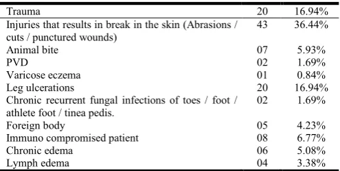
Table 3. Precipitating causes / Risk factors
Trauma 20 16.94%
Injuries that results in break in the skin (Abrasions / cuts / punctured wounds)
43 36.44%
Animal bite 07 5.93%
PVD 02 1.69%
Varicose eczema 01 0.84%
Leg ulcerations 20 16.94%
Chronic recurrent fungal infections of toes / foot / athlete foot / tinea pedis.
02 1.69%
Foreign body 05 4.23%
Immuno compromised patient 08 6.77%
Chronic edema 06 5.08%
Lymph edema 04 3.38%
Table 4. Site wise distribution of cellulities
Lower extremity 87(66 U/L 21L/L) 73.72
Upper extremity 21 (19 U/L 3 B/L) 17.71
Axilla 01 0.84
Facial 01 0.84
Scalp 01 0.84
Scrotum 03 2.54
Anterior abdominal wall 03 2.54
Neck 01 0.84
Table 5. Clinical Features
Symptoms
Swelling 89 75.42
Pain 108 91.52
Redness 41 34.74
Discharge 34 28.81
Fever 50 42.37
Table 6. Clinical Signs
General Examination
Fever 50 42.37
Tachycardia 02 1.69
Hypotension 10 8.47
Local Examination
Colour changes 62 52.54
Local rise of temperature 88 74.57
Tenderness 83 70.33
Erythema 72 61.01
Blisters 15 12.71
Raised glossy tight stretched skin 34 28.81
Fluctuations 08 6.78
Dilated veins 03 2.54
Eczema 03 2.54
[image:2.595.38.293.467.601.2]Cracks 05 4.23
Table 7. Treatment
A) Medical Treatment 28 23.72
B) Surgical Treatment 90 76.27
A) Medical Treatment = 28
-Rest and limb elevation -Antibiotics
-Analgesics
[image:2.595.43.284.638.752.2]a conservative treatment and 90 patients were managed with surgical treatment.
B) Surgical Treatment
Releasing incision and drainage 24 20.33
Debridment and skin grafting 46 38.98
Releasing I & D along with debridment 7 5.93
Debridment and flap 1 0.84
Amputation 11 9.32
Above knee amputation 01 0.84
Below knee amputation 02 1.69
Disarticulation of fingers / toes 08 6.78
Foreign body removal 02 1.69
Mortality -03
DISCUSSION
Prospectively 118 collected cellulities case patient 71-males, 31 females, with the male female ratio (2.02: 1) where included in the study conducted at department of general surgery, Al-Ameen Medical College, over a period of 2010 – 2012. Table-1 shows demographic data of the patient. Median age 46.41% with average age, 9 months to 85 years and standard deviation 20.25 was observed in our study. Study conducted by Sigridur et al. showed the median age of the participants was 66.5 years (interquartile range 48.8 – 77.0) (Sigridur Bjornsdottir et al., 2005). Table 2 shows associated diseases /medical conditions. In our patients, 47 case, 39.83 % where associated with medical disease (concomitant diseases). Patients with co-morbid conditions that predispose them to infection are also at increased risk of cellulitis (CREST, 2005). Study conducted by Sigridur et al. shows cardiovascular 55 endocrine or metabolic 18, Gastro intestinal 7, neurological 8 rheumatic or orthopedic 19, psychiatric 12, pulmonary 7, urinary 10 and other 1(Sigridur Bjornsdottir et al., 2005).
In our study 47 cases 39.83% where associated with medical diseases. Among them Diabetes Mellitus -15, Anemia – 07, Hypertension – 05, Obesity – 06, Renal failure-07, COPD-03, IHD-02, Hepatomegally-01, Blood disorders-01. People at risk of cellulitis included those with trauma or disrupted area of the skin or other medical conditions. Several factors appeared to strongly affect cellulitis (Mary Eagle, 2007). In our study we observed precipitating causes / risk factors. Trauma-20, injuries that results break in the skin (abrasion / cut/ punctured wounds)-43, animal bite-07, PVD-02, varicose eczema-01, leg ulceration-02, chronic recurrent fungal infections of toes / foot / athlete foot / tinea pedis -02, foreign body-05, immuno compromised patient-08, chronic edema-06, lymph edema-04.
There are many risk factors for cellulitis (Mary Eagle, 2007; El-daher, 1996; Hughes et al., 2001; Stalbow, 2004).
Injuries or trauma that result in a break in the skin.
Insect bites and stings, and animal or human bites.
Chronic recurrent fungal infection of the fingers and toes, such as athlete’s foot or tinea pedis.
Peripheral arterial disease
Varicose eczema
Leg ulceration
Diabetes mellitus
Lymphatic insufficiency, poor lymphatic drainage, chronic edema and lymphoedema.
Liver disease such as chronic hepatitis or cirrhosis.
Obesity
Chronic skin disorders such as eczema psoriasis, which result in breaks or dry, flaky skin that provide an entry point for bacteria.
Infectious diseases that causes skin lesion such as chicken pox.
Infections related to surgical procedures.
Burns.
Foreign objects in the skin eg : IV Cannula, drainage tubes, percutaneous endoscopic gastrostomy feeding tubes and orthopedic pin.
Infection of the bones beneath the skin.
Weakened immune system or immuno suppressive or cortico steroid therapy may lead to more vulnerability to infection (Mary Eagle, 2007; Stalbow, 2004).
Any break in the skin integrity can allow bacteria to enter the skin and cause infection, which can spread and lead to cellulitis (Pauline Beldon, 2011). The young healthy individual who develops cellulitis after trauma differs from the middle aged patients, whose co-morbid conditions may predispose him or her to recurrent attacks, with or without apparent site of pathogen entry. Predisposing factors do not pursue causes bacterial cellulitis. The infection originate from the entry of pathogen through a disruption of the cutaneous barrier that may or may not be identified on physical examination (Sigridur Bjornsdottir et al., 2005).
All chronic leg ulcers contain bacteria (Gilchrist, 1999; Cox and Lawence, 1998) suggested that cellulitis secondary to leg ulceration may be caused by a large variety of organism which are known to colonize chronic wounds, including Streptococci,
Staphylococci, Pseudomonas, SPP. Bacteroides SPP (Cox, 1998) and other lesser common varieties (CREST,
2005). The role of various predisposing factors such as previous history of cellulitis, leg edema and Saphenectomy in the pathogenesis of cellulitis has not been elucidated. Although these factors do not directly cause infections, they probably facilitate it, development by impairing local defense mechanism (Sigridur Bjornsdottir et al., 2005). Cellulitis can occur anywhere on the body but is frequently encountered on the lower leg, ankles and arms (Linda Rafter, 2011; Bickley, 2003). In our study lower extremity-87, upper extremity-21, axilla-01, facial-01, scalp-01, scrotum-03, anterior abdominal wall-03, neck-01.
The lower limb is the most common for cellulitis to occur, often through a break in the skin due to an existing leg ulcer or an injury. The development of cellulitis in the lower limb may also be attributed to athlete foot. Injuries that have not been treated appropriately (especially on the hands and feet but even on the head) may be prone to severe infection that can result in cellulitis (Pauline Beldon, 2011). In our study we observed clinical symptoms were swelling-89, pain-108, redness-41, discharge-34, fever-50 and observed clinical signs on general examination were fever-80, tachycardia-02, hypotension-10 and local examination finding were colour changes-62, local rise of temperature-88, tenderness-83, erythema-72,
15, raised glossy tight stretched skin-34, fluctuation-08, dilated veins-03, eczema-03, cracks-05. Following an episode of lower limb cellulitis, around 7% of patient develop chronic edema and some persistent leg ulceration. Around 29% of patients develop recurrence, within a mean of three years, with venous insufficiency being the most common causes (CREST, 2005). It is important to avoid a misdiagnosis, especially because as mentioned above other conditions such as dermatitis eczema, thrombo-phlebitis (inflammation of vein) or venous hypertension (Dupuy et al., 1999) can all be easily mistaken for cellulitis (Nazarko, 2009).
A deep vein thrombosis may present as a swollen, painful leg, but will not have the painful spreading erythema (redness). Similarly, chronic edema of the lower legs may present with blistering in severe cases, but again there will be no signs of infection (Hunter et al., 2002). Bilateral concurrent cellulitis is extremely uncommon occasionally if the patient has more than one wound, cellulitis may be present bilaterally (Mary Eagle, 2007). Cellulitis may be triggered by one or more bacteria, commonly Haemolytic Streptococci and G Streptococci, Streptococcus pyogenes and Staphylococcus aureus (Baxter and McGregor, 2001; Cooper and White, 2009).
Patients with cellulitis may present with inflammation heat, redness and pain (Nowa, 2000). Initially the inflammation is localized, but increases as the infection progresses. When this occurs, the patient can be systemically unwell and the skin appears to be tight and glossy (Price, 2009). Table -1 outlines the four clinical classes of cellulitis (CREST, 2005). The common symptoms of cellulitis which may result in skin changes affecting its colour sensation and temperature are:
Redness of the skin, presents as either red streaking or broad area of redness. It may be difficult to diagnose cellulitis from observation in people with darkened skin., therefore it is important to recognize other clinical symptoms as they present. Swelling usually has a rapid spreading ascent up the lower leg starting from the foot but can commence from the calf. Raised swollen, tight glossy stretched appearance of the skin. Often a clear demarcation line of pale skin against the red raised, swollen, tight glossy stretched appearance of the infected area this may be difficult to identify in the skin with darken pigmentation. Pain or tenderness, area of heat (hot / warm) tender erythematous swelling in tissues surrounding an existing wound. A tender swollen limb with generalized erythema may be accompanied by pyrexia / fever above 38 degree (Gilchrist, 1999). Flue like symptoms before cellulitis develops and as the condition spreads to the body via the blood, then fever and chills can result. Elevated white cell count indicating bacterial infection.
Draining or leaking of yellow clear fluid or pus from the skin. Over time the area of painful redness tends to expand (dry cellulitis) as the infection and resulting tissue damage spreads rapidly.
Particularly if not promptly treated small red dots may appear on top of the dry cellulitis and less commonly small blisters may form and burst (wet cellulitis) (Mary Eagle, 2007).
Clinical classes of cellulitis (Linda Rafter, 2011)
Class –I
Patients have no signs of systemic toxicity, or uncontrolled co morbidities and can be managed with oral anti microbial on an out patient basis.
Class-II
Patient systemically ill or systemically well, but have a co morbidity such as peripheral vascular disease, chronic venous insufficiency or morbid obesity which complicate or delays resolution of infection.
Class-III
Patient may have a significant systemic complication such as acute confusion tachycardia tachypnoea, hypertension or be unstable. Systemically well but have a cormorbidity that may interfere with response to therapy or have threatening infection due to vascular compromise.
Class-IV
Patient develops sepsis syndrome or severe life threatening infection such as necrotizing fasciitis (Linda Rafter, 2011). It is often difficult for a clinician to identify cellulitis from presenting symptoms, such as redness alone, therefore spreading erythema is considered a more accurate indicator of infection when accompanied by other symptoms such as raised temperature or increase in a pain levels (Mary Eagle, 2007).
Pathogens associated with clinical scenario – Diabetes – Staphylococcus aureus, group B Streptococci
Anaerobes Gram- Negative bacilli.
Cirrhosis – campylobacter fetus, Coliforms, vibrio, vulnificus, Capnocytophaga canimorsus.
Neutropenia – Pseudomonas aurginosa .
Human bite – Ephimella corrodens, cat bite – Pasteurella multocida.
Dog bite – P-Multocida, C-canimorsus. Rat bite- Streptobacillus monoliformis. Hot tub exposures – P. aurgenosa.
Fresh water Laceration : Aeromonas hydrophila. Fish tank exposure – Mycobacterium marinum. IV drug use : MRSA, P aurgenosa.
In our study 28 patient underwent medical treatment, in that rest with limb elevation, antibiotics and analgesic.
Surgical treatment includes
Releasing incision and drainage-24 patients, debridment and skin grafting-46, releasing I & D along with -07, Debridment and Flap – 01, Amputation-01, Above knee amputation -01, Below knee amputation-02, disarticulation of finger / toes – 08 patients, foreign body removal-02.
Treatment of cellulitis
antibiotic sensitive to streptococci and staphylococci may be intravenously or oral antibiotics. Common treatment is a combination of benzyl penicillin (unless there is a contraindication by allergy) and a broad spectrum antibiotic such as flucloxacillin antibiotics may be given intravenously initially and than orally once they begin to take effect (Beldon and Burton, 2005). Clinician must relieve pain and discomfort because without this relief, the patient will not be able to tolerate other treatment.
It is important that efficacy of pain relief is checked by regularly asking about his / her pain status and documenting pain score using relevant tool such as visual analogue scale, verbal rating scale, numerical rating scale (Taylor, 2010). Clinician should ensure that affected part of the body, often lower limb is elevated so that the whole limb is supported and the patient perform dorsiflexion exercise to aid reduction of any edema (Hoffman, 2007).
As mentioned above, it is important to remember that cellulitis, is not restricted to the lower limbs, the patients hands, head, neck or any other part of the body may be affected by cellulitis if the infection is severe enough to spread (Pauline Beldon, 2011). If the cellulitis is wet, a wound swab obtained in order to determine both the causative organism (Holzapel, 1999) and sensitivity to antibiotic therapy in order to ensure rapid, clinically effective treatment (CREST, 2005). Blister should always be de-roofed as if they are left in situ the pressure from the blister may cause underlying ulceration (Pauline Beldon, 2011). Wet cellulitis (where there is fluid such as exudates associated with blistering) is often treated with potassium permanganate solution. The limb is soaked 10-15 minutes and then dried thoroughly simple non-adherent dressing may then be applied. However, if the limb is producing copious amounts of exudates, than a more absorbent dressing such as foam, alginate, fibrous dressing or hydration response dressing should be used (Pauline Beldon, 2011). High bioavailability makes parenteral administration of antimicrobials largely unnecessary for mild, uncomplicated cellulitis, parental therapy is usually reserved fro moderate to severe infections that my require hospitalization (CREST, 2005).
Topical treatment is an option for mild impetigo. Patients with mild, uncomplicated cellulitis who are not at high risk for MRSA should receive oral antibiotics active against both staphylococcus and streptococci. Abscess require drainage. Oral antibiotic are sufficient to treat mild MRSA infection. Patients unable to tolerate oral antibiotics of those with severe cellulitis and systemic toxicity who are at risk for MRSA should receive parenteral antibiotics active against MRSA and streptococci. Antimicrobials should be adjusted to focus on culture identified pathogen prompt surgical evaluation is indicated for patients with evidence of necrotizing fasciitis (CREST, 2005). One study suggests that abscess less than 5 cm in diameter may be effectively treated by incision and drainage without systemic antimicrobial therapy, but other studies show no correlation between the size of abscesses and the need for antibiotic therapy (CREST, 2005). Evidence of gangrenous or necrotizing infection requires immediate and thorough debridment. This is especially time of streptococcal necrotizing fascities. Expert consensus from the use of
intravenous immunoglobins to treat streptococcal necrotizing fasciitis, although studies are conflicting. Friable fascia and dark muscle that do not bleed or twitch requires debridment, which should be continued until viable tissue is reached. Multiple debridments are often required for patients with necrotizing fasciitis or myonecrosis. Delay of surgical debridement increases mortality (CREST, 2005). Extension of the redness and increase in the severity of the symptom may signal a deeper ,more serious infection of the inner layer of the skin ,once below the upper layer of the skin ,bacteria can spread rapidly entering lymph nodes and blood stream in rare cases streptococcal infection can spread to deeper layer of the fascial lining around muscles resulting in necrotizing fascitis .this represents an extreme emergency requiring urgent hospital treatment (Rowland, 2002).
Conclusion
As cellulitis is a common surgical emergency practitioner should be trained to recognize the symptoms and signs to process the treatment pathway early.And to prevent pain and distress of the patient in order to decrease in morbidity and mortality with improved quality of life.
Author contribution
Study conception and design: Dr. Azmath, Dr. Nishikant N
Gujar
Drafting of manuscript and critical revision: Dr Mansour A.
Binfayed
Supervision: Dr. Sajid Ahmed Mudhol
Acquisition of data: Dr. Shiraz Ahamed Sharief, Dr. Sachin,
Dr. Vipin Balachandran, Dr. Mohammed KhasifHabeeb
Acknowledgments
The authors deeply acknowledge Dr .B .S.Patil, Dean of Al ameen Medical College, Bijapur for granting permission to publish this study and we are very much thankful to Dr. Saleem Dhundasi, Vice Principle of Al Ameen Medical College, Bijapur and Dr.Satish Rashinkar Medical Superintendent, Al Ameen Hospital, Bijapur for their valuable support in conduction of this study. And we also thankful to our House surgeons Dr. Rubby kristy and Dr.Vidhya for helping this study.
REFERENCES
Baxter, H. and McGregor, 2001. Understanding and managing cellulitis.nurs Standard 15(44):50-6.
Beldon, P. and Burton, F. 2005. Management guidelines for lower limb cellulitis.Wounds UK1(3):16-22.
Bickley, L. S. 2003. Bali’s Guide to physical Examination and History-taking. Lippincott, Williams and Wilkins, London.
Carratala, J., Roson, B. and Fernandez-sabe, N. et al. 2003. factors associated with complications and mortality in adult patients hospitalized for infectious cellulitis. Eur J Clin Microbiol Infect Dis, 22:151-7
Cooper, R. and White, R. 2009. Cutaneous infections in lymphoedema. J lymphoedema 4(1);44-8.
Cox, N. and Lawence, C. M. 1998. Diagnostic problems in Dermatology. Mosby, London:146-7.
CREST, 2005 Guidelines on The management of cellulitis in adults ,CREST, available online at www.crestni.org.uk (assessed date January 2014).
Drug Therapeutic Bulletin 2003. Dilemmas when managing cellulitis. Drugs Ther Bull., 41(6):43-6 (assessed date January 2014).
Dupuy, A., Benchikhi, R. and Roujeau, J. et al 1999. Risk factors for erysipelas of the leg (cellulitis):case control study . Br Med J., 318:1591-4.
El-daher, N. and Magnussen, C. R. 1996. Skin and soft tissue infections: Outpatient management and indications for Hospitalisation.consultant 36(12):2563-6.
Eriksson, B., Jorup-Ronstrom, C., Karkkonen, K. and Sjoblom, A. C. 1996. Holm SE.Erysipelas: clinical and bacteriological spectrum and seriological aspects.clin Infect Dis, 23:1091-8.
Gilchrist, B. 1999. Wound infection. In:Wound Management Theory and practice. Eds, Miller M., Glover D. Nursing Times Books. Emap Health care ltd, Dorchester.
Hoffman, D. 2007. The autolytic debridment of venous leg ulcers.Wound Essentials 2:68-73.
Holzapel, L. Jacquet-franacllon, T. and Rahmani, J. et al. 1999 Microbiological evaluation of infected wounds of the extremities in 214 adults. J Accident Emergency Med.,
16:32-4.
Hughes, E., van Onselen, J., eds, 2001. Dermatology Nursing: A practical Guide. Churchill Livingstone, London:207. Hunter, J., Savin, J. and Dahl, M. 2002. Clinical
Dermatology.3rd Edition. Blackwell sciences ltd, oxford, London.
Linda Rafter. Management of acute cellulitis with the Eclypse Boot. Wounds UK , 2011,vol 7;62-68 .
Mary Eagle, Understanding cellulitis of lower limb, Wound Essentials. Volume 2, 2007:34-44.
National Health Executive, 2006. public Expenditure statistical Analysis. Available online at; www.nationalhealthexecutive.com/news-woundcare-silent-epidemic.htm
Nazarko, L. 2009. The accurate diagnosis and treatment of lipodermatosclerosis. Br J Nurs., 18(12):715-8.
Nowa, K. T. and Handford, A. 2000. Essentials of Pathophysiology; concepts and applications for health care Professionals.2nd edn. McGraw-Hill Boston MA.
Pauline Beldon. The Assessment, Diagnosis and Treatment of cellulitis. Wound Essentials Volume 6.2011:60-68. Price, N. 2009. Management of cellulitis after insect bites.
Emergency Nurse, 17(7):24-7.
Rowland, B. 2002. Gale Encyclopedia of Medicine, The Gale Group inc Gale Detroit, USA.
Sigridur Bjornsdottir et al. Risk factors for acute cellulitis of the lower limb: A prospective case control study.
Clinical infectious diseases, 2005:41:1416-22.
Stalbow, J. 2004. Preventing cellulitis in Older people with Persistent lower limb oedema. Br J Nurs., 13(12):725-32. Taylor, A. 2010. Principles of pain assessment. Wound
Essentials 5:104-10.