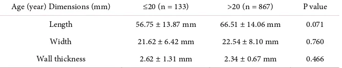
Anatomy of the Gall Bladder in the Ultrasound about 1000 Cases
Full text
Figure


Related documents
The paper is discussed for various techniques for sensor localization and various interpolation methods for variety of prediction methods used by various applications
Мөн БЗДүүргийн нохойн уушгины жижиг гуурсанцрын хучуур эсийн болон гөлгөр булчингийн ширхгийн гиперплази (4-р зураг), Чингэлтэй дүүргийн нохойн уушгинд том
19% serve a county. Fourteen per cent of the centers provide service for adjoining states in addition to the states in which they are located; usually these adjoining states have
Field experiments were conducted at Ebonyi State University Research Farm during 2009 and 2010 farming seasons to evaluate the effect of intercropping maize with
Aptness of Candidates in the Pool to Serve as Role Models When presented with the candidate role model profiles, nine out of ten student participants found two or more in the pool
Results suggest that the probability of under-educated employment is higher among low skilled recent migrants and that the over-education risk is higher among high skilled
Date of review, Publication details (Title, Authors, Reference), Database, Methodology (interview, case study, report, survey etc.,), Sample Population, Target
This essay asserts that to effectively degrade and ultimately destroy the Islamic State of Iraq and Syria (ISIS), and to topple the Bashar al-Assad’s regime, the international
