Journal of the American Chemical Society is published by the American Chemical Society. 1155 Sixteenth Street N.W., Washington, DC 20036
Published by American Chemical Society. Copyright © American Chemical Society. However, no copyright claim is made to original U.S. Government works, or works produced by employees of any Commonwealth realm Crown government in the course of their duties.
Communication
A synthetic replicator drives a propagating reaction-diffusion front
Ilaria Bottero, Jürgen Huck, Tamara Kosikova, and Douglas Philp
J. Am. Chem. Soc., Just Accepted Manuscript • DOI: 10.1021/jacs.6b03372 • Publication Date (Web): 13 May 2016
Downloaded from http://pubs.acs.org on May 17, 2016
Just Accepted
A synthetic replicator drives a propagating reaction-diffusion front
Ilaria Bottero, Jürgen Huck, Tamara Kosikova and Douglas Philp*
School of Chemistry and EaStCHEM, University of St Andrews, North Haugh, St Andrews, Fife KY16 9ST, UK
ABSTRACT: A simple synthetic autocatalytic replicator is capable of establishing and driving the propagation of a reaction-diffusion front within a 50 µL syringe. This replicator templates its own synthesis through a 1,3-dipolar cycloaddition reaction between a nitrone compo-nent, equipped with a 9-ethynylanthracene optical tag, and a maleimide. Kinetic studies using NMR and UV-Vis spectroscopies confirm that the replicator forms effi-ciently and with high diastereoselectivity and this repli-cation process brings about a dramatic change in optical properties of the sample – a change in the color of the fluorescence in the sample from yellow to blue. The ad-dition of a small amount of the pre-formed replicator at a specific location within a microsyringe, filled with the reaction building blocks, results in the initiation and propagation of a reaction-diffusion front. The realization of a replicator capable of initiating a reaction-diffusion front provides a platform for the examination of inter-connected replicating networks under out-of-equilibrium conditions involving diffusion processes.
The spontaneous generation of stationary patterns and propagating fronts in chemical systems1 has intrigued scientists for generation. Such phenomena are ubiqui-tous in nature2 and the physical processes behind their appearance and stability have been studied extensively3 and are now relatively well understood. Propagating reaction-diffusion fronts have received significant atten-tion in this respect. Frequently, one or more oscillatory or autocatalytic processes are found at the core of these systems. Front generation is initiated when an autocata-lyst is added at a discrete location in an expanse of reac-tant, initially at uniform concentration and the ensuing reaction generates wave fronts, which propagate out-ward from the initial reaction zone. In almost all4 of the examples reported to date, the autocatalysis is based on inorganic chemistry, although, more recently, a small number of examples based on RNA5 and DNA6 have been described. Self-replication7 represents a niche of autocatalytic behavior in which a structurally complex template is capable of recognizing the building blocks
necessary for its own formation and catalyzing their re-action to form an exact copy of itself. We, and others, have described the use of such systems in instructable networks8, as tools for dynamic systemic resolution9 and in the construction10 of mechanically-interlocked mole-cules. Although all of these systems display the non-linear kinetic characteristics of autocatalytic systems to a greater or lesser extent, in general, they have been stud-ied under well-stirred batch reactor conditions. The con-sequence of this reaction format places a fundamental limit on the level of complexity and emergence that can be generated by such system. In order to create diverse emergent behavior there is a need to study self-replicating systems under out-of-equilibrium conditions and propagating reaction-diffusion fronts could provide an ideal vehicle for such studies. Here, we report the design and implementation of a molecular replicating system capable of generating and sustaining a propagat-ing reaction-diffusion front.
Previously, we have described11 an efficient synthetic replicator based on the general design shown in Figure 1a. Reaction of nitrone 1a with maleimide 2 in CDCl3 at
–10 °C results in the rapid, autocatalytic formation of the cycloadduct 3a. Cycloadduct 3a is a very efficient template for its own formation – it is capable of acceler-ating the reaction between 1a and 2 up to 125 × through a ternary complex [1a•2•3a] and the structure of this complex ensures that only the trans diastereoisomer of
3a is formed during this process. Conventionally, we have monitored the kinetics of replication processes by NMR spectroscopy. In a reaction diffusion format, this reaction requires an alternative method to monitor the progress12 of the reaction. Ideally, we desired an optical signature of replication. Therefore, we designed nitrone
1b, bearing a 9-ethynylanthracene tag. RM1 calculations (Figure 1b) indicated that this nitrone, in partnership with maleimide 2 was capable of furnishing template 3b
through a ternary complex [1b•2•3b]. This complex should permit the formation of only the trans diastereo-sisomer of 3b. TD-DFT calculations (see Supporting Information) indicated that a significant change in the 350 to 400 nm region of the UV-Vis spectrum could be expected on conversion of nitrone 1b to cycloadduct 3b Page 1 of 11
ACS Paragon Plus Environment Journal of the American Chemical Society
as a result of the presence of the 9-ethynylanthracene unit. We synthesized nitrone 1b using standard methods and this compound forms yellow-colored solutions in CDCl3, which exhibit an intense yellow fluorescence
(Figure 1a). Pleasingly, the conversion of 1b into 3b
resulted in a very significant color change – the yellow fluorescence of 1b being replaced (Figure 1a) by the blue fluorescence of 3b.
Figure 1. (a) A self-replicating template 3a is constructed by the reaction of nitrone 1a with maleimide 2 through in the catalytically-active [1a•2•3a] ternary complex. Re-placement of the substituent R affords replicator 3b, which incorporates a 9-ethynylanthracene optical tag, derived from nitrone 1b. The formation of cycloadduct 3b, mediat-ed by ternary complex [1b•2•3b], is now associated with a change in fluorescence from yellow (1b) to blue (3b). (b) Calculated (RM1) structure of the transition state leading to
3b from the ternary complex [1b•2•3b],
Next, we conducted a series of kinetic experiments in-volving the reaction of 1b and 2 in CDCl3 at 0 and
20 °C, monitoring the production of cycloadduct 3b by 500 MHz 1H NMR spectroscopy. The results of these experiments and subsequent fitting of the experimental data at 20 °C to the appropriate kinetic model are shown in Figures 2a and 2b. These kinetic experiments reveal
that replicator 3b is an excellent template for its own formation – the ternary complex [1b•2•3b] generates an effective molarity13 (EM) of 16.2 M for the cycloaddi-tion reaccycloaddi-tion (for details, see Supporting Informacycloaddi-tion). This value for the ternary complex EM is broadly simi-lar to those determined previously9e,10b,11 for similar rep-licators and indicates that the incorporation of the 9-ethynylanthracene optical probe has essentially no effect on the functioning of the replicator.
Figure 2. (a) Concentration (red circles) and rate (black dotted line) vs time profile for the formation of 3b from nitrone 1b and maleimide 2 as determined by 500 MHz 1H NMR spectroscopy ([1b] = [2] = 10 mM, 20 °C, CDCl3).
The red line shows the fit of the appropriate kinetic model to these data. (See Supporting Information) (b) The appear-ance of the resonappear-ance associated with the formation of the
trans-3b cycloadduct over time in the 500 MHz 1H NMR spectrum of a reaction mixture containing 1b and 2 ([1b] = [2] = 10 mM, 20 °C, CDCl3). (c) Selected UV-Vis spectra
recorded during the reaction of 1b and 2 ([1b] = [2] = 10 mM, 20 °C, CDCl3), showing the disappearance of
nitrone 1b (346 nm band) and simultaneous appearance of cycloadduct 3b (297 nm band). (d) Comparison of absorb-ance at 297 nm vs time. The blue circles show data deter-mined experimentally from the spectra in Figure 2c. The red line shows the absorbance at 297 nm computed using the concentrations of 3b determined from the best fit of the appropriate kinetic model to the NMR data in Figure 2a.
Having established that 3b was indeed capable of tem-plating its own formation, we next sought to establish that the color change that is observed during this reac-tion is a signature of the autocatalytic replicareac-tion pro-cesses. Accordingly, we monitored the formation of 3b
using UV-Vis spectroscopy (Figure 2c) under identical conditions to those employed in the NMR kinetic
exper-ACS Paragon Plus Environment
[image:3.612.60.290.139.505.2] [image:3.612.319.554.149.405.2]iments. As expected, the UV-Vis spectra recorded during the reaction show the disappearance of a band corre-sponding to nitrone 1b at 346 nm with the concomitant appearance of a band at 297 nm corresponding to cy-cloadduct 3. In order to relate this data to the kinetic data derived from NMR spectroscopy, we reconstructed the reaction profile at 297 nm by computing the expected absorbance at this wavelength from the concentrations of the components of the reaction mixture determined from the best fit our kinetic model to the NMR data. The excellent agreement (Figure 2d) between the calculated and observed reaction profiles provides compelling evi-dence that the color change that is observed during this reaction is indeed the signature of the replication of 3b.
Figure 3. (a) Graphical representation and (b) photograph of the experimental setup employed for investigation of the propagating reaction-diffusion front initiated by replicator
3b in two 50 µL gas tight syringes. The upper syringe in each case represents the control experiment comprising the nitrone 1b and maleimide 2 only. ([1b] = [2] = 5 mM, 20 °C, CDCl3). The lower syringe is seeded with ca. 2 µL
of a solution of 3b. ([1b] = [2] = 5 mM, [3b] = 10 mM, 20 °C, CDCl3).
Normally, propagating reaction-diffusion fronts are ob-served in reactions that are initiated on flat plates or within capillary tubes. Since CDCl3 is a relatively
vola-tile solvent, we chose to investigate whether 3b was ca-pable of supporting a propagating reaction-diffusion front within a 50 µL gas tight syringe of internal diame-ter 1.03 mm. Figure 3 illustrates our experimental setup. Two syringes were placed side-by-side in a specially constructed stand housed within a controlled environ-ment where the temperature was regulated at 20 °C. One syringe was filled with a 5 mM solution of nitrone 1b
and maleimide 2 in CDCl3. The second syringe was
pre-pared identically with the exception that approximately 2 µL of a 10 mM solution of replicator 3b was drawn into the end of the syringe after it was filled with the solution of 1b and 2.
We envisaged that the syringe containing only 1b and 2
would change color uniformly as replicator 3b was
formed. In the other syringe, the presence of 3b would initiate the replication process and the diffusion of the replicator thus formed would establish a reaction-diffusion front that would propagate along the syringe, being observed as the progression of a blue band along the initially yellow syringe.
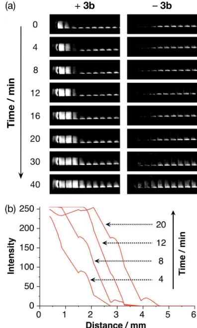
Figure 4. (a) Processed grayscale images, acquired over time with the syringes illuminated using a 365 nm UV lamp, of the template-initiated reaction-diffusion experi-ment (+3b, left column) and the control experiment (–3b, right column). (b) Smoothed profiles of the grayscale im-ages from the reaction-diffusion experiment (+3b) over time showing the progression of the reaction diffusion front over 20 minutes.
The syringes were illuminated using a 365 nm UV lamp and a color image was captured every two minutes using a digital camera. Processing of these images (see Sup-porting Information) afforded the data shown in Figure 4a. It is evident from these images that replicator 3b has, established (Figure 4a, +3b) a propagating reaction-diffusion front within the syringe to which it was added initially. By contrast, no such feature is evident within the control syringe (Figure 4a, –3b). As an additional control, we examined the diffusion of 3b in the absence of an autocatalytic reaction. Approximately 2 µL of a 10 mM solution of replicator 3b was drawn into the end of a syringe filled with a 5 mM solution of nitrone 1b
only in CDCl3. In this case, the blue color of replicator
Page 3 of 11
ACS Paragon Plus Environment Journal of the American Chemical Society
[image:4.612.338.536.118.447.2] [image:4.612.61.282.214.386.2]3b disappears as a result of diffusion processes within the syringe, leaving the syringe visually indistinguisha-ble from one that had been filled with nitrone 1b only. No optical signature of a propagating reaction-diffusion front is observed in this case. Selected images were pro-cessed further (see Supporting Information) to compute the profile (Figure 4b) of the propagating front at a se-quence of time points. These data clearly show the pro-gression of the reaction-diffusion front mediated by rep-licator 3b along the syringe. In many cases, reaction-diffusion fronts propagate at constant linear or radial velocity. However, in this case, the progression14 of the front slows and will eventually stall as nitrone 1b and maleimide 2 are depleted throughout syringe as a result of the background rate of the cycloaddition reaction forming 3b being significant on the timescale of the ex-periment.
Here, we have described the first example of a propagat-ing reaction-diffusion front that is initiated and driven by a synthetic replicator. The work reported here represents a proof-of-principle. The successful implementation of a replicator-driven reaction-diffusion front, mediated by an autocatalytic replicator of defined structure and with specific interactions and catalytic relationships with oth-er similar replicators, opens up15 a number of exciting possibilities. This reaction format will allow us to ex-plore networks8a of replicators under conditions and out-comes that lie far from the constraints16 imposed by well-stirred batch reactors. These studies are currently underway in our laboratory.
ASSOCIATED CONTENT
Electronic Supplementary Information (ESI) available: experimental procedures, details of kinetic measurements and fitting, computational modeling of UV spectra, details of UV-Vis and fluorescence analyses and methods and analyses for the reaction-diffusion experiments.
AUTHOR INFORMATION
Corresponding Author
Funding Sources
The financial support for this work was provided by EPSRC (Grants EP/E017851/1 and EP/K503162/1). No competing financial interests have been declared.
ACKNOWLEDGMENT
We thank EPSRC for postgraduate studentship awards to JH (EP/ E017851/1) and TK (EP/K503162/1).
REFERENCES
(1) (a) Jee, E.; Bánsági Jr., T.; Taylor, A. F.; Pojman, J. A. Angew. Chem. Int. Ed. 2016, 55, 2127. (b) Showalter, K.; Epstein, I. R. Chaos
2015, 25, 097613. (c) Vanag, V. K.; Epstein, I. R. Int. J. Dev. Biol.
2009, 53, 673. (d) Vanag, V. K.; Epstein, I. R. Chaos 2008, 18,
026107. (e) Epstein, I. R.; Pojman, J. A.; Steinbock, O. Chaos 2006, 16, 037101. (f) Mikhailov, A. S.; Showalter, K. Phys. Rep. 2006, 425, 79. (g) Taylor, A. F.; Britton, M. M. Chaos 2006, 16, 037103. (h) Johnson, B. R.; Scott, S. K. Chem. Soc. Rev. 1996, 265. (i) Pojman, J. A. Frontal Polymerization. In Polymer Science: A Comprehensive Reference, Matyjaszewski, K.; Möhler, M.; Eds.; Elsevier B.V., Amsterdam, 2012, pp 3018–3020.
(2) (a) Kondo, S.; Miura, T. Science 2010, 329, 1616. (b) Volpert, V.; Petrovskii, S. Phys. Life Rev. 2009, 267. (c) Camazine, S.; Deneubourg, J.-L.; Franks, N. R.; Sneyd, J.; Theraulaz, G.; Bonabeau, E. Self-Organization in Biological Systems. In Princeton studies in complexity, Anderson, P. W.; Epstein, J. M.; Foley, D. K.; Levin, S. A.; Nowak, M. A.; Eds.; Princeton University Press, 2003. (d) Hess, B. Naturwissenschaften 2000, 87, 199. (e) Ball, P. The Self-made Tapestry: Pattern Formation in Nature; Oxford University Press Inc., New York, 2001.
(3) (a) Chemical waves and patterns, Kapral, R.; Showalter, K.; Eds.; Kluwer Academic Publishers, 2012. (b) Grzybowski, B. A.; Chemistry in Motion, John Wiley & Sons, Ltd., 2009. (c) Epstein, I. R.; Pojman, J. A. An introduction to nonlinear chemical dynamics: oscillations, waves, patterns, and chaos; Oxford University Press,
1998. (d) Cross, M. C.; Hohenberg, P. C. Rev. Mod. Phys. 1993, 65, 851.
(4) Boga, E.; Peintler, G.; Nagypál, I. J. Am. Chem. Soc. 1990, 112, 151.
(5) (a) McCaskill, J. S.; Bauer, G. J. Proc. Natl. Acad. Sci. U. S. A.
1993, 90, 4191. (b) Bauer, G. J.; McCaskill, J. S.; Otten, H. Proc. Natl. Acad. Sci. U. S. A. 1989, 86, 7937.
(6) (a) Zadorin, A. S.; Rondelez, Y.; Galas, J. C.; Estévez-Torres, A. Phys. Rev. Lett. 2015, 114, 1. (b) Padirac, A.; Fujii, T.; Estévez-Torres, A.; Rondelez, Y. J. Am. Chem. Soc. 2013, 135, 14586.
(7) (a) Bissette, A. J.; Fletcher, S. P. Angew. Chemie Int. Ed. 2013, 52, 12800. (b) Huck, J.; Philp, D. In Supramolecular Chemistry: From Molecules to Nanomaterials; John Wiley & Sons, Ltd., 2012; Volume 4, pp 1415–1446. (c) Plasson, R.; Brandenburg, A.; Jullien, L.; Bersini, H. Artif. Life, 2011, 17, 219. (d) Vidonne, A.; Philp, D. European J. Org. Chem. 2009, 2009, 593. (e) Patzke, V.; von Kiedrowski, G. ARKIVOC 2007, 46, 293. (f) Paul, N.; Joyce, G. F. Curr. Opin. Chem. Biol. 2004, 8, 634.
(8) (a) Kassianidis, E.; Pearson, R. J.; Wood, E. A.; Philp, D. Faraday Discuss. 2010, 145, 235. (b) Ashkenasy, G.; Ghadiri, M. R. J. Am. Chem. Soc. 2004, 126, 11140. (c) Yao, S.; Ghosh, I.; Zutshi, R.; Chmielewski, J. Nature 1998, 396, 447. (d) Sievers, D.; von Kiedrowski, G. Chem. Eur. J. 1998, 4, 629.
(9) (a) Dadon, Z.; Samiappan, M.; Wagner, N.; Ashkenasy, G. Chem. Commun. 2012, 48, 1419. (b) Carnall, J. M. A.; Waudby, C. A.; Belenguer, A. M.; Stuart, M. C. A.; Peyralans, J. J.-P.; Otto, S. Science 2010, 327, 1502. (c) Nguyen, R.; Allouche, L.; Buhler, E.; Giuseppone, N. Angew. Chem. Int. Ed. 2009, 48, 1093. (d) Xu, S.; Giuseppone, N. J. Am. Chem. Soc. 2008, 130, 1826. (e) Sadownik, J. W.; Philp, D. Angew. Chem. Int. Ed. 2008, 47, 9965.
(10) (a) Vidonne, A.; Kosikova, T.; Philp, D. Chem. Sci. 2016, 7, 2592. (b) Kosikova, T.; Hassan, N. I.; Cordes, D. B.; Slawin, A. M. Z.; Philp, D. J. Am. Chem. Soc. 2015, 137, 16074. (c) Vidonne, A.; Philp, D. Tetrahedron 2008, 64, 8464.
(11) Kassianidis, E.; Philp, D. Angew. Chem. Int. Ed. 2006, 45, 6344.
(12) For an example of an optical readout amplified by autocatalysis, see Mohapatra, H.; Schmid, K. M.; Phillips, S. T. Chem. Commun. 2012, 48, 3018.
(13) (a) Page, M. I.; Jencks, W. P. Proc. Natl. Acad. Sci. U. S. A.
1971, 68, 1678. (b) Page, M. I. Chem. Soc. Rev. 1973, 2, 295. (c) Kirby, A. J. Adv. Phys. Org. Chem. 1980, 17, 183.
(14) In certain cases, convection can affect wave front propagation. For a discussion of convective effects on chemical waves, see Pojman, J. A.; Epstein, I. R. J. Phys. Chem. 1990, 94, 4966.
(15) Merkin, J. H.; Poole, A. J.; Scott, S. K.; Masere, J.; Showalter, K. J. Chem. Soc., Faraday Trans. 1998, 94, 53.
(16) (a) Kosikova, T.; Mackenzie, H.; Philp, D. Chem. Eur. J.
2016, 22, 1831. (b) Sadownik, J.; Philp, D. Org. Biomol. Chem. 2015, 13, 10392.
ACS Paragon Plus Environment
Page 5 of 11
ACS Paragon Plus Environment Journal of the American Chemical Society
6
Table of Contents artwork
T
im
e
Distance
[A•B•T] Replicator-initiated propagating
reaction-diffusion front
ACS Paragon Plus Environment
(a)
(b)
3b
1b
2
Page 7 of 11ACS Paragon Plus Environment Journal of the American Chemical Society
(c) (a)
5.90 5.85
O N
N R2
O
R3
O
H H
R1
H
(b)
δH
UV-Vis
(d)
Overlay
ACS Paragon Plus Environment
(b)
5 μL
Propagation of an autocatalytic reaction under reaction-diffusion conditions
Autocatalytic template 3b Nitrone 1b and maleimide 2 (a)
10 20 30 40 μL
3b
5 μL
Page 9 of 11
ACS Paragon Plus Environment Journal of the American Chemical Society
0
4
8
12
16
20
30
40
(b)
Time / min
ACS Paragon Plus Environment
Ti
m
e
Distance
[A•B•T]
Template-directed propagating reaction-diffusion front
Page 11 of 11
ACS Paragon Plus Environment Journal of the American Chemical Society
![Figure 1. (a) A self-replicating template placement of the substituent R affords replicator by the reaction of nitrone 3a is constructed 1a with maleimide 2 through in the catalytically-active [1a•2•3a] ternary complex](https://thumb-us.123doks.com/thumbv2/123dok_us/8982951.394611/3.612.319.554.149.405/replicating-template-placement-substituent-replicator-constructed-maleimide-catalytically.webp)