American Journal of Plant Sciences, 2019, 10, 1325-1349 http://www.scirp.org/journal/ajps
ISSN Online: 2158-2750 ISSN Print: 2158-2742
DOI: 10.4236/ajps.2019.108096 Aug. 23, 2019 1325 American Journal of Plant Sciences
Difference in Nitrogen Starvation-Inducible
Expression Patterns among Phylogenetically
Diverse Ammonium Transporter Genes in the
Red Seaweed Pyropia yezoensis
Chengze Li
1, Inori Ariga
1, Koji Mikami
2*1Graduate School of Fisheries Sciences, Hokkaido University, Hakodate, Japan 2Faculty of Fisheries Sciences, Hokkaido University, Hakodate, Japan
Abstract
Nitrogen deficiency induces senescence and the expression of genes encoding ammonium transporters (AMTs) in terrestrial plants where the AMT family is subdivided into AMT1 and AMT2 subfamilies. Nitrogen starvation in the red seaweed Pyropia yezoensis causes senescence-like discoloration. In this study, we identified five genes in P. yezoensis encoding AMT domain-containing proteins, which were phylogenetically categorized into the AMT1 subfamily. We also found a gene encoding a Rhesus protein (Rh) that was related to, but diverged from, AMTs. Moreover, our phylogenetic analysis showed that AMT domain-containing proteins from micro- and macro-algae belonged to either the AMT1 or Rh subfamily, indicating the absence of AMT2 in algae. Gene expression analyses revealed the presence of gametophyte- and sporo-phyte-specific AMT1 genes that were up-regulated transiently and continual-ly, respectivecontinual-ly, under nitrogen-deficient conditions. In addition, up-regulated sporophyte-specific gene expression was suppressed when nitrogen was re-supplied. Accordingly, an expansion of the ancient AMT gene has produced AMT1 functional variants differing in temporal and nitrogen starvation-in- ducible expression patterns during the life cycle of P. yezoensis. These find-ings help elucidate the unique nutrition starvation responses involving func-tionally diverse AMT1 and Rh subfamilies in red seaweed.
Keywords
Ammonium Transporter, Discoloration, Nitrogen, Pyropia yezoensis
1. Introduction
Ammonium (NH4+) is a major nitrogen source for higher plants and algae [1]
How to cite this paper: Li, C.Z., Ariga, I. and Mikami, K. (2019) Difference in Ni-trogen Starvation-Inducible Expression Pat-terns among Phylogenetically Diverse Am-monium Transporter Genes in the Red Seaweed Pyropia yezoensis. American Jour-nal of Plant Sciences, 10, 1325-1349.
https://doi.org/10.4236/ajps.2019.108096
Received: July 4, 2019 Accepted: August 20, 2019 Published: August 23, 2019
Copyright © 2019 by author(s) and Scientific Research Publishing Inc. This work is licensed under the Creative Commons Attribution International License (CC BY 4.0).
http://creativecommons.org/licenses/by/4.0/
DOI: 10.4236/ajps.2019.108096 1326 American Journal of Plant Sciences
[2][3] and is used in the biosynthesis of nitrogen-containing compounds such as amino acids and nucleic acids [4][5]. The influx of extracellular NH4+ into
cells is mediated by ammonium transporters (AMTs) [6]. In plants, AMTs are encoded by a multigene family comprising the AMT1 and AMT2 subfamilies [2] [7]. AMT1s are responsible for high-affinity NH4+ transport in rice (Oryza
sa-tiva), wheat (Triticum aestivum) and many other plant species [5] [7] [8] [9] [10]. Genes encoding AMT1s show different expression patterns. For instance, in Arabidopsis thaliana, AtAMT1; 1 is expressed in the roots and leaves [4] and the expression of AtAMT1; 2/1; 3 and AtAMT1; 5 is mostly restricted to roots [11], while AMT1; 4 shows pollen-specific expression [12]. In addition, AtAMT1; 1 and AtAMT1; 3 are induced by nitrogen starvation, whereas AtAMT1; 2 ex-pression is insensitive to nitrogen deficiency [13]. A recent study showed that the protein encoded by AtAMT2; 1 transfers NH4+ from roots to shoots [14],
but the physiological functions of AMT2s are less understood than those of AMT1s.
In the red seaweed Pyropia yezoensis, nitrogen limitation induces severe dis-coloration in the gametophytic thallus due to a 20% - 50% reduction of the pig-ment content [15]. Discoloration in thallus decreases its quality as a food [16] [17]. This discoloration can be rescued by increasing the nitrogen concentration in the medium [15][16]. Of the various nitrogen sources, NH4+ is
preferential-ly used over NO3−, urea, and other organic nitrogen sources in P. yezoensis[15]
[16], suggesting the importance of AMTs for nitrogen homeostasis in this spe-cies. Genomic and transcriptomic analyses have identified several AMT genes in algae, including the green algae Chlamydomonas reinhardtii[3][18] and Volvox carteri [19], the red algae Galdieria sulphuraria [20] and Porphyra umbilicalis
[21], and the diatom Cylindrotheca fusiformis[22]. Of these, expression of one of the AMT genes in Po. umbilicalis and CfAMT1 from Cylindrotheca fusiformis
was up-regulated under low-nitrogen conditions [22] [23]. In contrast, AMT genes in P. yezoensis are currently restricted to PyAMT1, whose expression is also induced under nitrogen-deficient conditions.
The P. yezoensis life cycle consists of gametophyte and sporophyte genera-tions [24][25]. The nitrate transporter gene PyNRT2shows gametophyte-specific expression [26]. Among three urea transporter genes (PyDUR3.1, PyDUR3.2, and PyDUR3.3) in P. yezoensis, PyDUR3.3exhibits sporophyte-specific expres-sion that is nutrient deficiency independent; however, nutrient deficiency-inducible expression was observed for PyDUR3.1 and PyDUR3.2, although they have expressed generation independently and gametophyte specifically, respectively [26] [27]. The expression of PyAMT1 is gametophyte specific and is regulated both temporally and by nitrogen deficiency stress [15]. Notably, nitrogen defi-ciency-inducible expression of PyAMT1 is strongly suppressed by addition of
4
NH+ compared to urea and other amino acid compounds [15].
These findings led us to hypothesize the presence of an AMT gene family in P.
demon-C. Z. Li et al.
DOI: 10.4236/ajps.2019.108096 1327 American Journal of Plant Sciences
strated that there are indeed multiple AMT1 genes in P. yezoensis, with diversity in both phylogenetic relationships with other plant and algal AMTs and expres-sion patterns during the life cycle and under nitrogen-deficient conditions
2. Materials and Methods
2.1. Algal Samples and Culture Conditions
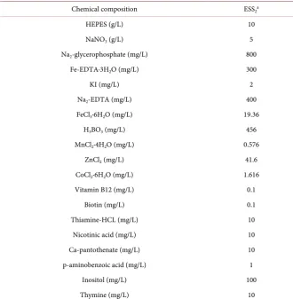
Gametophytes, conchosporangia and sporophytes of Pyropia yezoensis (strain U-51) were maintained in sterilized artificial seawater (SEALIFE; Marinetech, Tokyo, Japan) enriched with ESS2 containing NaNO3 as a nitrogen source,
vita-mins, and trace metal elements (Table S1) [28]. The algae were grown under 60 μmol photons·m−2·s−1 light in a short-day photoperiod (10 h light/14 h dark) at
15˚C with air filtered through a 0.22-μm filter (Whatman; Maidstone, UK). The culture medium was changed weekly. For nitrogen starvation experiments, ga-metophytes, conchosporangia and sporophytes were treated with artificial sea-water without ESS2 (free of a nitrogen source) for a week. Algal materials were
sampled daily after starting the starvation treatment to measure gene expression.
2.2. Quantification of Photosynthetic Pigments
Gametophytes and sporophytes were treated with N-free (ESS2-free) seawater for
3, 5 or 7 days to observe discoloration. For recovery from discoloration, game-tophytes and sporophytes discolored for a week were transferred into seawater supplied with 500 μM of NH4Cl, NaNO3, or urea and then cultured for a further
week. Discolored and recovered samples (0.1 g fresh weight per sample) were used to calculate chlorophyll a (Chl a) contents according to Seely et al. [29] and phycoerythrin (PE) and phycocyanin (PC) contents as described by Beer and Eshel [30].
2.3. Identification and Characterization of AMTs
Unigenes annotated as putative AMTs were selected from our transcriptome analyses of P. yezoensis [31], and their identity was confirmed by comparison of predicted amino acid sequences with those of known AMTs by a BLAST search (https://blast.ncbi.nlm.nih.gov/Blast.cgi) after identification of full-length open reading frames (ORFs) with the ORF finder
(https://www.ncbi.nlm.nih.gov/orffinder/). The ProtParam tool
(https://web.expasy.org/protparam/) was used to predict the molecular weights,
theoretical isoelectric point (pI), and grand average of hydropathicity (GRAVY). The location of the ammonium transporter (AMT) domain was identified with Pfam (http://pfam.xfam.org/search#tabview=tab0), and transmembrane helices in the conserved AMT domain were predicted using a SMART search
DOI: 10.4236/ajps.2019.108096 1328 American Journal of Plant Sciences
2.4. Phylogenetic Analysis
AMTs used for the phylogenetic analysis were obtained from GenBank, genome and EST databases and our unpublished transcriptome analyses are listed in Table S2 with their accession numbers and gene IDs. These included AMTs from Streptophyta (Arabidopsis thaliana, https://www.arabidopsis.org; Physco-mitrella patens, https://genome.jgi.doe.gov/Phypa1_1/Phypa1_1.home.html), Rho-dophyta (Porphyra umbilicalis,
https://phytozome.jgi.doe.gov/pz/portal.html#; Porphyra purpurea,
https://www.ncbi.nlm.nih.gov/sar/SRX100230;Porphyridium purpureum,
http://cyanophora.rutgers.edu/porphyridium/; Cyanidioschyzon merolae,
http://merolae.biol.s.u-tokyo.ac.jp; Galdieria sulphuraria,
http://plants.ensembl.org/Galdieria_sulphuraria/Info/Index), Chlorophyta
(Chlamydomonas reinhardtii, https://genome.jgi.doe.gov/Chlre4/Chlre4.home.html;
Volvox carteri f. nagariensis, https://www.uniprot.org/proteomes/UP000001058), and Heterokontophyta (Phaeodactylum tricomutum,
https://genome.jgi.doe.gov/Phatr2/Phatr2.home.html; Cylindrotheca fusiformis,
https://www.uniprot.org/uniprot/?query=cylindrotheca+fusiformis&sort=score). A neighbor-joining phylogenetic tree was constructed with MEGA 7 software
(https://www.megasoftware.net) using ClustalW to align the AMT amino acid
sequences.
2.5. Total RNA Extraction and cDNA Synthesis
Total RNA was separately extracted from gametophytes, conchosporangia and sporophytes using the RNeasy Plant Mini Kit (Qiagen, Hilden, Germany) and then treated with DNase (TURBO DNA-free TM kit, Invitrogen, Carlsbad, USA) to remove genomic DNA contamination. Then, first-stand complementary DNA (cDNA) was synthesized from 300 ng of total RNA with the PrimeScript 1st strand cDNA Synthesis Kit (TaKaRa Bio, Kusatsu, Japan) according to the man-ufacturer’s instructions. Before being used as a template in quantitative PCR (qPCR) analyses, the quality of the cDNA was evaluated by amplification of the
P. yezoensis 18S rRNA gene with its gene primer set (Table S3) [27] via PCR reactions with Phusion high-fidelity DNA polymerase with GC buffer (Biolabs, Massachusetts, USA) according to the manufacturer’s instructions. The thermal cycling parameters consisted of an initial denaturation step 98˚C for 30 s, and 30 cycles of 98˚C for 10 s, 60˚C for 30 s and 72˚C for 20 s, and a final extension step 72˚C for 5 mins.
2.6. Gene Expression Analysis
Primers for qPCR were designed using Primer Premier 5 software
electropho-C. Z. Li et al.
DOI: 10.4236/ajps.2019.108096 1329 American Journal of Plant Sciences
resis. Primer sets that amplified DNA bands with expected sizes were employed for the qPCR. qPCR was carried out in a total volume of 20 μl containing 10 μl of 2 × SYBR Premix Ex Taq GC, 0.4 μl of ROX Reference Dye, 2 μl of cDNA tem-plate, and 0.4 μl (10 μM) of each primer, using the SYBR Premix Ex Taq GC kit (Takara Bio, Kusatsu, Japan). The thermal cycling parameters consisted of 95˚C for 5 min and 40 cycles of 94˚C for 30 s, 60˚C for 30 s and 72˚C for 20 s. The dissociation curve was generated by heating from 60 to 95˚C to check for specificity of amplification using an Applied Biosystems 7300 real-time PCR system (Life Technologies, Carlsbad, USA). The data were examined with one-way ANOVA, and the level of significance was defined at P < 0.05.
3. Results
3.1. A multiplicity of
P
.
yezoensis
AMT Genes
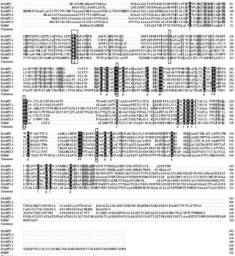
Based on the functional annotation in our P. yezoensis transcriptome analysis [31], we identified six unigenes (CL1839, CL3739, Unigene15210, CL1882. Con-tig5, CL1882. Contig6 and Unigene24155) as candidate P. yezoensis AMT (PyAMT) genes, in addition to the known PyAMT1 [15]. These six additional unigenes contained predicted open reading frames (ORFs) encoding 484, 654, 589, 616, 690, and 522 amino acid products (Figure 1) with molecular masses of 51.2, 67.8, 58.67, 63.68, 72.06, and 53.78 kDa, respectively, and all contained the conserved AMT domain (Table S4). In addition, CL1839, CL3739, Unigene15210, CL1882. Contig5, CL1882. Contig6 and Unigene24155showed 55.04%, 32.42%, 38.07%, 27.77%, 25.36%, and 16.88% identity to PyAMT1, respectively. Based on these findings, we concluded that all of the candidates are likely to be AMTs. However, the product of Unigene 24155 had 12 predicted transmembrane (TM) helixes in contrast to the products of the other candidate genes, which had 11 TM helixes (Table S5), suggesting a structural difference in Unigene24155 from the other genes. Other structural characteristics of these gene products, includ-ing a total number of atoms and theoretical pI, are listed in Table S4.
The crystal structure of the ammonium transporter protein in Escherichia coli
(AmtB) revealed that two phenylalanine residues, Phe107 and Phe215, block the hydrophobic NH4+ conduction pore, and two highly conserved histidine
resi-dues, His168 and His318, maintain the shape of the central pore involved in
4
NH+ transport [32] [33]. As shown in Figure 1, these four sites were highly
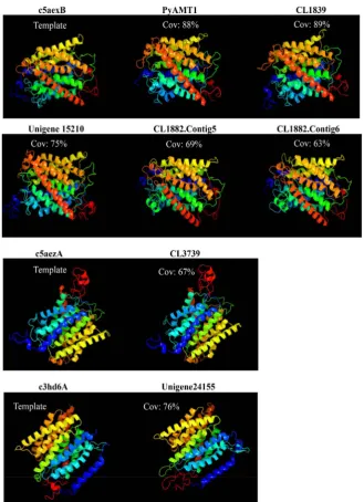
conserved in P. yezoensis AMTs. In addition, in consistent with Kakinuma et al. [15], a tripeptide sequence, Phe-Gly-Phe (Tyr/Asn), indicating AMT identity, was found in all of the PyAMTs (Figure 1). Moreover, three-dimensional struc-tures predicted in silico for all of P. yezoensis AMT domain-containing proteins were similar to those of known AMTs. Indeed, crystal structures of 5 PyAMTs (PyAMT1, CL1839, Unigene 15210, CL1882. Contig5, CL1882. Contig6) struc-turally resembled that of the AMT template c5aexB (Saccharomyces cerevisiae
DOI: 10.4236/ajps.2019.108096 1330 American Journal of Plant Sciences
Figure 1. Conservation of AMT identity among six PyAMT1s and PyRh. Amino acid residues conserved in all seven proteins and in six proteins are highlighted by black (with white characters) and gray backgrounds, respectively. The boxes indicate the Phe-Gly-Phe (Tyr/Asn) triplet conserved in AMTs, as well as phenylalanine and histidine residues corresponding to residues in-volved in NH4+ binding and transport in E. coli EcAmtB. The amino acid numbers are indicated on the right.
protein, Rh, structurally related to AMT) (Figure 2). Taken together with cha-racteristics in primary sequences (Figure 1), these findings highly suggested functional NH4+-transport and Rh activities of AMT domain-containing
C. Z. Li et al.
[image:7.595.208.538.71.526.2]DOI: 10.4236/ajps.2019.108096 1331 American Journal of Plant Sciences
Figure 2. High degree of similarity in three-dimensional structures between AMT do-main-containing proteins from P. yezoensis and known AMTs. Templates c5aexB and c5aezA indicate three-dimensional structures of AMTs (MEP2s) from Saccharomyces ce-revisiae and Candida albicans, respectively, while c3hd6A represents a three-dimensional structure of the human rhesus protein (Rh). Cov means the coverage percent of AMT domain-containing proteins to the corresponding templates.
3.2. Phylogenetic Classification of
P
.
yezoensis
AMTs into AMT1
and Rhesus Protein Subfamilies
DOI: 10.4236/ajps.2019.108096 1332 American Journal of Plant Sciences
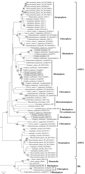
we considered these unigenes to encode AMT1s and designated them as PyAMT1.2 (CL1839), PyAMT1.3 (CL3739), PyAMT1.4 (Unigene15210), PyAMT1.5 (CL1882. Contig5), and PyAMT1.6 (CL1882. Contig6). Unigene24155 shared 27.56% and 24.70% identity with CrRh1 and CrRh2 from the green alga Chlamydomonas reinhardtii, respectively, but only 16.88% with PyAMT1. Our analysis placed this protein in the Rh clade, which is phylogenetically divergent from both AMT1 and AMT2 clades (Figure 3). Rh proteins are homologues of AMT proteins that were first identified in human erythroid cells [34][35]. The predicted 12 trans-membrane helixes of the Unigene24155 protein is in accordance with Rh pro-teins of other organisms [36][37]. Thus, we designated Unigene24155 as PyRh.
Our phylogenetic analysis also indicated that algal AMTs were mostly classi-fied into the AMT1 subfamily, which is distantly related to the AMT2 subfamily clade. In addition, AMT1s from red algae, green algae, diatoms, and land plants formed independent clades. Thus, an ancient algal AMT1 may have existed prior to the divergence of red and green algae, although the origin of AMT2s is un-clear.
The six PyAMT1s were subdivided into three different Rhodophyta clades, each of which contained pairs of PyAMT1s: PyAMT1 and PyAMT1.2, PyAMT1.3 and PyAMT1.4, and PyAMT1.5 and PyAMT1.6 (Figure 3). In addition, the three Rhodophyta clades also contained pairs of AMT1s from the red seaweed
Porphyra umbilicalis: The PyAMT1/1.2 clade with Pum0126s0003.1 (OSX77964.1), Pum1775s0001.1 (OSX69172.1), Pum0463s0020.1 (OSX72158.1), and Pum016- 5s0019.1 (OSX77025.2); the PyAMT1.3/1.4 clade with Pum0027s0002.1 (OSX80- 976.1) and Pum1656s0001.1 (OSX69292.1); and the PyAMT1.5/1.6 clade with Pum0022s0083.1 (OSX81363.1) (Figure 3). Moreover, AMT1s from Chlorophy-ta were separated into three different clades, independent from each other and from the three clades of Rhodophyta. Thus, expansion and divergence of the an-cient algal AMT1 gene into three groups occurred independently in red and green algae after their establishment.
3.3. Differences in Temporal Expression Patterns of
PyAMT
1 and
PyRh
Genes during the Life Cycle
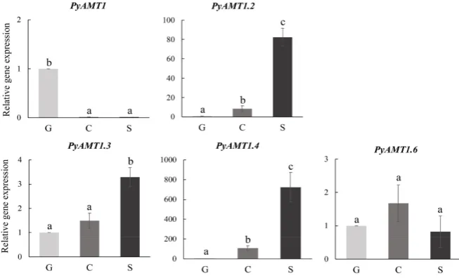
Figure 4 shows the relative transcript abundance of PyAMT1 and PyRh genes in the gametophyte, conchosporangium and sporophyte tissues under normal growth conditions. When life cycle specificity of gene expression was compared between the gene pairs found in the three phylogenetic clades, PyAMT1 was specifically expressed in the gametophyte, while the expression of PyAMT1.2 was found in both the sporophyte and conchosporangium. By contrast, PyAMT1.3 and
PyAMT1.4 exhibited the same sporophyte-dominant expression pattern. PyAMT1.6 was expressed constitutively, whereas transcripts of PyAMT1.5 were not detect-able at any stage (data not shown). Thus, two of the gene pairs in the same clade did not share the same expression pattern. We did not find evidence of PyRh
C. Z. Li et al.
[image:9.595.233.522.69.655.2]DOI: 10.4236/ajps.2019.108096 1333 American Journal of Plant Sciences
DOI: 10.4236/ajps.2019.108096 1334 American Journal of Plant Sciences
Figure 4. Differences in temporal expression patterns of PyAMTs in the life cycle of P.
yezoensis. The relative mRNA levels of PyAMT1s were normalized with the reference gene 18S rRNA. Error bars indicate the standard deviation of triplicate experiments (n = 3), and different letters on bars indicate significant differences at P < 0.05 tested with ANOVA. G, gametophyte; C, conchosporangium; S, sporophyte.
3.4. Induction of and Recovery from Discoloration
Gametophyte and sporophyte tissues maintained in the ESS2-containing
seawa-ter were transferred to seawaseawa-ter without ESS2 and cultivated for an additional 3,
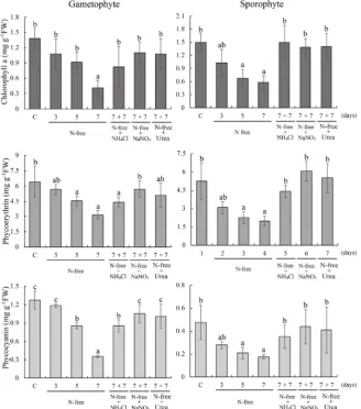
5 or 7 days. As results, discoloration was initially observed after 3 days and gradually strengthened until 7 days in both gametophytes and sporophytes (Figure 5). Correspondingly, the contents of photosynthetic pigments Chl a, PE and PC were decreased respectively in gametophytes from 1.37 to 0.40, 6.42 to 3.18, and 1.27 to 0.35 mg∙g−1 FW and in sporophyte from 1.49 to 0.58, 5.24 to
1.99, and 0.48 to 0.18 mg∙g−1 FW (Figure 6). To examine recovery from
discolo-ration, 7-day-discolored gametophytes and sporophytes were treated with nutri-tion-deficient medium containing 500 μM NH4Cl, NaNO3, or urea for a week.
As shown in Figure 5, discoloration was recovered visibly, which was supported by the increase in the contents of Chl a, PE and PC to the levels corresponding to those in non-discolored gametophytes and sporophytes (Figure 6). These find-ings suggested essential roles of the AMT activity for recovery from discolora-tion in P. yezoensis.
3.5. Diversity in the Nutrition Starvation-Inducible Pattern of
PyAMT
1 Subfamily Genes during the Life Cycle
C. Z. Li et al.
[image:11.595.224.527.74.589.2]DOI: 10.4236/ajps.2019.108096 1335 American Journal of Plant Sciences
DOI: 10.4236/ajps.2019.108096 1336 American Journal of Plant Sciences
Figure 6. Changes in the contents of chlorophyll a, phycoerythrin and phycocyanin in discolored and recovered P. yezoensis. Culture conditions were identical to those in Fig-ure 5. C means control samples. Error bars indicate the standard deviation of triplicate experiments (n = 3), and different letters on bars indicate significant differences at P < 0.05 tested with ANOVA.
conchosporangia and sporophytes. Moreover, the PyAMT6 expression gradually increased in gametophyte tissue, and transient expression of PyAMT6 was ob-served in conchosporangia and sporophytes. Transcripts of PyAMT1.5 and PyRh
remained undetectable in all tissues evaluated, even under nitrogen-deficient con-ditions (data not shown).
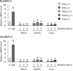
We further examined the expression of the PyAMT1genes to determine the effects of nitrogen recovery on their expression. For these experiments, we se-lected PyAMT1.2 and PyAMT1.4, whose expression continually increased in sporophytes under nitrogen deficiency. When discolored sporophytes produced by 7-day culture in the ESS2-less medium were transferred to ESS2-less medium
containing 500 μM NH4Cl and further cultured for 3 days, the expression of the
C. Z. Li et al.
[image:13.595.212.541.67.501.2]DOI: 10.4236/ajps.2019.108096 1337 American Journal of Plant Sciences
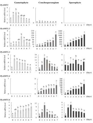
Figure 7. Differences in nitrogen starvation-induced expression patterns among PyAMT1
genes. Samples of gametophytes, conchosporangia, and sporophytes treated with ESS2-free seawater were collected every day for 7 days to examine the expression of the
PyAMT1 genes by qRT-PCR. The relative mRNA levels were normalized with the ref-erence gene 18S rRNA. Error bars indicate the standard deviation of triplicate experi-ments (n = 3), and different letters on bars indicate significant differences at P < 0.05 tested with ANOVA. G, gametophyte; C, conchosporangium; S, sporophyte. The num-bers on the X-axis indicate the duration of culture under nitrogen-deficient conditions (days).
NaNO3or 500 μM urea (Figure 8). These results were consistent with the
pre-vious observation [15] that PyAMT1 gene expression is down-regulated by the addition of inorganic and organic nitrogen sources.
4. Discussion
A NH4+ is an important nitrogen source that is transported via AMTs, which
DOI: 10.4236/ajps.2019.108096 1338 American Journal of Plant Sciences
Figure 8. Suppression of the nitrogen starvation-induced expression of PyAMT1 genes via recovery with different nitrogen sources. Sporophyte samples grown in ESS2-less seawater for 7 days (∆N) were treated with seawater containing 500 μM of NH4Cl, NaNO3, or urea for 3 days. Sporophytes cultured in nitro-gen-supplied seawater were collected every day for 3 days, and the expression of
PyAMT1.2 and PyAMT1.4 was examined by qRT-PCR (upper and lower, re-spectively). The relative mRNA levels were normalized with the reference gene
18S rRNA. Error bars indicate the standard deviation of triplicate experiments (n = 3), and different letters on bars indicate significant differences at P < 0.05 tested with ANOVA. S, sporophytes cultured in ESS2-containing seawater. The numbers on the X-axis indicate the duration of culture under nitrogen-deficient conditions (days).
AMT1, AMT2 and Rh [38]. Although the structure, expression patterns and physiological roles of AMT genes have been well studied in animals and land plants [2][13][34][39], algal AMT genes remain poorly understood. Here, we report the presence of the AMT1 andRh gene subfamilies in P. yezoensis and the diversity in their expression patterns.
Our phylogenetic analysis demonstrated the diversity of the AMT1 subfamily, consisting of independent phylum-specific clades (Figure 3). Land plants have their own AMT1 subfamily, with five genes in Arabidopsis[4][11][12][13], at least 10 genes in rice [9], and 23 genes in wheat [8]. Although AMT1s from land plants formed a single clade, it was separated from the algal AMT1 subfamily, in which the unicellular green algae C. reinhardtii and the red seaweed Po.
C. Z. Li et al.
DOI: 10.4236/ajps.2019.108096 1339 American Journal of Plant Sciences
analysis indicated that all of the AMTs in Po. umbilicalis are AMT1 subfamily members (PuAMT1s) as are other algal genes (Figure 3). Moreover, the six
PyAMT1s and seven PuAMT1s were subdivided into three groups (Figure 3). The existence of multiple independent AMT1 clades in P. yezoensis and Po.
umbilicalis is the distinguishing characteristic of AMT1s from Bangiales, since AMT1s from land plants formed a single clade (Figure 3). These findings imply that an ancient red algal AMT gene may have diversified into three genes prior to the separation of Pyropia and Porphyra, and then further diversification oc-curred independently for each of the three genes in these species.
Similar to Rhodophyta, AMT1s of Chlorophyta were divided into three groups, although their phylogenetic positions were different from the Rhodophyta clades (Figure 3). This result is in agreement with the report that three subfamilies of CrAMT1s have been established in Chlamydomonas[3]. Therefore, a two-step diversification of algal AMT1s has been proposed: An early expansion of the an-cient algal gene into three variants prior to the divergence of Chlorophyta and Rhodophyta and a late duplication of each of the three variants after the diver-gence of the green and red lineages.
The multiplicity of AMT1 genes in Chlorophyta and Rhodophyta points to functional divergence of these genes in seaweeds. This hypothesis is supported by our gene expression analyses indicating differences in temporal and nitrogen stress-inducible expression patterns for the PyAMT1 genes (Figure 7 and Figure 8). PyAMT1, PyAMT1.3, and PyAMT1.4 commonly exhibited a transient in-crease in their expression under nitrogen-deficient conditions, although differ-ences were observed in their life cycle stage-specific expression. Thus, it seems that functional divergence in PyAMT1s might allow for functional specialization over a range of NH4+ concentrations, which would enable P. yezoensis to react
appropriately to a wide range of NH4+ concentrations in the environment. In
the future, functional analysis of each PyAMT1 focused on NH4+ uptake under
nitrogen deficiency conditions should help us to understand their transport ca-pacity and how P. yezoensis responds and adapts to nitrogen deficiency stress during its life cycle.
DOI: 10.4236/ajps.2019.108096 1340 American Journal of Plant Sciences
different from that in land plants, although loss of photosynthetic pigments in discoloration is common.
The discoloration in P. yezoensis thalli was rescued and the mRNA level of
PyAMT1 was down-regulated by an increase in the concentrations of not only inorganic but also organic nitrogen sources [15]. Similarly, nitrogen deficien-cy-inducible discoloration and the expression of PyAMT1.2 and PyAMT1.4 in the sporophyte were also strongly repressed after addition of NH4Cl, NaNO3,
and urea in the culture medium (Figure 8). Since inorganic and organic nitro-gen sources are metabolized into NH4+ to assimilate the nitrogen into cellular
components [46] [47], NaNO3and urea might increase the intracellular NH4+
contents and similarly affect the expression of PyAMT1 genes. These findings indicated that discoloration in P. yezoensis during nitrogen starvation is induced by the decreased extracellular nitrogen content, although leaf senescence in land plants occurs as a result of nitrogen reallocation between different tissues [48] [49].
Another distinguishing characteristic of algal NH4+ transporters is the
pres-ence of Rh proteins, such as PyRh in P. yezoensis and contig2015.11 in Porphy-ridium purpureum (Figure 3), which contain the conserved AMT domain but are distantly related to the AMT1 and AMT2 subfamilies [36][50]. Rh split from
AMT in archaeal species and coexists in microbes and invertebrates, but not in fungi, vascular plants, and vertebrates [50]. To date, Rh has not been reported in algae except for CrRh1 and CrRh2 from the green alga C. reinhardtii[37][51]; our findings reveal the presence of Rh in red algae (Figure 3). Although the ex-pression of CrRh1 and CrRh2 is regulated by CO2 [37] [51], the expression of
PyRh was not detected during the life cycle in P. yezoensis nor under the nitro-gen-deficient conditions (data not shown). Thus, little is known about the phy-siological functions of Rh proteins in algae.
In conclusion, algal NH4+ transporters are divided into the AMT1 and Rh
subfamilies. The AMT1 subfamily of P. yezoensis consists of three groups con-taining genes whose expression patterns differ temporally and are nitrogen defi-ciency-dependent during the life cycle. These findings are novel for algal NH4+
transporters, and future work should elucidate the functions of each member of the AMT1 and Rh subfamilies, which could help clarify the unique algal strate-gies of response and acclimation to nitrogen deficiency stress by phylogenetically independent and diverse AMT1s and Rhs during the life cycle.
Acknowledgements
We are grateful to Mr. Masahiro Suda and Mr. Ryunosuke Irie for their sup-porting for laboratory culture of P. yezoensis gametophytes and sporophytes. Chengze Li was supported by the Ministry of Education, Culture, Sports, Science and Technology of Japan and by the China Scholarship Council.
Conflicts of Interest
C. Z. Li et al.
DOI: 10.4236/ajps.2019.108096 1341 American Journal of Plant Sciences
References
[1] Nicolaus, W., Sonia, G., Alain, G. and Frommer, W.B. (2000) The Molecular Physi-ology of Ammonium Uptake and Retrieval. Current Opinion in Plant Biology, 3, 254-261.
[2] Couturier, J., Montanini, B., Martin, F., Brun, A., Blaudez, D. and Chalot, M. (2007) The Expanded Family of Ammonium Transporters in the Perennial Poplar Plant.
New Phytologist, 174, 137-150. https://doi.org/10.1111/j.1469-8137.2007.01992.x [3] González-Ballester, D., Camargo, A. and Fernández, E. (2005) Ammonium
Trans-porter Genes in Chlamydomonas: The Nitrate-Specific Regulatory Gene Nit2 Is In-volved in Amt1; 1 Expression. Plant Molecular Biology, 56, 863-878.
https://doi.org/10.1007/s11103-004-5292-7
[4] Gazzarrini, S., Lejay, L., Gojon, A., Ninnemann, O., Frommer, W.B. and Wiréna, N. (1999) Three Functional Transporters for Constitutive, Diurnally Regulated, and Starvation-Induced Uptake of Ammonium into Arabidopsis Roots. The Plant Cell, 11, 937-947. https://doi.org/10.1105/tpc.11.5.937
[5] Ludewig, U., Neuhauser, B. and Dynowski, M. (2007) Molecular Mechanisms of Ammonium Transport and Accumulation in Plants. FEBS Letters, 581, 2301-2308. https://doi.org/10.1016/j.febslet.2007.03.034
[6] D’Apuzzo, E., Rogato, A., Simon-Rosin, U., Alaoui, H.E., Barbulova, A., Betti, M., et al. (2004) Characterization of Three Functional High-Affinity Ammonium Trans-porters in Lotus Japonicus with Differential Transcriptional Regulation and Spatial Expression. Plant Physiology, 134, 1763-1774.
https://doi.org/10.1104/pp.103.034322
[7] Wittgenstein, N., Le, C.H., Hawkins, B.J. and Ehlting, J. (2014) Evolutionary Classi-fication of Ammonium, Nitrate, and Peptide Transporters in Land Plants. BMC Evolutionary Biology, 14, 1-17. https://doi.org/10.1186/1471-2148-14-11
[8] Li, T., Liao, K., Xu, X., Gao, Y., Wang, Z., Zhu, X., et al. (2017) Wheat Ammonium Transporter (AMT) Gene Family: Diversity and Possible Role in Host-Pathogen In-teraction with Stem Rust. Frontiers in Plant Science, 8, 1637.
https://doi.org/10.3389/fpls.2017.01637
[9] Li, C., Tang, Z., Wei, J., Qu, H., Xie, Y. and Xu, G. (2016) The OsAMT1.1 Gene Functions in Ammonium Uptake and Ammonium-Potassium Homeostasis over Low and High Ammonium Concentration Ranges. Journal of Genetics and Ge-nomics, 43, 639-649. https://doi.org/10.1016/j.jgg.2016.11.001
[10] Zhang, F., Liu, Y., Wang, L., Bai, P., Ruan, L., Zhang, C., et al. (2018) Molecular Cloning and Expression Analysis of Ammonium Transporters in Tea Plants ( Ca-mellia sinensis (L.) O. Kuntze) under Different Nitrogen Treatments. Gene, 658, 136-145. https://doi.org/10.1016/j.gene.2018.03.024
[11] Yuan, L., Loque, D., Kojima, S., Rauch, S., Ishiyama, K., Inoue, E., et al. (2007) The Organization of High-Affinity Ammonium Uptake in Arabidopsis Roots Depends on the Spatial Arrangement and Biochemical Properties of AMT1-Type Transpor-ters. The Plant Cell Online, 19, 2636-2652. https://doi.org/10.1105/tpc.107.052134 [12] Yuan, L., Graff, L., Loqué, D., Kojima, S., Tsuchiya, Y.N., Takahashi, H., et al.
(2009) AtAMT1; 4, a Pollen-Specific High-Affinity Ammonium Transporter of the Plasma Membrane in Arabidopsis. Plant and Cell Physiology, 50, 13-25.
https://doi.org/10.1093/pcp/pcn186
DOI: 10.4236/ajps.2019.108096 1342 American Journal of Plant Sciences
Journal, 48, 522-534. https://doi.org/10.1111/j.1365-313X.2006.02887.x
[14] Giehl, R.F.H., Laginha, A.M., Duan, F., Rentsch, D., Yuan, L. and Wiren, N. (2017) A Critical Role of AMT2; 1 in Root-to-Shoot Translocation of Ammonium in Ara-bidopsis. Molecular Plant, 10, 1449-1460.
https://doi.org/10.1016/j.molp.2017.10.001
[15] Kakinuma, M., Nakamoto, C., Kishi, K., Coury, D.A., Amano, H. (2017) Isolation and Functional Characterization of an Ammonium Transporter Gene, PyAMT1, Related to Nitrogen Assimilation in the Marine Macroalga Pyropia yezoensis
(Rhodophyta). Marine Environmental Research, 128, 76-87. https://doi.org/10.1016/j.marenvres.2016.08.007
[16] Amano, H., Noda, H. (1987) Effect of Nitrogenous Fertilizers on the Recovery of Discoloured Fronds of Porphyra yezoensis. Botanica Marina, 30, 467-473.
https://doi.org/10.1515/botm.1987.30.6.467
[17] Sakaguchi, K., Ochiai, N., Park, C.S., Kakinuma, M. and Amano, H. (2002) Evalua-tion of DiscoloraEvalua-tion in Harvested Laver Porphyra yezoensis and Recovery after Treatment with Ammonium Sulfate Enriched Seawater. Nippon Suisan Gakkaishi, 69, 399-404. https://doi.org/10.2331/suisan.69.399
[18] Merchant, S.S., Prochnik, S.E., Vallon, O., Harris, E.H., Karpowicz, S.J., Witman, G.B., et al. (2007) The Chlamydomonas Genome Reveals the Evolution of Key An-imal and Plant Functions. Science, 318, 245-251.
https://doi.org/10.1126/science.1143609
[19] Prochnik, S.E., Umen, J., Nedelcu, A.M., Hallmann, A., Miller, S.M., Nishii, I., et al. (2010) Genomic Analysis of Organismal Complexity in the Multicellular Green Al-ga Volvox carteri. Science, 329, 223-226. https://doi.org/10.1126/science.1188800 [20] Schonknecht, G., Chen, W.H., Ternes, C.M., Barbier, G.G., Shrestha, R.P., Stanke,
M., et al. (2013) Gene Transfer from Bacteria and Archaea Facilitated Evolution of an Extremophilic Eukaryote. Science, 339, 1207-1210.
https://doi.org/10.1126/science.1231707
[21] Brawley, S.H, Blouin, N.A., Ficko-Blean, E., Wheeler, G.L., Lohr, M., Goodson, H.V., et al. (2017) Insights into the Red Algae and Eukaryotic Evolution from the Genome of Porphyra umbilicalis (Bangiophyceae, Rhodophyta). Proceedings of the National Academy of Sciences, 114, 6361-6370.
https://doi.org/10.1073/pnas.1703088114
[22] Hildebrand, M. (2005) Cloning and Functional Characterization of Ammonium Transporters from the Marine Diatom Cylindrotheca fusiformis (Bacillariophy-ceae). Journal of Phycology, 41, 105-113.
https://doi.org/10.1111/j.1529-8817.2005.04108.x
[23] Eriksen, R.L. and Klein, A.S. (2018) Organism-Environment Interactions and Dif-ferential Gene Expression Patterns among Open-Coastal and Estuarine Populations of Porphyra umbilicalis Kützing (Rhodophyta) in the Northwest Atlantic. Fisheries and Aquatic Sciences, 21, 28. https://doi.org/10.1186/s41240-018-0103-2
[24] Takahashi, M. and Mikami, K. (2017) Oxidative Stress Promotes Asexual Repro-duction and Apogamy in the Red Seaweed Pyropia yezoensis. Frontiers in Plant Science, 8, 62. https://doi.org/10.3389/fpls.2017.00062
[25] Adams, E., Mikami, K. and Shin, R. (2017) Selection and Functional Analysis of a
Pyropia yezoensis Ammonium Transporter PyAMT1 in Potassium Deficiency.
Journal of Applied Phycology, 29, 2617-2626. https://doi.org/10.1007/s10811-017-1196-1
C. Z. Li et al.
DOI: 10.4236/ajps.2019.108096 1343 American Journal of Plant Sciences
Molecular Analysis of Physiological Responses to Changes in Nitrogen in a Marine Macroalga, Porphyra yezoensis (Rhodophyta). Cell Biology and Toxicology, 24, 629-639. https://doi.org/10.1007/s10565-007-9053-7
[27] Kakinuma, M., Suzuki, K., Iwata, S., Coury, D.A., Iwade, S. and Mikami, K. (2015) Isolation and Characterization of a New DUR3-Like Gene, PyDUR3.3, from the Marine Macroalga Pyropia yezoensis (Rhodophyta). Fisheries Science, 82, 171-184. https://doi.org/10.1007/s12562-015-0947-7
[28] Takahashi, M., Saga, N. and Mikami, K. (2010) Photosynthesis-Dependent Extra-cellular Ca2+ Influx Triggers an Asexual Reproductive Cycle in the Marine Red Ma-croalga Porphyra yezoensis. American Journal of Plant Science, 1, 1-11.
https://doi.org/10.4236/ajps.2010.11001
[29] Seely, G.R., Duncan, M.J. and Vidaver, W.E. (1972) Preparative and Analytical Ex-traction of Pigments from Brown Algae with Dimethyl Sulfoxide. Marine Biology, 12, 184-188. https://doi.org/10.1007/BF00350754
[30] Beer, S. and Eshel, A. (1985) Determining Phycoerythrin and Phycocyanin Concen-trations in Aqueous Crude Extracts on Red Algae. Marine and Freshwater Research, 36, 785-792. https://doi.org/10.1071/MF9850785
[31] Mikami, K., Li, C., Irie, R. and Hama, Y. (2019) A Unique Life Cycle Transition in the Red Seaweed Pyropia yezoensis Depends on Apospory. Communication Biolo-gy, 2, 299. https://doi.org/10.1038/s42003-019-0549-5
[32] Soupene, E., Chu, T., Corbin, R.W., Hunt, D.F. and Kustu, S. (2002) Gas Channels for NH3: Proteins from Hyperthermophiles Complement an Escherichia coli Mu-tant. Journal of Bacteriology, 184, 3396-3400.
https://doi.org/10.1128/JB.184.12.3396-3400.2002
[33] Zheng, L., Kostrewa, D., Berneche, S., Winkler, F.K. and Li, X.D. (2004) The Me-chanism of Ammonia Transport Based on the Crystal Structure of AmtB of Esche-richia coli. Proceedings of the National Academy of Sciences, 101, 17090-17095. https://doi.org/10.1073/pnas.0406475101
[34] Marini, A.M., Urrestarazu, A., Beauwens, R. and André, B. (1997) The Rh (Rhesus) Blood Group Polypeptides Are Related to NH4+ Transporters. Trends in
Biochemi-cal Sciences, 22, 460-461. https://doi.org/10.1016/S0968-0004(97)01132-8
[35] Huang, C.H. and Peng, J. (2005) Evolutionary Conservation and Diversification of Rh Family Genes and Proteins. Proceedings of the National Academy of Sciences, 102, 15512-15517. https://doi.org/10.1073/pnas.0507886102
[36] Nakhoul, N.L. and Hamm, L.L. (2004) Non-Erythroid Rh Glycoproteins: A Putative New Family of Mammalian Ammonium Transporters. Pflugers Archiv—European Journal of Physiology, 447, 807-812. https://doi.org/10.1007/s00424-003-1142-8 [37] Soupene, E., King, N., Field, E., Liu, P., Niyogi, K.K., Huang, C.H., et al. (2002)
Rhesus Expression in a Green Alga Is Regulated by CO2. Proceedings of the
Nation-al Academy of Sciences, 99, 7769-7773. https://doi.org/10.1073/pnas.112225599 [38] Michele, R.D., Loque, D., Lalonde, S. and Frommer, W.B. (2012) Ammonium and
Urea Transporter Inventory of the Selaginella and Physcomitrella Genomes. Front Plant Science, 3, 62. https://doi.org/10.3389/fpls.2012.00062
[39] Suzuki, A., Komata, H., Iwashita, S., Seto, S., Ikeya, H., Tabata, M., et al. (2017) Evolution of the RH Gene Family in Vertebrates Revealed by Brown Hagfish ( Epta-tretus atami) Genome Sequences. Molecular Phylogenetics Evolution, 107, 1-9. https://doi.org/10.1016/j.ympev.2016.10.004
Planta-DOI: 10.4236/ajps.2019.108096 1344 American Journal of Plant Sciences
rum, 138, 256-267. https://doi.org/10.1111/j.1399-3054.2009.01336.x
[41] Meng, S., Peng, J.S., He, Y.N., Zhang, G.B., Yi, H.Y., Fu, Y.L., et al. (2016) Arabi-dopsis NRT1.5 Mediates the Suppression of Nitrate Starvation-Induced Leaf Senes-cence by Modulating Foliar Potassium Level. Molecular Plant, 9, 461-470.
https://doi.org/10.1016/j.molp.2015.12.015
[42] Till, I., Anna, M.Z., Marion, K. and Peter, D. (2006) A Salvage Pathway for Phytol Metabolism in Arabidopsis. The Journal of Biological Chemistry, 281, 2470-2477 https://doi.org/10.1074/jbc.M509222200
[43] Gomez, F., Carrión, C., Costa, M., Desel, C., Kieselbach, T., Funk, C., et al. (2019) Extra-Plastidial Degradation of Chlorophyll and Photosystem I in Tobacco Leaves Involving “Senescence-Associated Vacuoles”. ThePlant Journal, 99, 465-477. https://doi.org/10.1111/tpj.14337
[44] Edward, H. and Amasino, R.M. (2001) Nutrients Mobilized from Leaves of Arabi-dopsis thaliana during Leaf Senescence. Journal of Plant Physiology, 158, 1317-1323. https://doi.org/10.1078/0176-1617-00608
[45] Reed, R. (1990) Solute Accumulation and Osmotic Adjustment. Cambridge Univer-sity, New York.
[46] Xu, G., Fan, X. and Miller, A.J. (2012) Plant Nitrogen Assimilation and Use Effi-ciency. Annual Review of Plant Biology, 63, 153-182.
https://doi.org/10.1146/annurev-arplant-042811-105532
[47] Imamura, S., Terashita, M., Ohnuma, M., Maruyama, S., Minoda, A., Weber, A.P.M., et al. (2010) Nitrate Assimilatory Genes and Their Transcriptional Regula-tion in a Unicellular Red Alga Cyanidioschyzon merolae: Genetic Evidence for Ni-trite Reduction by a Sulfite Reductase-Like Enzyme. Plant and Cell Physiology, 51, 707-717. https://doi.org/10.1093/pcp/pcq043
[48] Gregersen, P.L., Holm, P.B. and Krupinska, K. (2008) Leaf Senescence and Nutrient Remobilisation in Barley and Wheat. Plant Biology, 10, 37-49.
https://doi.org/10.1111/j.1438-8677.2008.00114.x
[49] Diaz, C., Lemaitre, T., Christ, A., Azzopardi, M., Kato, Y., Sato, F., et al. (2008) Ni-trogen Recycling and Remobilization Are Differentially Controlled by Leaf Senes-cence and Development Stage in Arabidopsis under Low Nitrogen Nutrition. Plant Physiology, 147, 1437-1449. https://doi.org/10.1104/pp.108.119040
[50] Peng, J. and Huang, C.H. (2006) Rh Proteins vs Amt Proteins: An Organismal and Phylogenetic Perspective on CO2 and NH3 Gas Channels. Transfusion Clinique et
Biologique, 13, 85-94. https://doi.org/10.1016/j.tracli.2006.02.006
[51] Soupene, E., Inwood, W. and Kustu, S. (2004) Lack of the Rhesus Protein Rh1 Im-pairs Growth of the Green Alga Chlamydomonas reinhardtii at High CO2.
C. Z. Li et al.
[image:21.595.207.538.88.426.2]DOI: 10.4236/ajps.2019.108096 1345 American Journal of Plant Sciences
Table S1. Chemical compositions of ESS2.
Chemical composition ESS2a
HEPES (g/L) 10
NaNO3 (g/L) 5
Na2-glycerophosphate (mg/L) 800
Fe-EDTA∙3H2O (mg/L) 300
KI (mg/L) 2
Na2-EDTA (mg/L) 400
FeCl3∙6H2O (mg/L) 19.36
H3BO3 (mg/L) 456
MnCl2∙4H2O (mg/L) 0.576
ZnCl2 (mg/L) 41.6
CoCl2∙6H2O (mg/L) 1.616
Vitamin B12 (mg/L) 0.1
Biotin (mg/L) 0.1
Thiamine-HCL (mg/L) 10
Nicotinic acid (mg/L) 10
Ca-pantothenate (mg/L) 10
p-aminobenzoic acid (mg/L) 1
Inositol (mg/L) 100
Thymine (mg/L) 10
aFinal concentration of ESS
DOI: 10.4236/ajps.2019.108096 1346 American Journal of Plant Sciences
Table S2. Land plant and algal AMTs and Rhs used for the phylogenetic analysis.
Species Gene symbol/Annotation Accession No. Division AAs
Aquilegia coerulea PIA32185.1 Streptophyta 468
Aquilegia coerulea PIA65513.1 Streptophyta 475
Aquilegia coerulea PIA59506.1 Streptophyta 489
Aquilegia coerulea PIA35542.1 Streptophyta 479
Aquilegia coerulea PIA31609.1 Streptophyta 477
Aquilegia coerulea PIA31140.1 Streptophyta 485
Aquilegia coerulea PIA45105.1 Streptophyta 481
Arabidopsis thaliana AMT1.1 AEE83287.1 Streptophyta 501 Arabidopsis thaliana AMT1.2 AAD38253.1 Streptophyta 514 Arabidopsis thaliana AMT1.3 AEE76886.1 Streptophyta 498 Arabidopsis thaliana AMT1.4 AEE85527.1 Streptophyta 504 Arabidopsis thaliana AMT1.5 AEE76885.1 Streptophyta 496 Arabidopsis thaliana AMT2 AEC09519.1 Streptophyta 475 Chlamydomonas reinhardtii ammonium transporter XP_001702082.1 Chlorophyta 432 Chlamydomonas reinhardtii ammonium transporter XP_001692671.1 Chlorophyta 542 Chlamydomonas reinhardtii ammonium transporter XP_001693464.1 Chlorophyta 499 Chlamydomonas reinhardtii ammonium transporter XP_001697504.1 Chlorophyta 539 Chlamydomonas reinhardtii ammonium transporter AAS90602.1 Chlorophyta 579 Chlamydomonas reinhardtii ammonium transporter XP_001695208.1 Chlorophyta 610 Chlamydomonas reinhardtii ammonium transporter XP_001692923.1 Chlorophyta 481 Chlamydomonas reinhardtii AMT1.2 AAM94623.2 Chlorophyta 542 Chlamydomonas reinhardtii Rh1 XP_001695464.1 Chlorophyta 574 Chlamydomonas reinhardtii Rh2 AAM19664.1 Chlorophyta 638 Chondrus crispus unnamed protein product XP_005710198.1 Rhodophyta 458 Cyanidioschyzon merolae ammonium transporter XP_005536541.1 Rhodophyta 373 Cylindrotheca fusiformis AMT1 AAV70489.1 Heterokontophyta 511 Cylindrotheca fusiformis AMT-like AAV70490.1 Heterokontophyta 511 Ectocarpus siliculosus ammonium transporter CBN77717.1 Heterokontophyta 494 Emiliania huxleyi ammonium transporter XP_005785348.1 Heterokontophyta 462 Galdieria sulphuraria ammonium transporter EME32138.1 Rhodophyta 502 Galdieria sulphuraria ammonium transporter EME30100.1 Rhodophyta 604
Homo sapiens Rh (CE) NP_065231.3 Mammalia 417
Homo sapiens Rh (D) NP_001269800.1 Mammalia 493
Phaeodactylum tricornutum ammonium transporter XP_002176480.1 Heterokontophyta 540
C. Z. Li et al.
DOI: 10.4236/ajps.2019.108096 1347 American Journal of Plant Sciences
Continued
Physcomitrella patens ammonium transporter XP_001786055.1 Bryophyta 495 Physcomitrella patens ammonium transporter XP_001785500.1 Bryophyta 505 Physcomitrella patens ammonium transporter XP_001758611.1 Bryophyta 511 Physcomitrella patens ammonium transporter XP_001778521.1 Streptophyta 563 Physcomitrella patens ammonium transporter XP_001770054.1 Streptophyta 498 Physcomitrella patens ammonium transporter XP_001762803.1 Streptophyta 475 Physcomitrella patens ammonium transporter XP_001754238.1 Streptophyta 493 Physcomitrella patens ammonium transporter XP_001752514.1 Streptophyta 490 Physcomitrella patens ammonium transporter XP_001781204.1 Streptophyta 495 Physcomitrella patens ammonium transporter XP_001754816.1 Streptophyta 494
Porphyra purpurea Contig05572 Rhodophyta 455
Porphyra purpurea Contig8812 Rhodophyta 487
Porphyra purpurea Contig05687 Rhodophyta 489
Porphyra purpurea Contig2135 Rhodophyta 485
Porphyra umbilicalis Pum0027s0002.1 (AMT2.2) OSX80976.1 Rhodophyta 411 Porphyra umbilicalis Pum0165s0019.1 (AMT2.1) OSX69292.1 Rhodophyta 644 Porphyra umbilicalis Pum0463s0020.1 (AMT1.2) OSX72158.1 Rhodophyta 545 Porphyra umbilicalis Pum0126s0003.1 (AMT1.4) OSX77964.1 Rhodophyta 480 Porphyra umbilicalis Pum0165s0019.1 (AMT1.1) OSX77025.1 Rhodophyta 485 Porphyra umbilicalis Pum1775s0001.1 (AMT1.3) OSX69172.1 Rhodophyta 428 Porphyra umbilicalis Pum0022s0083.1 (AMT3) OSX81363.1 Rhodophyta 616
Porphyridium purpureum contig3836.1 Rhodophyta 562
Porphyridium purpureum contig2277.5 Rhodophyta 517
Porphyridium purpureum contig2333.1 Rhodophyta 492
Porphyridium purpureum contig2015.11 Rhodophyta 444
Pyropia yezoensis AMT1 BAV55983.1 Rhodophyta 483
Pyropia yezoensis AMT1.2 MK537329 Rhodophyta 484
Pyropia yezoensis AMT1.3 MK537330 Rhodophyta 654
Pyropia yezoensis AMT1.4 MK537331 Rhodophyta 589
Pyropia yezoensis AMT1.5 MK537332 Rhodophyta 616
Pyropia yezoensis AMT1.6 MK537333 Rhodophyta 690
Pyropia yezoensis Rh MK537334 Rhodophyta 522
Sus scrofa Rh protein BAB84562.1 Mammalia 423
DOI: 10.4236/ajps.2019.108096 1348 American Journal of Plant Sciences
Table S3. Primer sequences used for quantitative PCR analyses.
Primer name Sequence (5'-3') temperature (Annealing ˚C) Product size (bp)
Q-Py18S-F CGACCGTTTACTGTGAAG
58 160
Q-Py18S-R GACAATGAAATACGAATGCC Q-PyAMT1-F TGTGGTTTGGGTGGTACGG
60 169
Q-PyAMT1-R GCAGCTTGATGACAATGAGGG Q-PyAMT1.2-F GTGGCCGAGGAAGGGATT
59 132
Q-PyAMT1.2-R AGCGTGCGGACAAAGAAC Q-PyAMT1.3-F TCCAATCCTTGCTTTTCA
60 98
Q-PyAMT1.3-R GTTCCCGCTTGCTCCATA Q-PyAMT1.4-F GGGCGGGTTTATCTTGTG
58 162
Q-PyAMT1.4-R GTCTGCTGGACGGTGAGG Q-PyAMT1.5-F CCGAGTATGGGTGGTTGG
60 152
Q-PyAMT1.5-R GGTGGGTAGGCGGTGAAG Q-PyAMT1.6-F GCGGGCATGGAAGGACAG
57 145
Q-PyAMT1.6-R AAGGCGGGGAAGGAGGGA Q-PyRh-F AAGCGAGAAGACGGAGCA
62 194
Q-PyRh-R TGGAAGACATTCAGAACG
Table S4. Physical and chemical characteristics of PyAMTs and PyRh.
Name Amino acids numbers Molecular weight Total number of atoms Isoelectric point (pI) Grand average of hydropathicity (GRAVY)
Ammonium transporter domain locationa
Accession number
PyAMT1 483 50.98 7164 4.8 0.637 40 - 449 BAV55983.1
PyAMT1.2 484 51.2 7202 4.79 0.555 38 - 448 MK537329
PyAMT1.3 654 67.8 9576 6.16 0.518 75 - 505 MK537330
PyAMT1.4 589 58.67 8227 5.98 0.543 53 - 486 MK537331
PyAMT1.5 616 63.68 8934 5.08 0.501 51 - 473 MK537332
PyAMT1.6 690 72.06 10149 9.21 0.39 52 - 474 MK537333
PyRh 522 53.78 7540 5.5 0.543 30 - 431 MK537334
[image:24.595.57.541.430.584.2]C. Z. Li et al.
[image:25.595.205.539.88.314.2]DOI: 10.4236/ajps.2019.108096 1349 American Journal of Plant Sciences
Table S5. Locations of transmembrane (TM) helices in PyAMT1s and PyRha.
TM helices PyAMT1b PyAMT1.2 PyAMT1.3 PyAMT1.4 PyAMT1.5 PyAMT1.6 PyRh
1 41 - 63 40 - 62 7 - 29 5 - 27 29 - 47 34 - 48 21 - 43 2 76 - 98 75 - 97 77 - 99 52 - 74 51 - 73 52 - 74 63 - 85 3 118 - 140 117 - 139 120 - 142 86 - 108 85 - 107 86 - 108 92 - 114 4 147 - 169 146 - 168 157 - 176 132 - 154 140 - 162 140 - 162 129 - 151 5 184 - 206 183 - 205 183 - 204 159 - 181 167 - 189 169 - 191 158 - 180 6 218 - 240 217 - 239 224 - 246 201 - 223 215 - 226 214 - 236 190 - 207 7 262 - 284 261 - 283 267 - 289 244 - 263 255 - 277 249 - 271 227 - 244 8 296 - 318 290 - 309 324 - 343 306 - 328 290 - 312 291 - 313 259 - 281 9 322 - 344 319 - 341 350 - 372 341 - 374 327 - 349 325 - 347 294 - 311 10 351 - 373 346 - 368 377 - 394 389 - 411 370 - 392 375 - 397 315 - 337 11 402 - 424 401 - 423 452 - 474 414 - 430 424 - 446 421 - 443 349 - 371
12 399 - 412
aThe number indicating positions of TM helices is based on assignment of the start methionine residue as 1 in